Detection of polyamino acids using trimethincyanine dyes

a technology of trimethincyanine and polyamino acids, which is applied in the direction of fluid pressure measurement, liquid/fluent solid measurement, peptides, etc., can solve the problems of high signal loss, inability to visualize by the naked eye, and electrophoretically separated polyamino acids, etc., to achieve easy stripping of easy to detect polyamino acids in a relatively short period of tim

Inactive Publication Date: 2006-09-21
SIGMA ALDRICH CO LLC
View PDF13 Cites 9 Cited by
  • Summary
  • Abstract
  • Description
  • Claims
  • Application Information

AI Technical Summary

Benefits of technology

[0011] Among the various aspects of the present invention is the provision of a method for detecting polyamino acids. This method utilizes trimethincyanine dyes that interact non-covalently with polyamino acids to produce an optically detectable dye/polyamino acid complex. Using the method of the present invention, polyamino acids can be detected on a variety of electrophoretic media or in solution. The trimethincyanine dyes used in the method of the present invention are relatively easy, safe, and economical to synthesize, and they are capable of detecting polyamino acids in a relatively rapid

Problems solved by technology

Polyamino acids which have been electrophoretically separated on an electrophoretic medium such as, for example, an agarose or polyacrylamide gel, typically cannot be visualized by the naked eye.
However, a lengthy destaining process may result in a relatively higher signal loss.
While Coomassie Blue staining is relatively inexpensive and easy to use, the Coomassie Blue staining procedure generally requires a relatively longer staining and destaining time compared to other methods, and provides results in a relatively narrow dynamic range.
Moreover, once the electrophoretic medium, or more specifically the electrophoresis gel, has been stained with Coomassie Blue, the gel typically cannot undergo further electrophoretic transfer procedures (e.g., electrophoretically blotting the gel to a membrane matrix) for immunoassays such as, for example, Western blotting.
Silver staining requires a fixing step and, similar to Coomassie Blue staining, the process is relatively time-consuming and the resulting product yields a relatively narrow linear response.
Moreover, the silver staining procedure necessitates the use of various toxic, unstable, and expensive solutions, therefore silver staining is often disfavored due to associated material handling issues.
Finally, the silver staining procedure is often difficult to control, especially during the developing step, therefore obtaining reproducibility is often relatively difficult.
However, these dyes occasionally form undesirable precipitates on the gels, they tend to be unsuitable for staining proteins in isoelectric focusing gels, and they show reduced sensitivity when staining proteins on 2-D gels.
), which is difficult to do without destroying the gel.
Additionally, to the extent

Method used


Image

  • Detection of polyamino acids using trimethincyanine dyes
  • Detection of polyamino acids using trimethincyanine dyes
  • Detection of polyamino acids using trimethincyanine dyes

Examples


example 1

Determining the Spectroscopic Properties of Suitable Trimethincyanine Dyes

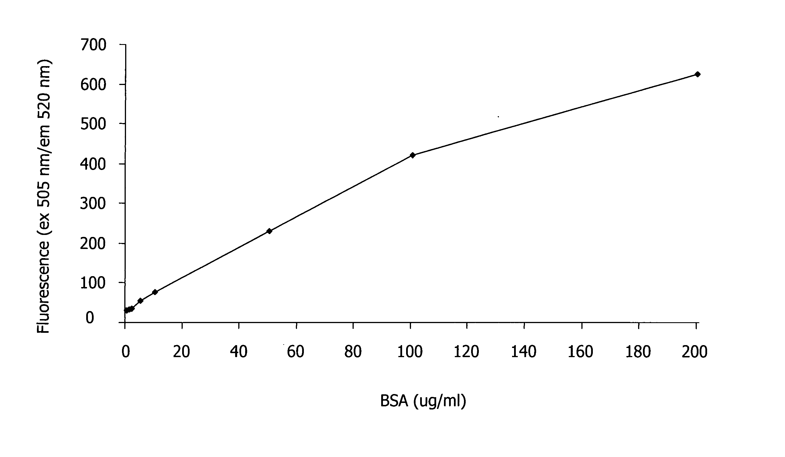
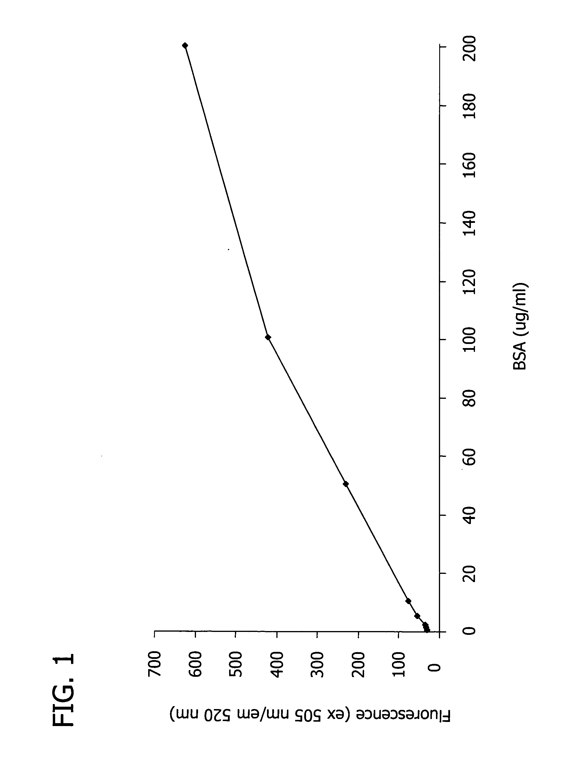
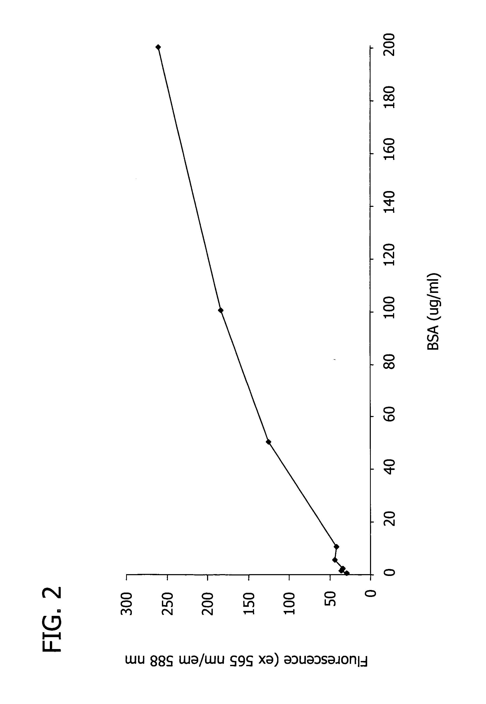
[0127] In this Example, the spectroscopic properties of the trimethincyanine dyes listed in Table 1 were measured, and the theoretical efficiency of the dyes was estimated.

[0128] First, two solutions of each trimethincyanine dye listed in Table 1 were created by adding the trimethincyanine dye to separate solutions of (i) 0.05 M Tris-HCl buffer and (ii) 0.05 M Tris-HCl buffer with 0.05% SDS and 0.2 mg / ml bovine serum albumin (BSA). The concentration of the trimethincyanine dye in each solution was about 1×10⁻⁵ M. The fluorescence of each solution was excited at the maximum wavelength of the fluorescence excitation spectrum and recorded. The efficiency of the trimethincyanine dyes was measured by comparing the spectral-luminescent value of the solution containing dye, buffer, SDS, and BSA (i.e., solution (ii)) with the solution containing only dye and buffer (i.e., solution (i)).

[0129] Table 2 illustrates th...

example 2

Detection of Polyamino Acids on an SDS-PAGE Gel

[0130] In this Example, a sample was electrophoretically separated on an SDS-PAGE gel, the gel was combined with a trimethincyanine dye listed in Tables 1 and 2, and the polyamino acids present in the sample were optically detected with a blue light transilluminator.

[0131] First, a sample buffer (0.0625 M Trizma-HCl (pH 6.75), containing 2% SDS, 5% 2-mercaptoethanol, 10% glycerol and 0.001% bromophenol blue) was prepared according to the method of Lämmli (Nature 227, 680-685 (1970)). To 1 ml of the sample buffer was added 1-10 mg of a mixture of three proteins, bovine serum albumin (BSA) (67 kD), ovalbumin (29 kD), and carboanhydrase (29 kD) (available from Sigma-Aldrich Co., St. Louis, Mo.) to form the sample. The sample was incubated in boiling water for 60 seconds. A portion of the sample was then diluted 20 to 2000 times in sample buffer and loaded onto a 10-20% precast Novex-Gel (EC61352, Invitrogen, Carlsbad, Calif.). A protein ...

example 3

Detection of Polyamino Acids on an SDS-PAGE Gel with an Additional Fixation Step

[0134] In this Example, a sample was analyzed according to the procedure described in Example 2; however, in this Example, following electrophoresis, the gel was incubated in 5% trichloroacetic acid for 30 minutes, and then soaked in a 0.05% SDS solution for 30 minutes.

[0135] Following this additional fixation step, the gel was stained with a staining solution (˜50 ml). The staining solution was prepared by diluting 5000 times in sodium acetate buffer (pH 4.5) a stock solution (5 mg in 1 ml DMSO) of Dye I.D. No. U from Tables 1 and 2.

[0136] The gel was illuminated with an ultraviolet screen and photographed according to the procedure described in Example 2. LOD reached 5-10 ng / band for each of the three proteins in the sample.


PUM

PropertyMeasurementUnit
Fractionaaaaaaaaaa
Fractionaaaaaaaaaa
Fractionaaaaaaaaaa

Abstract

The present invention is generally directed to a method for detecting polyamino acids. More specifically, the present invention is directed to a method for detecting polyamino acids using trimethincyanine dyes that interact non-covalently with polyamino acids to produce an optically detectable dye/polyamino acid complex.

Description

CROSS-REFERENCE TO RELATED APPLICATIONS [0001] This application is a non-provisional of U.S. Provisional Patent Application Ser. No. 60 / 649,257 filed Feb. 1, 2005. The entire text of which is hereby incorporated herein by reference.FIELD OF THE INVENTION [0002] The present invention is generally directed to a method for detecting polyamino acids. More specifically, the present invention is directed to a method for detecting polyamino acids using trimethincyanine dyes that interact non-covalently with polyamino acids to produce an optically detectable dye / polyamino acid complex. BACKGROUND OF THE INVENTION [0003] Detection and subsequent analysis or quantification of polyamino acids is an important step in many applications commonly used in life sciences research. Primarily, polyamino acids are detected and analyzed using known techniques such as separating the polyamino acids by gel electrophoresis or by the electrophoretic transfer of gels containing separated polyamino acids to me...

Claims


Application Information

Patent Timeline
IPC IPC(8): G01N33/00B01D57/02G01N27/00
CPCC09B23/0008C09B23/06C09B23/12G01N27/44726
Inventor KOVALSKA, VLADYSLAVAKRYVOROTENKO, DMYTROLOSYTSKYY, MYKHAYLONORDING, PIERRERUECK, ALEXANDERSCHOENENBERGER, BERNHARDYARMOLUK, SERGIYWAHL, FABIAN
Owner SIGMA ALDRICH CO LLC
Why Eureka
Try Eureka