

Bone repairing material using a chondrocyte having the potential for hypertrophy and a scaffold
- Summary
- Abstract
- Description
- Claims
- Application Information
AI Technical Summary
Benefits of technology
Problems solved by technology
Method used
Image

Examples
example 1
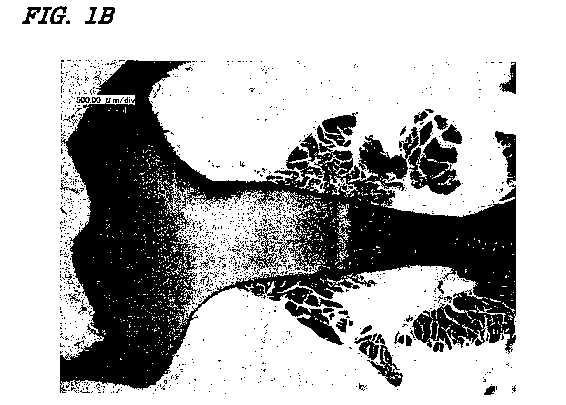
Summary of Example 1 and Comparative Examples 1A-1C
[0217] Osteogenesis was more prominent after the subcutaneous implantation of composite material using chondrocytes having the potential for hypertrophy and a biocompatible scaffold into rats, compared with the pelleted chondrocytes having the potential for hypertrophy alone. In contrast, when the chondrocytes having the potential for hypertrophy or hydroxyapatite were implanted alone, osteogenesis was not observed. These results suggest that the composite material of the present invention can be used to treat bone deficits which are too large to treat with conventional composite materials.
example 2
Effect of Subcutaneous Implantation of a Composite Material Using Chondrocytes having the Potential for Hypertrophy Derived from Sterna and a Biocompatible Scaffold
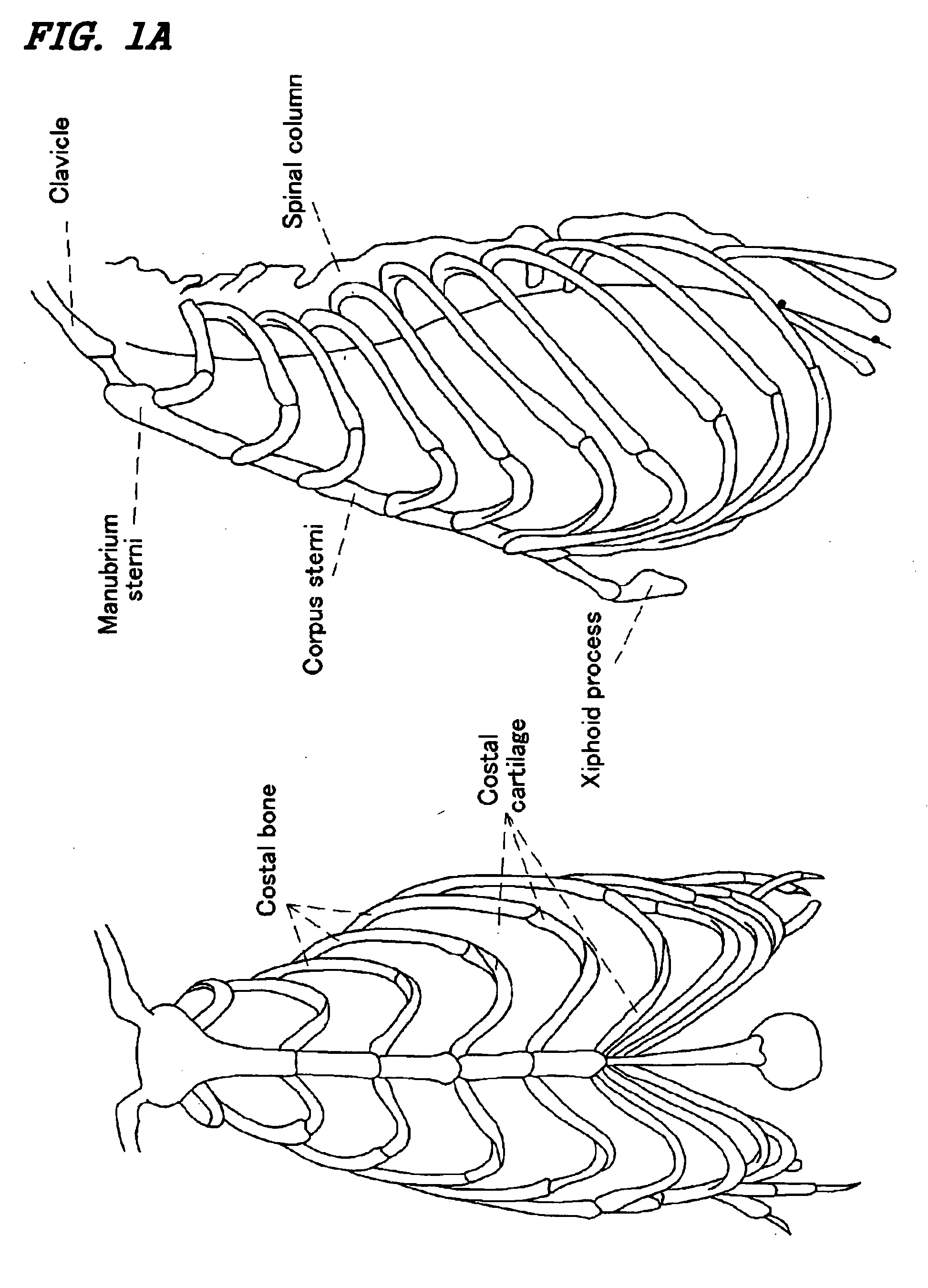
(Preparation of Chondrocytes having the Potential for Hypertrophy from Sterna)
[0218] Male rats (Wistar) that were 4-8 weeks old were sacrificed using chloroform. The rats' chests were shaved using a razor and their whole bodies immersed in Hibitane (10-fold dilution) to be disinfected. The rats' chests were incised and the processus xiphoideus junction located in the inferior portion of the corpus sterni and other regions were removed from sterna aseptically. The translucent growth cartilage region was collected from the processus xiphoideus junction located in the inferior portion of the corpus sterni. These samples were sectioned and incubated in 0.25% trypsin-EDTA / D-PBS at 37° C., with stirring, for 1 hour. The sections were then washed and collected by centrifugation (1000 rpm (170×g)×5 min.) followed by incubation...
example 3
Effect of Implantation of a Composite Material Using Chondrocytes having the Potential for Hypertrophy Derived from Costa / Costal Cartilage and a Biocompatible Scaffold, into a Region of Bone Deficient
(Preparation and Identification of Chondrocytes having the Potential for Hypertrophy from Costa / Costal Cartilage)
[0248] A composite material using chondrocytes having the potential for hypertrophy and a biocompatible scaffold was prepared by a method as described in Example 1.
(Production of Region of Bone Deficient)
[0249] Male rats (Wistar) were anesthetized. Skin in the femoral ortibial region was ablated and soft tissue retracted to one side to expose a femoral or tibial region, or the scalp was ablated to a septically expose the skull. A trephine bar or disc was attached to a dental trephine and used to make perforated or dissecting bone deficiencies at a femoral or tibial region, or in the skull. The composite material prepared above was implanted into the newly-made bone defi...
PUM
| Property | Measurement | Unit |
|---|---|---|
| Temperature | aaaaa | aaaaa |
| Fraction | aaaaa | aaaaa |
| Volume | aaaaa | aaaaa |
Abstract
Description
Claims
Application Information