Wound healing monitoring and treatment

a monitoring device and wound technology, applied in the field of wound healing monitoring and treatment, can solve the problems of inability to determine the efficacy of treatment, and the inability of clinicians to be certain about the condition of the tissue being treated, so as to facilitate quality imaging

Inactive Publication Date: 2006-10-26
CARESTREAM HEALTH INC
View PDF43 Cites 157 Cited by
  • Summary
  • Abstract
  • Description
  • Claims
  • Application Information

AI Technical Summary

Benefits of technology

[0020] Briefly, according to one aspect of the present invention a polarization based diagnostic device for optically examining the medical condition of tissue comprises an illumination optical system, comprising a light source having one or more light emitters, and beam shaping optics, which together provide illumination light to the tissue being examined. An optical detection system comprises imaging optics and an optical detector array detects light from the tissue. Polarizing optics provided in both the illumination optical system and the optical detection system are crossed so as to pass orthogonal polarization states. Iterative rotational means are provided to rotate the orthogonal polarization states relative to the tissue being examined. Image enhancement means includes some combination of image processing, sequential multi-spectral illumination and imaging, and image focus control to facilitate quality imaging at varying depths within the tissue. A controller operates the light source, the detector array, the multi-spectral illumination and imaging, and the image focus control, as well as providing image processing of the captured images to aid the diagnostic process.

Problems solved by technology

In general, the healing of wounds, burns, and other injuries is an uncertain endeavor.
The clinician cannot be certain about the condition of the tissue being treated, the efficacy of treatments, and whether further treatments or a change in treatments is appropriate.
These wounds are particularly intractable for a variety of reasons, with age, nutrition, diabetes, infection, marginalized immune systems, and other factors; all contributing to the ongoing difficulties in healing.
In most cases, such wounds are chronic because the wound healing is stalled relative to one or more aspects of the process.
These traditional methods are disadvantaged by the time delay in evaluating tissue cultures or histology, which easily can be a week or two.
Additionally, these approaches are invasive, and actually cause further damage to the tissue.
These technologies are used for a variety of applications (mammography, brain scans, etc.), but are seldom used for examining soft tissue wounds, and can expose the patient to high energy radiation (x-rays, etc.).
While polarization microscopes work well for examining tissue, such systems are both large and expensive, and require tissue or cellular samples that can be examined in-vitro (on a slide) rather than in-vivo.
Although the fiber optic OCT systems can have a small probe for in-vivo testing, these systems are complicated and expensive, and not likely to be used by a clinician in wound assessment either in the field or in many clinical settings.
This system is designed to provide images of the micro-circulatory vascular network, and is not optimized to examine the collagen network present in the dermal layers of skin.
The Cytoscan system does not provide the proper optical wavelengths, high contrast polarizers, or polarization control to properly examine the collagen networks.
Professor Mester found that a specific range of exposure conditions stimulated cell growth and wound healing, while lesser doses were ineffective and larger doses were inhibitory.
The light therapy devices that are commercially available today are disadvantaged in that the clinician does not know either the optical dosage delivered (light into the tissue) or the effective dosage delivered (light-tissue interaction).
In part, the uncertainty is because many participants are not well educated in optics, and do not know how to measure light properly.
However, the uncertainty is also because the science of light therapy is complicated.
With these uncertainties, the clinician does not really know the efficacy of prior light applications relative to the response of the injured tissue, or whether further light application is appropriate, with what parameters, and for what effects.
fects. However, none or these prior art light therapy devices utilize diagnostic methods or devices that examine the structure and integrity of the extracellular matrix, in particular relative the collagen network, whether with polarized light, or by other

Method used


Image

  • Wound healing monitoring and treatment
  • Wound healing monitoring and treatment
  • Wound healing monitoring and treatment

Examples


Embodiment Construction

[0033] The following is a detailed description of the preferred embodiments of the invention, reference being made to the drawings in which the same reference numerals identify the same elements of structure in each of the several figures.

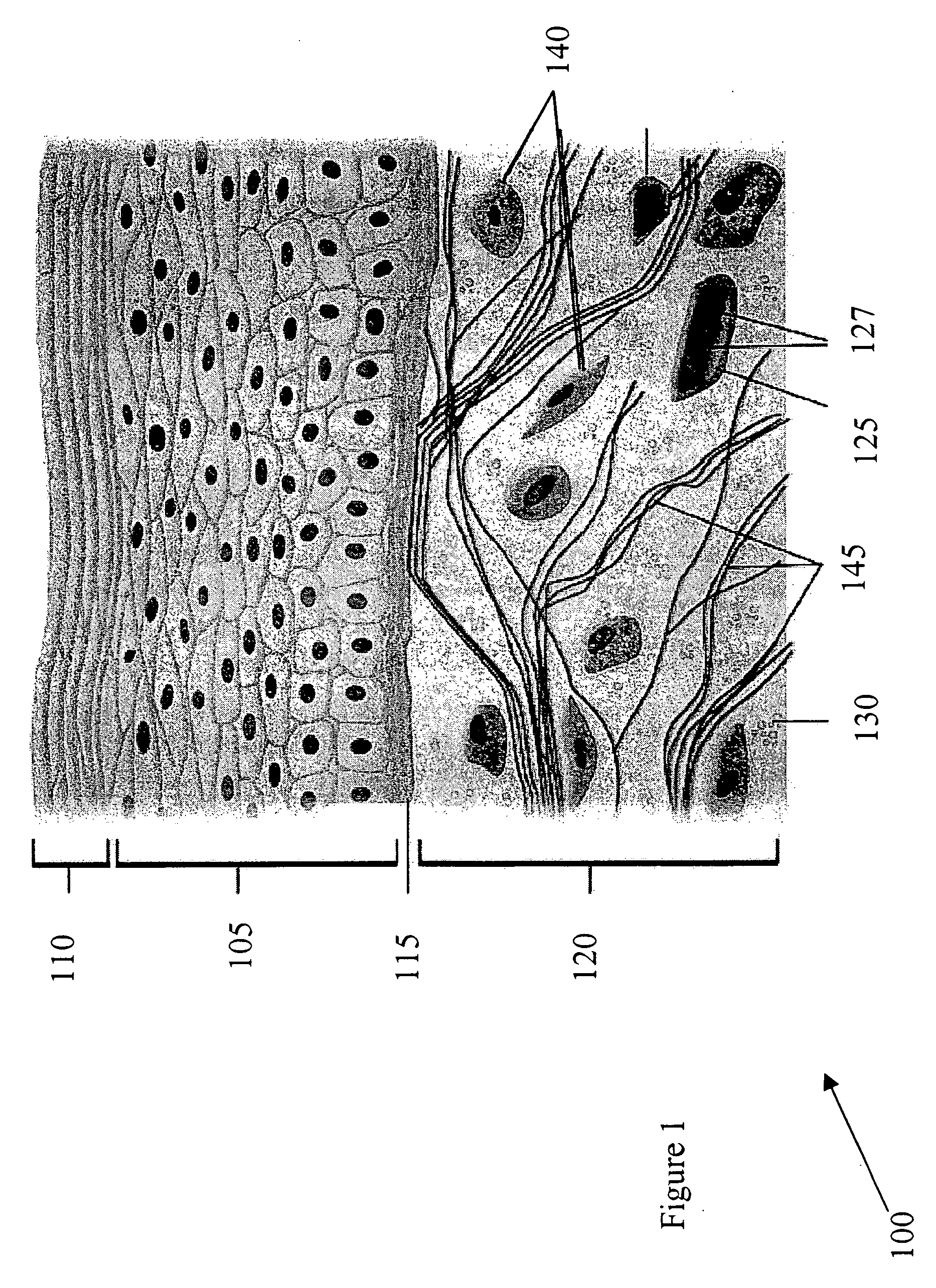
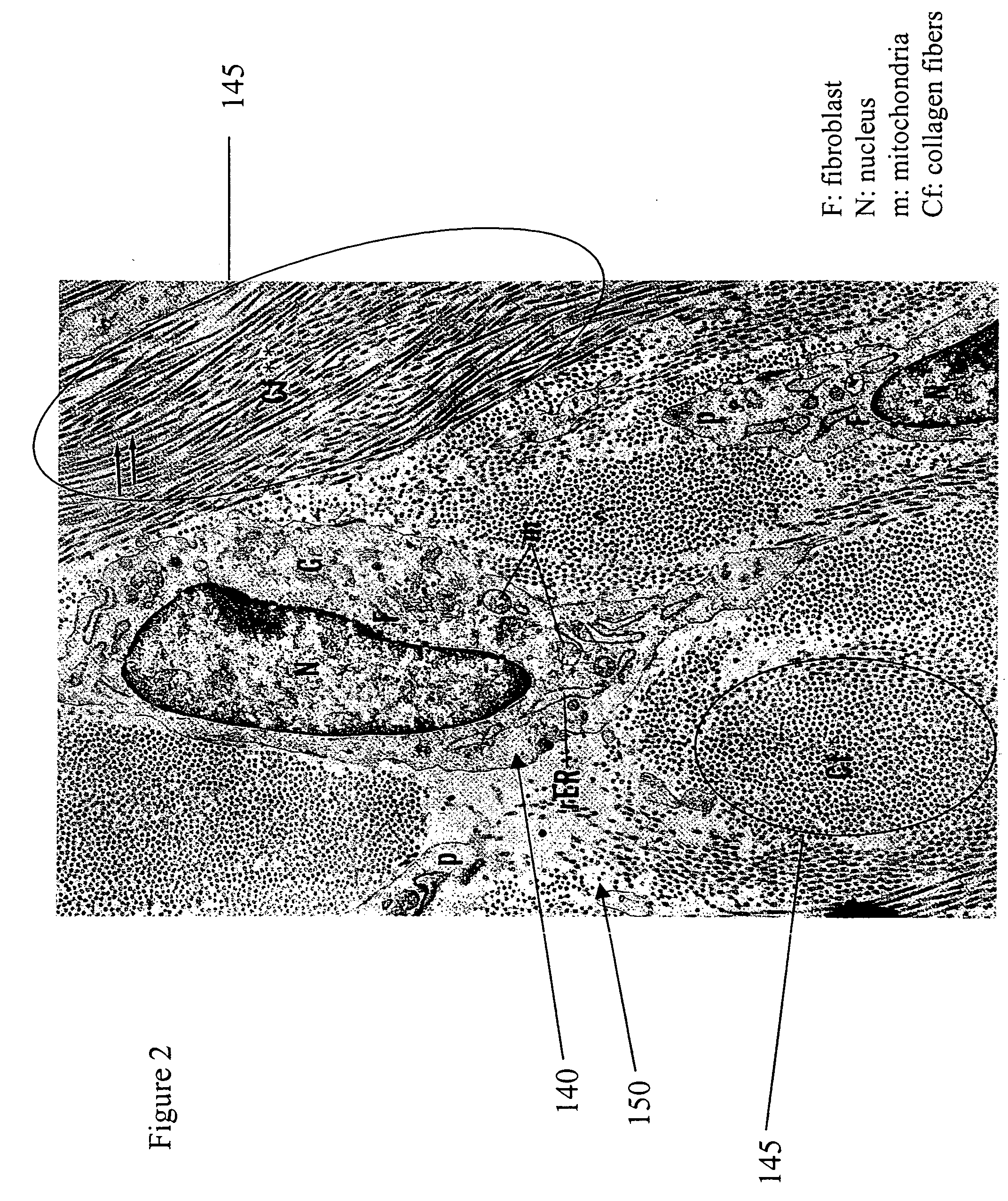
[0034] The present invention can be best appreciated within the context of the biology of normal, wounded, and healed skin, and in particular, with respect to the function of fibroblasts and collagen. Accordingly, FIG. 1 depicts the cross-sectional composition of skin. Skin 100 (or the integument) covers the entire external surface of the human body and consists of two mutually dependent layers, the epidermis 105 and the dermis, which rest on a fatty subcutaneous layer, the panniculus adiposus (not shown). The epidermis 105, which is the outer layer of skin, is made up of epithelial cells (also known as squamous cells or keratinocytes), basal cells, and melanocytes. The outermost layer of the epidermis 105 comprises layers of dead epithelial cells...


PUM


Abstract

A polarization based diagnostic device (200) for optically examining the medical condition of tissue (290) comprises an illumination optical system (205), comprising a light source (220) and beam shaping optics. An optical detection system (210) comprises imaging optics and an optical detector array, which detects light from the tissue. Polarizing optics provided in both the illumination optical system and the optical detection system are crossed and pass orthogonal polarization states. Iterative rotational means rotate the orthogonal polarization states relative to the tissue being examined. Image enhancement means includes image processing, sequential multi-spectral illumination and imaging, and image focus control to facilitate quality imaging at varying depths within the tissue. A controller (215) operates the light source, the detector array, the multi-spectral illumination and imaging, image focus control, and image processing.

Description

FIELD OF THE INVENTION [0001] The invention relates generally to a monitoring device for examining the state of tissues, and in particular a device that examines the condition of collagen structures within tissue using a light based diagnostic device in close proximity with the skin of a patient. The device of the invention is optimally used in cooperation with a light therapy device. BACKGROUND OF THE INVENTION [0002] In general, the healing of wounds, burns, and other injuries is an uncertain endeavor. The clinician cannot be certain about the condition of the tissue being treated, the efficacy of treatments, and whether further treatments or a change in treatments is appropriate. As a particular example, many chronic wounds, such as pressure ulcers or venous stasis ulcers linger for months or even years, often despite the various treatments being applied. These wounds are particularly intractable for a variety of reasons, with age, nutrition, diabetes, infection, marginalized imm...

Claims


Application Information

Patent Timeline
Patent Type & Authority Applications(United States)
IPC IPC(8): A61B6/00
CPCA61B5/0059A61B5/412A61B5/442A61N2005/073A61B5/445A61N5/0616A61B5/444
Inventor KURTZ, ANDREW F.
Owner CARESTREAM HEALTH INC
Why Eureka
Try Eureka