Method and apparatus for noninvasively monitoring parameters of a region of interest in a human body

a non-invasive monitoring and human body technology, applied in the direction of measuring/recording heart/pulse rate, using reradiation, sensors, etc., can solve the problems of fetal death, long-lasting periods of repeated respiratory difficulty, and inability to ensure fhr

Active Publication Date: 2006-11-02
OR NIM MEDICAL
View PDF13 Cites 87 Cited by
  • Summary
  • Abstract
  • Description
  • Claims
  • Application Information

AI Technical Summary

Benefits of technology

[0015] The main idea of the present invention consists of non-invasively monitoring the optical properties of a region of interest in a human (or animal) body utilizing the principles of ultrasound tagging of light, which in the present invention is aimed at distinguishing between optical responses of the region of interest in the selected volume (e.g., fetus, amniotic fluid, blood vessel) and the surroundings outside the region of interest; and / or significantly improving pulse oximetry based measurements.
[0028] a measurement unit comprising an optical unit having an illumination assembly configured to define at least one output port for illuminating light, and a light detection assembly configured to define at least one light input port for collecting light scattered from the illuminated body portion and to generate measured data indicative of the collected light; and an acoustic unit configured to generate acoustic waves of a predetermined ultrasound frequency range; the measurement unit being configured and operable to provide an operating condition such that the acoustic waves of the predetermined frequency range overlap with an illuminating region within the region of interest and substantially do not overlap with a region outside the region of interest, and that the detection assembly collects light scattered from the region of interest and light scattered from the region outside the region of interest, the measured data being thereby indicative of scattered light having both ultrasound tagged and untagged light portions, thereby enabling to distinguish between light responses of the region of interest and the region outside the region of interest;
[0039] According to yet another aspect of the invention, there is provided a method for use in noninvasive monitoring at least one parameter of a region of interest in a human body, the method comprising: operating an optical unit and an acoustic unit so as to provide that ultrasound waves of a predetermined frequency range and illuminating light overlap within the region of interest and substantially do not overlap with a region outside the region of interest, thereby producing measured data indicative of collected light including scattered light having ultrasound tagged and untagged light portions, thereby enabling extraction of a light response of the region of interest from all the other light portions in the collected light.
[0044] operating the optical and acoustic units, when in the optimal positioning, and generating measured data indicative of collected light including scattered light having ultrasound tagged and untagged light portions, thereby enabling extraction of a light response of the region of interest from all the other light portions in the collected light.
[0049] According to yet another aspect of the invention, there is provided a method for operating a monitoring system configured for noninvasive monitoring at least one parameter of a region of interest in a human body, which system comprises an optical unit and an acoustic unit configured to generate acoustic waves of a predetermined ultrasound frequency range, the method comprising: operating the monitoring system to provide an optimal positioning of the optical and acoustic units with respect to each other and with respect to the region of interest to satisfy an operating condition for measurements, said operating condition resulting in that the ultrasound waves of the predetermined frequency range overlap with illuminating light generated by the optical unit within the region of interest, while substantially not overlapping in a region outside the region of interest, and in that a detection assembly of the optical unit collects light scattered from the region of interest and from the region outside the region of interest, thereby obtaining measured data indicative of scattered light having ultrasound tagged and untagged light portions, and enabling extraction from said measured data a data portion indicative of a light response of the region of interest.

Problems solved by technology

However, a non-reassuring FHR is not always associated with the delivery of a compromised infant.
If the baby is delivered and the lungs are not mature, the baby may develop Respiratory Distress Syndrome (RDS), which can result either in fetal death or in long-lasting periods of repeated respiratory difficulty.
Although it is the clinical “gold standard” method, it remains a time-consuming process, has a large intralaboratory and interlaboratory coefficient of variation, and requires expertise.
In addition, the procedure of amniotic fluid drainage itself is invasive and suffers a small risk of abortion.
In cases where the fetus gasps during delivery, inhaling the sticky meconium into the upper respiratory tract results in partial airways obstruction.
However, in cases where the fetus head is tightly fitted in the pelvis, the amniotic fluid is not drained out resulting in misdiagnosis of the potential harmful outcome to the respiratory tract.

Method used


Image

  • Method and apparatus for noninvasively monitoring parameters of a region of interest in a human body
  • Method and apparatus for noninvasively monitoring parameters of a region of interest in a human body
  • Method and apparatus for noninvasively monitoring parameters of a region of interest in a human body

Examples


Embodiment Construction

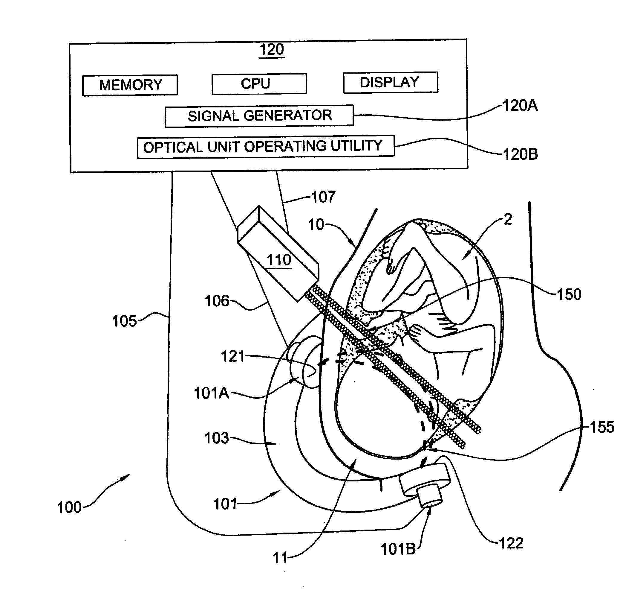
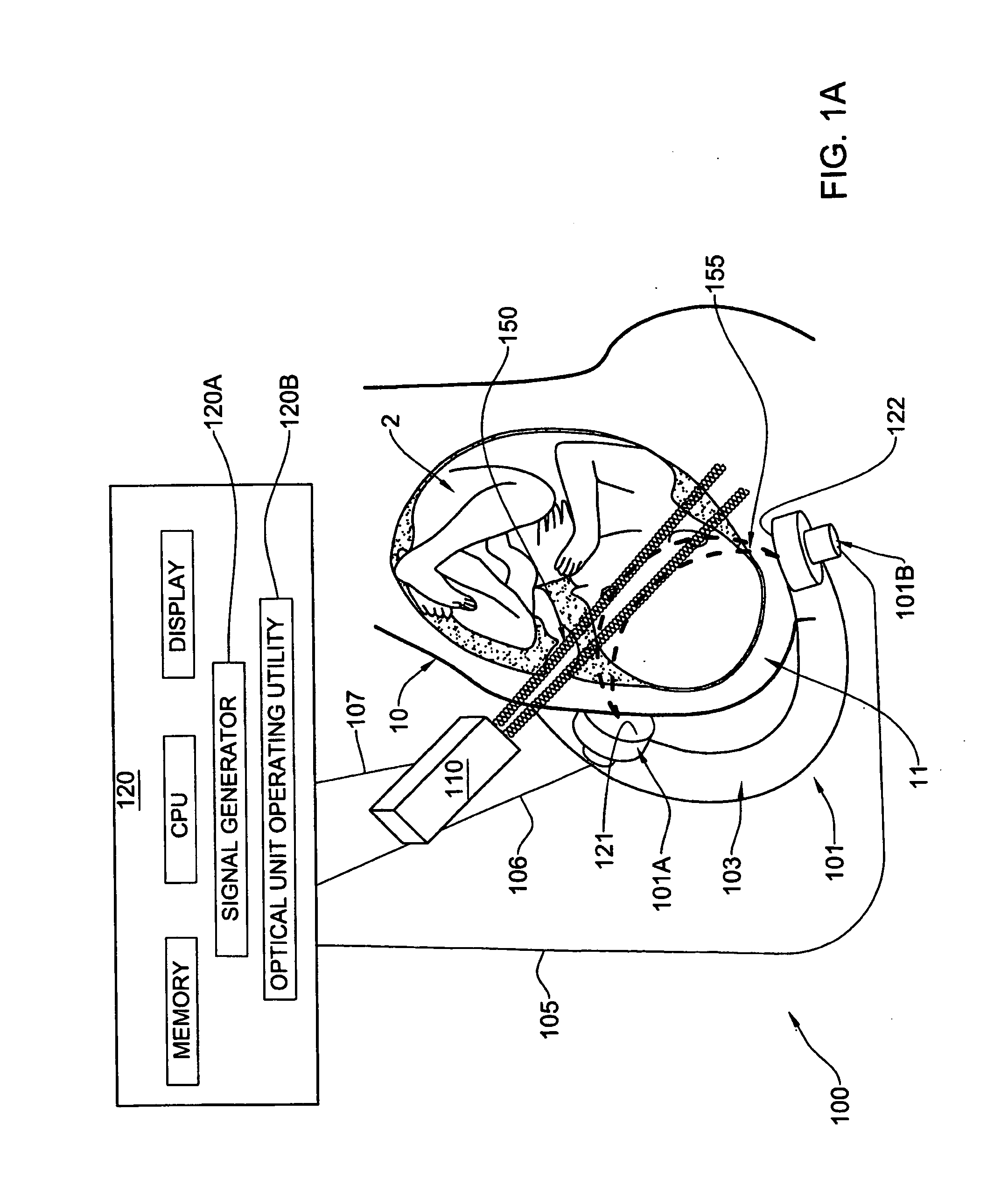
[0063] Referring to FIG. 1A, there is schematically illustrated a monitoring apparatus, generally designated 100, constructed and operated as a fetal oxygen saturation monitor according to the invention. It should, however, be understood that the apparatus configuration is suitable for measuring various other parameters of a fetus 2 (such as the concentration of an analyte in the fetal blood, or the perfusion of an analyte / metabolite in fetal or maternal tissues). It should be understood that the apparatus of the present invention may be used for monitoring blood or tissue parameters of a human being.

[0064] The apparatus 100 includes such main constructional parts as a measurement unit formed by an optical unit 101 including an illumination assembly 101A and a light detection assembly 101B; and an acoustic unit including a transducer arrangement 110. In the present example of FIG. 1A, the detection assembly includes a single detection unit. In this connection, it should be noted th...


PUM


Abstract

A method and system for noninvasive monitoring of at least one parameter of a region of interest in a human body. The system comprises a measurement unit and a control unit. The measurement unit comprises an optical unit having an illumination assembly (101A) and a light detection assembly (101B) to generate measured data indicative of collected light; and an acoustic unit (110) configured to generate acoustic waves of a predetermined ultrasound frequency range. The measurement unit provides an operating condition such that the acoustic waves overlap with an illuminating region within the region of interest and substantially do not overlap outside the region of interest. The measured data is indicative of scattered light having both ultrasound tagged and untagged light portions, enabling to distinguish between light responses of the region of interest and the region outside the region of interest.

Description

FIELD OF THE INVENTION [0001] This invention relates to a method and apparatus for noninvasive monitoring of parameters of a region of interest in a human body, such as oxygen saturation and / or concentration of analyte(s) in blood. BACKGROUND OF THE INVENTION [0002] Monitoring of the well-being of the fetus inside the uterus is very important and is carried periodically with respect to various parameters of the fetus. One of the important parameters to be monitored is oxygen saturation. Various techniques have been developed to enable noninvasive measurements of oxygen saturation. [0003] For example, U.S. Pat. No. 5,494,032 discloses an oximeter for reliable clinical determination of blood oxygen saturation in a fetus. This technique utilizes a multiple frequency light source which is coupled to an optical fiber. The output of the fiber is used to illuminate blood containing tissue of the fetus. The reflected light is transmitted back to the apparatus where the light intensities are...

Claims


Application Information

Patent Timeline
Patent Type & Authority Applications(United States)
IPC IPC(8): A61B5/00A61BA61B5/024A61B5/08
CPCA61B5/0097A61B5/02411A61B5/08A61B5/14542A61B5/7264A61B2562/0233A61B2562/043G01S15/8968A61B5/1464
Inventor BALBERG, MICHALPERY-SCHECHTER, REVITALOLSHANSKY, MICHAL
Owner OR NIM MEDICAL
Why Eureka
Try Eureka