
Method and apparatus for the detection of a bone fracture

a technology of bone fracture and detection method, applied in the field of ultrasonic detection system, can solve the problems of inability to reliably determine the accuracy of x-ray evaluation, and inability to detect abnormalities by laypeople, so as to increase the likelihood of detecting a true fracture, enhance sensitivity, and significant more simple

Inactive Publication Date: 2007-02-22
GOEPP JULIUS G +1
View PDF67 Cites 32 Cited by
  • Summary
  • Abstract
  • Description
  • Claims
  • Application Information

AI Technical Summary

Benefits of technology

[0036] As used in this specification, the term Fourier transform refers to a mathematical operation that results in the decomposition of a time series signal into harmonics of different frequencies and amplitudes. The Fourier transform itself is a substantially lengthy calculation to compute when analyzing real-time signals. For that reason, as used in this specification, the Fast Fourier Transform FFT refers to a simpler calculation which is substantially advantageous. FFT allows a sequence of time-domain samples to be efficiently converted into a frequency representation using a previously-specified discrete time window. The FFT generates the frequency power spectra, allowing the processor to monitor the relative magnitudes of various components of a signal under inspection. The processed signal may be exploited over time to detect small changes in the frequency content of the real-time signals that correspond on the one hand to normal structures and on the other to fractures and bone diseases.
[0039] The techniques described herein are advantageous because they are inexpensive and significantly more simple compared to prior art approaches. The techniques described herein are also advantageous because they increase the likelihood of detecting a true fracture (enhanced sensitivity) and decrease the likelihood of a false-positive identification (enhanced specificity), compared with prior art approaches. Additionally, the techniques of the invention are advantageous because they provide a range of alternatives, each of which is useful in appropriate situations and which may be used to cross-check one another for accuracy.

Problems solved by technology

In such cases, the X-ray evaluation was unnecessary.
There is currently no reliable method for an accurate determination by a layperson of the likelihood that an injury involves a fracture.
Other single-purpose, portable, and inexpensive ultrasound units are sold for layperson use, such as detecting and listening to fetal heart sounds, but such units are not intended for detecting abnormalities.
While all of these devices are useful in their intended applications of providing information about soft tissue structure and function, the characteristics of ultrasound make it unsuitable for high-quality diagnostic images of bone.
Thus, medical technology currently uses significantly more expensive, cumbersome, and potentially dangerous test methods, such as X-ray analysis, to identify acute structural changes in bone, such as those that appear in fractures or intrinsic bone lesions.
Simple application of any of these existing technologies is inadequate for the purpose described herein.
This degree of complexity would make such a device cumbersome and unreliable.
Several prior art devices have been designed to incorporate features of ultrasonography into the determination of bone structure and condition in patients either at risk for or with known fractures or bone diseases, but to date, no approach has addressed the simple detection of previously unidentified fractures or other bone lesions.
This is a useful technique for determining the degree of bone mineralization and degree of osteoporosis and hence, by implication, risk of future fracture, but it does not and is not intended to diagnose actual fracture in any bone.
The teachings of Chiabrera are deficient in that they cannot be modified to detect existing bone fractures.
For example, and as disclosed in U.S. Pat. No. 4,655,228 to Shimura (Ultrasonic Diagnosis Apparatus for Tissue Characterization) ultrasonic diagnostic devices are generally adapted to observe differences in soft-tissue morphology and are unsuitable for use with bone.
To date, there is no device that permits the simple detection, as opposed to diagnosis, of a bone fracture by a layperson.
The assembly of Hascoet is deficient in that it cannot be modified to be used by a layperson.
Moreover the device of Hascoet cannot be modified to obtain a hand-held device, nor can it be used for primary detection of a suspected fracture.

Method used


Image

  • Method and apparatus for the detection of a bone fracture
  • Method and apparatus for the detection of a bone fracture
  • Method and apparatus for the detection of a bone fracture

Examples


example 1

[0115] Two calcium impregnated tiles were placed next to one another such that a gap of approximately 5 mm was present between such tiles. This gap was then filed with Aquaflex brand ultrasound gel pad. Additional gel was placed over the tiles such that a substantially flat surface of gel was present over both tiles as well as the gap. Aquasonic brand coupling gel was placed over this surface. A Panametrics-NDT 20 MHz, 0.125″ ultrasonic transducer was placed in contact with the surface of the gel over the tile and moved from the starting tile, over the gap, and over the second tile. A JSR DPR300 Ultrasonic Pulser / Receiver was used to control the transducer. The received signal was transmitted from the transducer to a personal computer with the assistance of a DP308 Digitizer PCI interface card available from Acqiris. The results of this experiment are shown in FIG. 14.

[0116] As shown in FIG. 14, primary waveform 1402 is the reflected ultrasonic signal currently being sensed by the ...

example 2

[0117] An artificial bone manufactured by Sawbones was encased in Blue Phantom brand gel 1504. This gel is designed to closely approximate the average ultrasonic characteristics of human flesh. X-ray image 1506 shows an image of the bone 1508, an image of the gel 1504A, and an image of the bone fracture 1510. Ultrasonic transducer 1502 was placed on the surface of gel 1504 after coating gel 1504 with coupling medium (not shown). When the probe is placed over un-traumatized region 1512, a first signal was generated. When the probe is placed over traumatized region 1514, a second signal was generated. The frequency of the maximum return signal varied between approximately 9 and 10 MHz while the transducer was over un-traumatized region 1512. The frequency of the maximum return signal was consistently greater than 11.5 MHz while the transducer was disposed over traumatized region 1514. The threshold condition in the test device was configured such that that a maximum return signal less...


PUM


Abstract

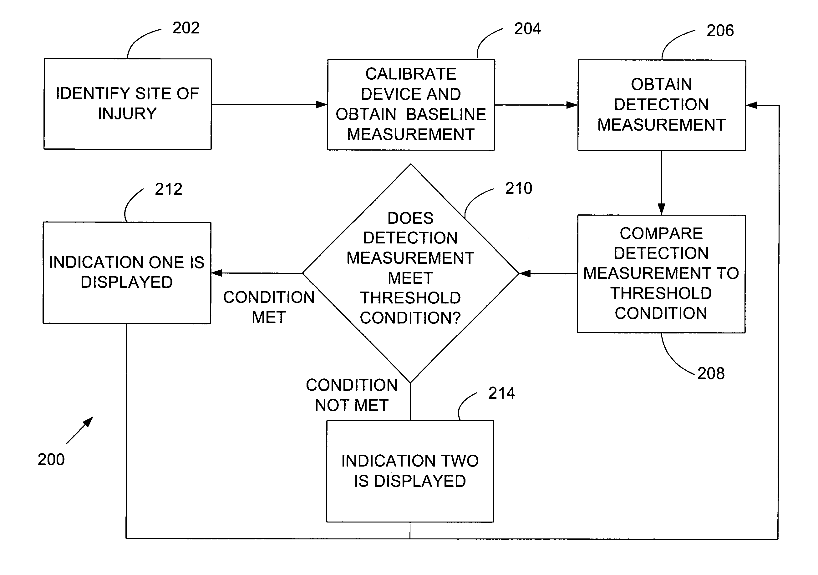
Disclosed in this specification is a device configured to detect fractures in a bone by reflecting waves off of the bone. Certain parameters of the reflected wave are compared to a threshold condition. When the threshold condition is met, a first indication is generated. When the threshold condition is not met, a second indication is generated. This device allows detection of bone fractures without requiring that the user of the device be skilled in image interpretation (e.g. interpreting x-ray or ultrasound images).

Description

CROSS-REFERENCE TO RELATED PATENT APPLICATIONS [0001] This application claims priority from applicant's co-pending patent application U.S. Ser. No. 60 / 704,990 (filed Aug. 2, 2005). The content of the aforementioned patent application is hereby incorporated by reference into this specification.FIELD OF THE INVENTION [0002] This invention relates to ultrasound detection systems, more specifically to a short-range and inexpensive ultrasound system for layperson use in detecting bone and / or tissue irregularities in an injured limb that may have a fracture or other abnormality. BACKGROUND OF THE INVENTION [0003] Hundreds of thousands of X-ray evaluations of injured bones are conducted each year in hospitals and clinics for the purpose of determining if a bone has been broken in an injury. The vast majority of these evaluations reveal normal bone, and the injury in such cases is labeled as a soft-tissue, usually trivial injury. In such cases, the X-ray evaluation was unnecessary. There is...

Claims


Application Information

Patent Timeline
IPC IPC(8): A61B8/00
CPCA61B5/6843A61B8/0875A61B8/4281A61B8/4455A61B5/7239
Inventor GOEPP, JULIUS G.HOYT, ZACHARY M.
Owner GOEPP JULIUS G
Learn More