
In vitro erythroid micronucleus assay for genotoxicity
a technology of erythroid micronucleus and genotoxicity, which is applied in the field of in vitro erythroid micronucleus assay for genotoxicity, can solve the problems of insufficient sensitivity of erythroid micronucleus, inability to accurately predict clinical outcome, and inability to accurately reflect in vivo response. , to achieve the effect of predicting clinical outcome more accurately and/or supply
- Summary
- Abstract
- Description
- Claims
- Application Information
AI Technical Summary
Benefits of technology
Problems solved by technology
Method used
Image

Examples
example 1
In Vitro Micronucleus Assay for Genotoxicity
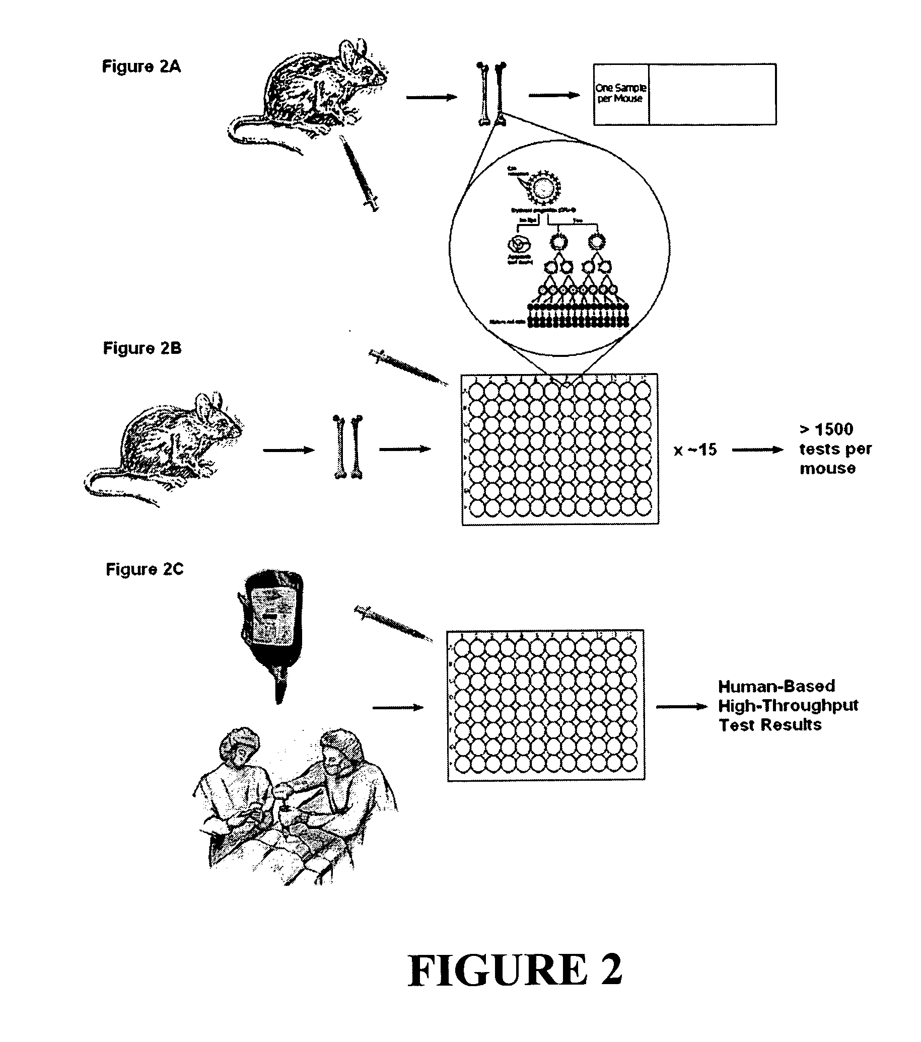
[0128] We have now demonstrated that culturing the lineage-marker negative (Lin−) fraction of the bone marrow (BM) from a single mouse under controlled conditions provides sufficient erythropoietic growth to test over 1500 conditions for a genotoxic response.
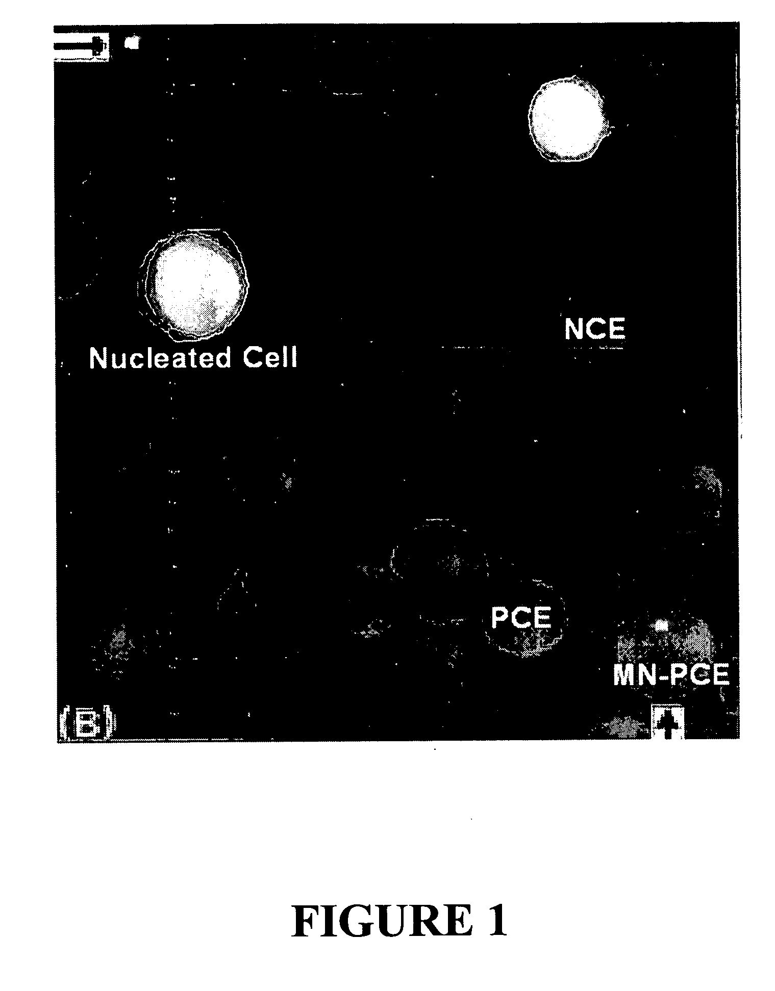
[0129]FIG. 1 shows a micrograph of a purified BM sample that has been stained with acridine orange (AO) and visualized using fluorescence microscopy. AO emits near 515 nm (green) when intercalated in double-stranded (ds) nucleic acid (mostly dsDNA) and near 630 nm (red) when intercalated in single-stranded nucleic acid (mostly RNA). Shown in the image are nucleated cells (green / yellow), NCEs (khaki / green because they are mostly devoid of nucleic acid), and PCEs (bright orange / red mostly due to the presence of ribosomes and mRNA). Also shown is a MN-PCE, which is a newly-formed erythrocyte that contains the remnants of prior genetic damage, either clastogenic or aneugenic, and which i...
example 2
Culture Conditions for Erythroid Growth
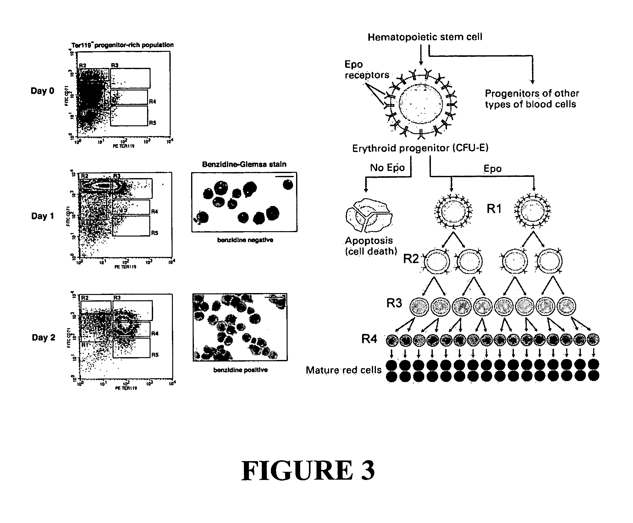
[0147] The flow cytometric techniques described in Example 1 were originally developed for E14.5 fetal liver, and developing an analogous culture system from adult erythroid progenitors required a modified starting population. While approximately 41 percent of R1 cells in FL are CFU-Es, R1 cells in BM contain the committed progenitors and differentiated progeny of a variety of hematopoietic lineages. Fortunately, a detailed knowledge of the cell-surface markers of murine CFU-Es exists (Terszowski, Waskow et al. 2004).
[0148] To develop improved culture technology, the potential of Ter-119− BM and Lin− BM, obtained from C57BL / 6J mice, to yield PCEs in culture was examined (see Tables I and II, and FIGS. 11, 12, 14, and 16). Initial studies revealed that either population, when cultured in the presence of Epo for approximately 72 hours, could be induced to undergo some degree of terminal erythropoiesis. However, Lin− mouse BM displayed greater s...
example 3
Effect of MGMT Expression on the BCNU Response
[0166] BCNU is an SN1 alkylating agent that can form adducts at several nucleophilic sites on DNA, including the O6 position of Guanine (Singer, B. et al. Nature 276, 85-88 (1978); Bodell, W. J. Chem Res Toxicol 12, 965-970(1999); Ludlum, D. B. Mutat Res 233, 117-126 (1990)). After this initial addition reaction, the alkyl group on the modified DNA base can react a second time to form an interstrand crosslink, but the alkyl group can also be removed by an alkyl transferase known as O6-methylguanine DNA methyltransferase (MGMT) to repair the DNA (Kohn, K. W Cancer Res 37, 1450-1454 (1977); Samson, L. & Cairns, J Nature 267, 281-283 (1977)). When MGMT− / − mice (on C57BL / 6J background) were treated with intermediate doses of BCNU by intraperitoneal injection and were then examined by the in vivo MN test 24 h later, it was found that the MN frequency in PCEs was significantly lower than that observed in wild-type C57BL / 6J mice (FIG. 17a). Ho...
PUM
| Property | Measurement | Unit |
|---|---|---|
| Time | aaaaa | aaaaa |
| Volume | aaaaa | aaaaa |
| Time | aaaaa | aaaaa |
Abstract
Description
Claims
Application Information