Methods for fusion polypeptide delivery into a cell

a polypeptide and cell technology, applied in the direction of peptide/protein ingredients, drug compositions, immunological disorders, etc., can solve the problem of limiting in vivo utility, achieve high cell or tissue specificity, minimize or avoid systemic side effects, and limit in vivo utility

Inactive Publication Date: 2007-05-10
FORHUMANTECH CO LTD
View PDF14 Cites 25 Cited by
  • Summary
  • Abstract
  • Description
  • Claims
  • Application Information

AI Technical Summary

Benefits of technology

[0012] Another preferred embodiment is the use of the cytoplasmic domain of CTLA-4 (SEQ ID NO:11) specific to the activated T cells as a cargo protein for PTD. Use of the PTD-ctCTLA-4 fusion protein overcomes the lack of tissue specificity of the cationic PTDs, which limits their in vivo utility.
[0013] Our invention enables the development of new therapeutic protei...

Problems solved by technology

Use of the PTD-ctCTLA-4 fusion protein overcomes the lack of tiss...

Method used


Image

  • Methods for fusion polypeptide delivery into a cell
  • Methods for fusion polypeptide delivery into a cell
  • Methods for fusion polypeptide delivery into a cell

Examples


example 1

Production of Expression Vectors

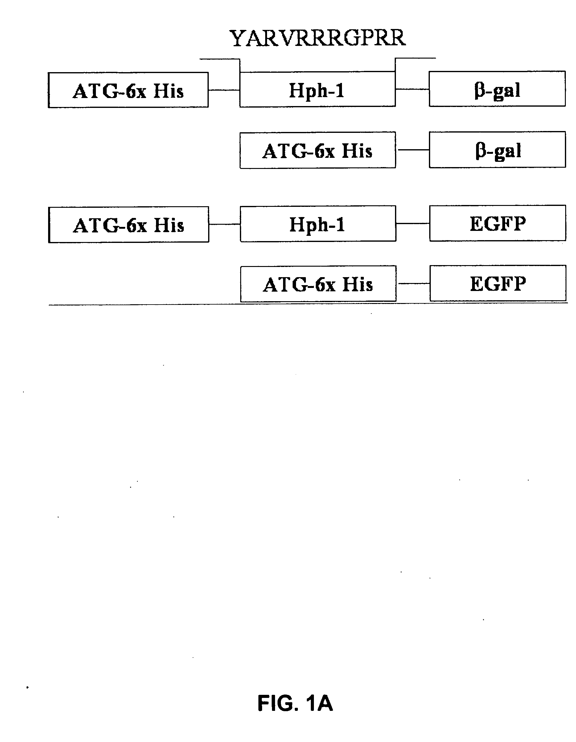
[0121] Based on common structural and functional features between TAT and uptake-targeting sequences which direct proteins from the cytosol to the intracellular organelles, an Hph-1 protein transduction domain (PTD) of 11 amino acids (YARVRRRGPRR) (SEQ ID NO:1) was identified from human transcriptional factor Hph-1. The terms “Hph-1-PTD” and “Hph-1” refer to this 11 amino acid sequence and are interchangeable.

[0122] Generation of Hph-1-PTD-ctCTLA-4, Hph-1-PTD-β-gal, Hph-1-PTD-ctCTLA-4-eGFP, and the corresponding genes not fused with Hph-1-PTD, was accomplished by generating PCR fragments and inserting each PCR fragment into the pRSET-B vector (Invitrogen) (FIG. 1A).

example 2

Preparation of the Recombinant Proteins

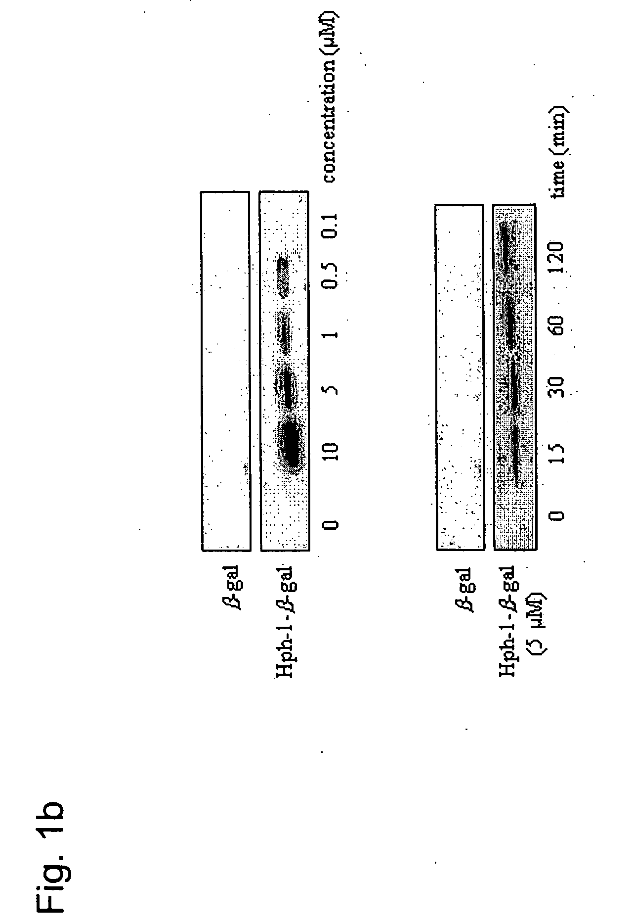
[0123] Site-directed mutagenesis was conducted to create the CTLA-4YF mutant. The fidelity of the reading frame was verified by sequencing. BL21 star (Novagen) transformed with each DNA construct was induced for 5 hours with 1 mM IPTG and sonicated in lysis buffer (6 M urea, 20 mM Tris-HCl, pH 8.0 and 500 mM NaCl). Lysates were clarified by centrifugation (20,000 r.p.m. for 30 min at 20° C.), adjusted to 10 mM imidazole (Tris-HCl, pH 8.0) and loaded on HisTrap chelating columns (Qiagen). Bound proteins were eluted with 50, 100, 250 mM or 3M imidazole in Tris-HCl buffer with >95% purity. The recombinant proteins contained in each fraction were desalted on PD-10 Sephadex G-25 (Amersham Pharmacia). The purified proteins were supplemented with 10% glycerol, aliquoted and flash-frozen at −80° C. (Becker-Hapak M., et al., Methods 24:247-256 (2001)).

example 3

Analysis of the In Vitro and In Vivo Transduction Efficiency of Hph-1-PTD

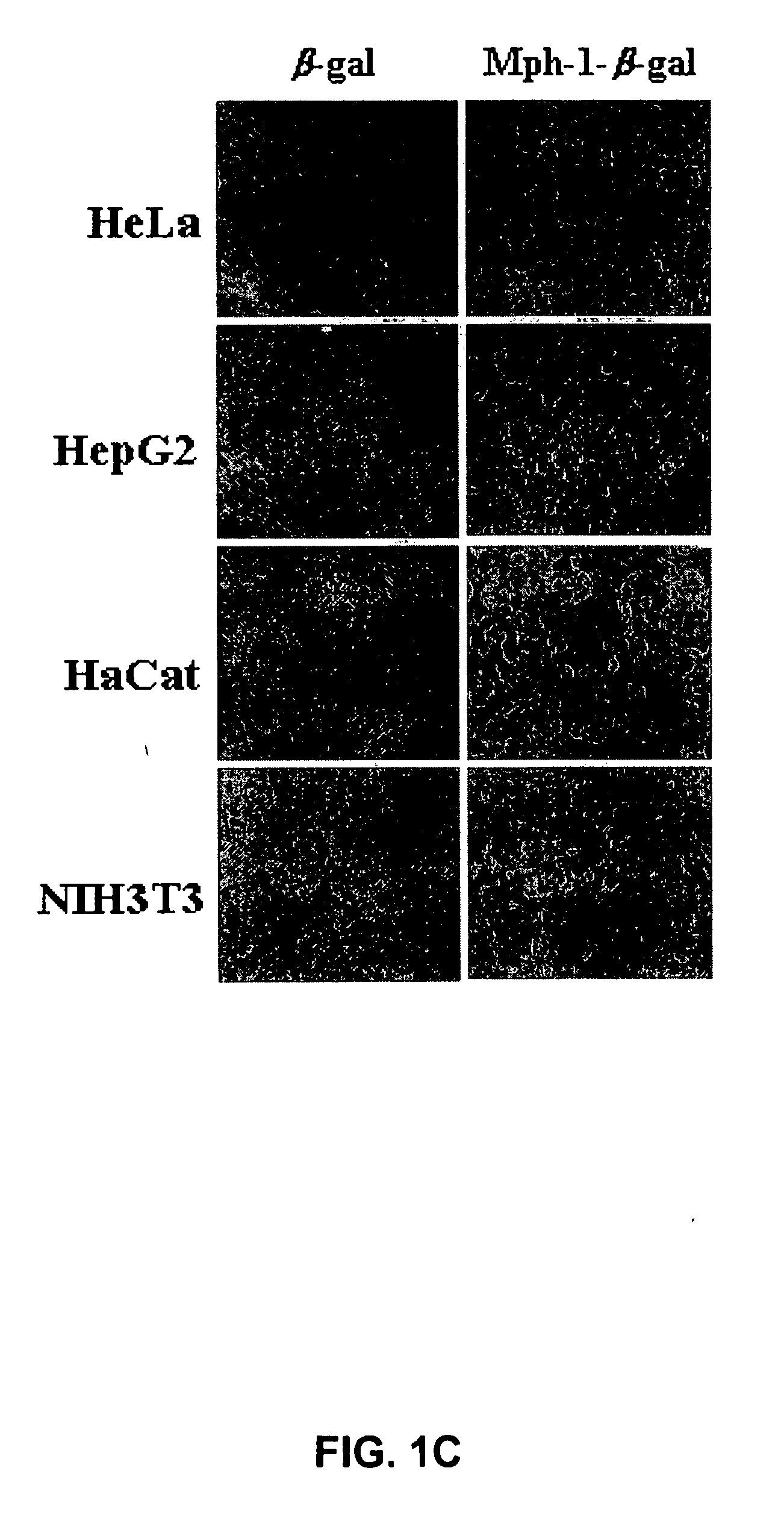
[0124] After the Hph-1-β-gal construct was transduced into each cell line for 1 hour, the cells were washed with PBS and fixed immediately. Then, the fixed cells were incubated with X-Gal solution for 1-3 hours at 37° C. The mice tissue samples from the liver, kidney, heart, lung, spleen and brain were isolated at 12 hours after i.p. injection of Hph-1-β-gal, sectioned (in 10- and 50-μm sections), and assayed using an X-Gal staining kit (Roche) (Schwarze S. R., et al., Science 285:1569-1572 (1999)). The image was captured by fluorescence microscopy (Nikon). Transduction capacity of Hph-1-β-gal was also analyzed by western blot analysis. Equal numbers of cells were solubilized in SDS-PAGE sample buffer and the proteins were resolved on a 12% gel, and blotted onto PVDF, which was probed with monoclonal antibody (1:1,000) to β-gal (Cell Signaling). Bound antibody was detected using horseradish peroxidase-conjugat...


PUM

PropertyMeasurementUnit
Fractionaaaaaaaaaa
Concentrationaaaaaaaaaa

Abstract

The present invention relates to methods for delivery of fusion polypeptides into cells. Methods are provided for local delivery of fusion polypeptides, e.g., through the skin, eye and the airway, to prevent allergic inflammation, airway hyper-responsiveness and to block T cell activation. Methods for delivery of fusion polypeptides to suppress graft rejection are also provided.

Description

CROSS REFERENCE TO RELATED APPLICATIONS [0001] This application claims the benefit of the filing date of U.S. Provisional Patent Application No. 60 / 733,183, filed Nov. 4, 2005, which is incorporated herein by reference in its entirety.STATEMENT REGARDING FEDERALLY-SPONSORED RESEARCH AND DEVELOPMENT [0002] Not applicable. BACKGROUND OF THE INVENTION [0003] 1. Field of the Invention [0004] The present invention relates to methods for delivery of a biomolecule transduction complex (BTC), such as a fusion polypeptide, into cells. The biomolecule transduction complex (BTC) comprises a protein transduction domain (PTD) and a molecule of interest, such as a polypeptide. [0005] 2. Background Art [0006] Generally, living cells are not permeable to macromolecules such as proteins and nucleic acids. The fact that only small-size molecules can permeate through the membrane of living cells at very low rates has restricted the research to develop drugs to cure, prevent, or diagnose diseases using...

Claims


Application Information

Patent Timeline
IPC IPC(8): A61K38/17
CPCC07K14/70521A61K38/00A61P11/06A61P17/00A61P19/02A61P25/00A61P29/00A61P37/02A61P37/06A61P37/08A61P43/00A61K38/16
Inventor LEELEE, SEUNG-KYOUCHOI, JE-MIN
Owner FORHUMANTECH CO LTD
Why Eureka
Try Eureka