Method to assay test substances

a technology of assay and test substance, applied in the field of membrane protein biogenesis, can solve the problems of optic detection and photobleaching methodology limiting the applicability of certain areas, and the applicability of the approach is not applicabl

Active Publication Date: 2007-05-17
THE JOHN HOPKINS UNIV SCHOOL OF MEDICINE
View PDF0 Cites 6 Cited by
  • Summary
  • Abstract
  • Description
  • Claims
  • Application Information

AI Technical Summary

Problems solved by technology

However, for proteins with limited or no glycosylation, this approach is not applicable.
However, the combination of optic detection and the photobleaching methodology limits its applicability in certain areas, especially evaluation of global surface expression and coupling the imaging signal to functionality of targeted molecules.

Method used


Image

  • Method to assay test substances
  • Method to assay test substances
  • Method to assay test substances

Examples


example 1

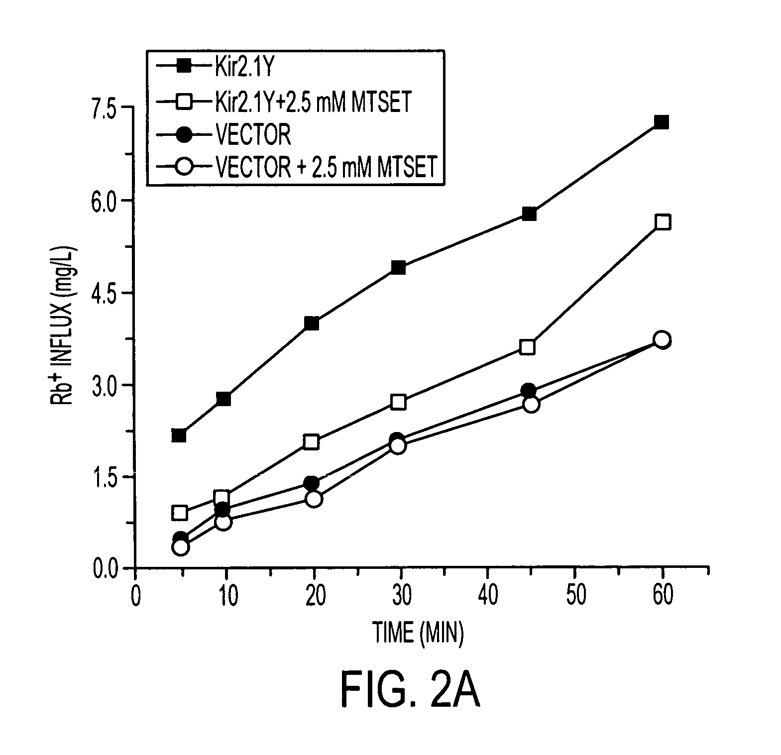
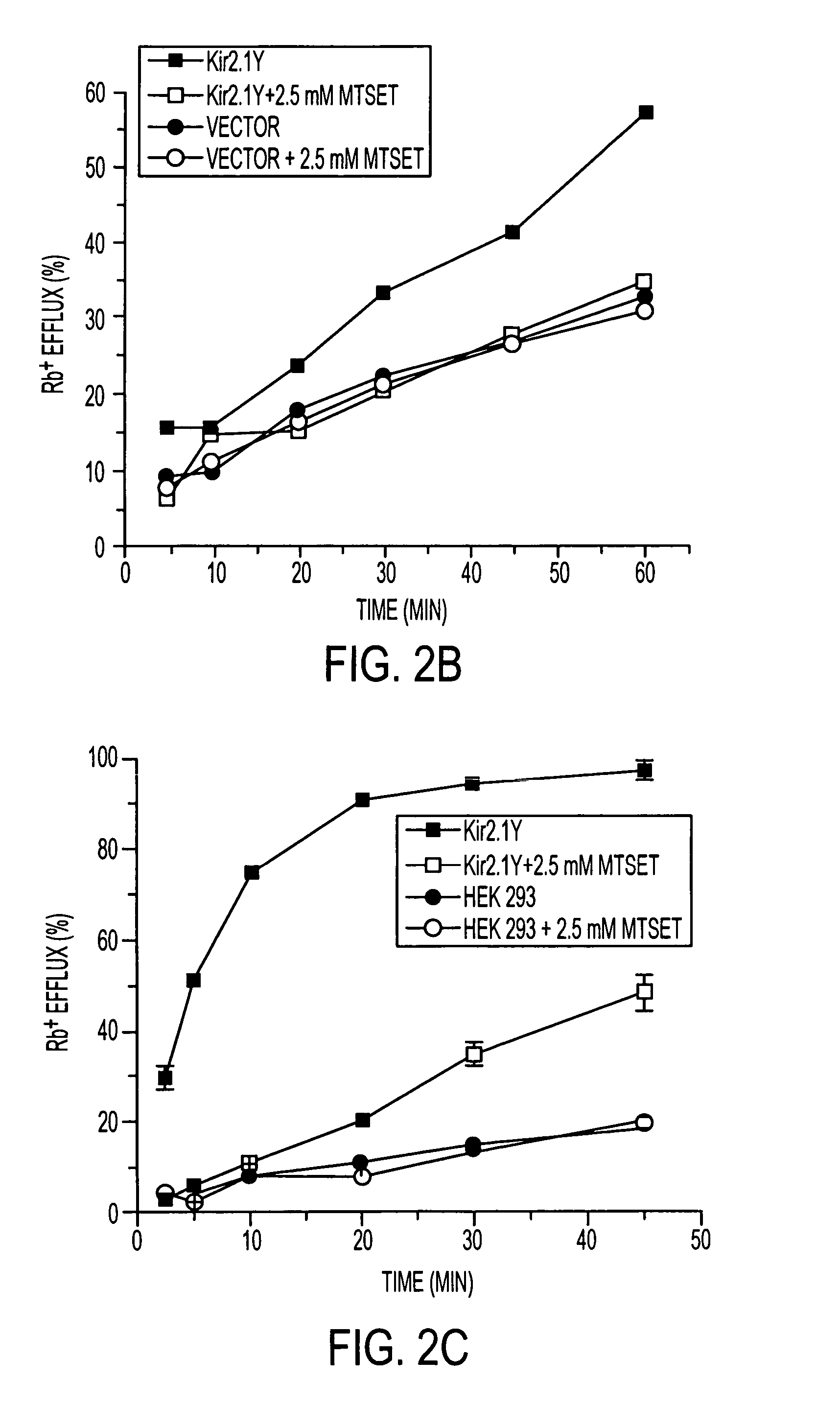
[0025] Materials. The methanethiosulfonate reagents, such as [2-(trimethylammonium)ethyl]methanethiosulfonate bromide (MTSET), were purchased from Toronto Research Chemicals (Downsview, ON, Canada). Brefeldin A (BFA), sodium butyrate, and RbCl were purchased from Sigma.

[0026] Transfection and Stable Cell Lines. Expression vectors were constructed using pcDNA3.1(+) (Invitrogen), and the Kir2.1 cDNA was cloned into HindIII and NotI sites. For the two Kir2.1 cDNA clones used in the study, one has the hemagglutinin (HA) epitope inserted at the first extracellular loop between M1 and P-loop to facilitate surface detection (13), referred to as Kir2.1. The other has both the HA epitope and a peptide, FRGRSWTY, fused at the C-terminus to facilitate surface expression, which is referred to as Kir2.1Y. The transfection into HEK 293 cells was carried out with FuGENE 6 (Roche Diagnostics). Stable clones were selected for neomycin resistance. Stable clones with high expression level of channel ...

example 2

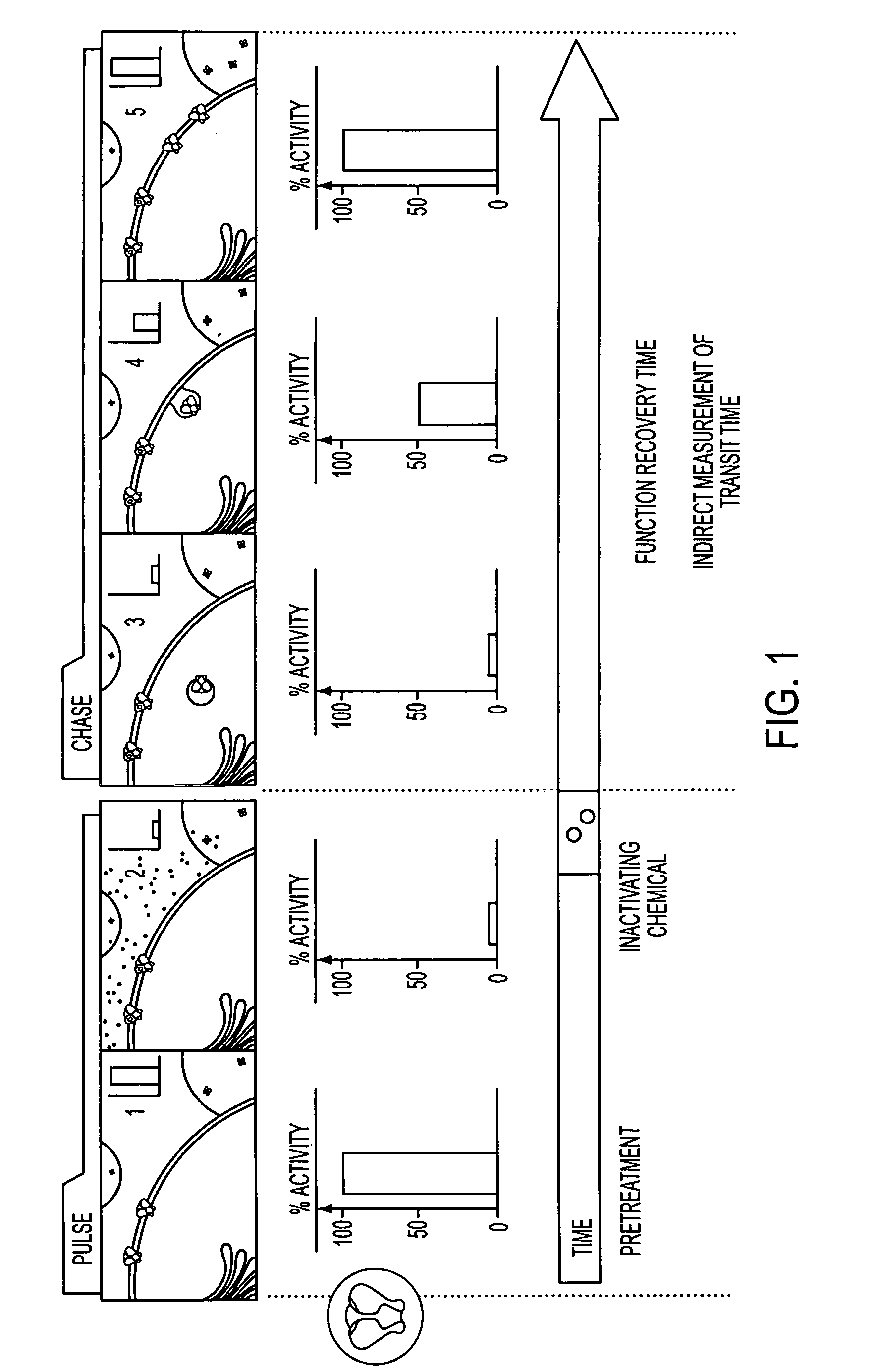
[0031] Kir2.1 encodes an inward rectifier K+ channel consisting of four subunits that align to form a centrally positioned hydrophilic pore to conduct K+ ions (14). One of the attractive features of the Kir2.1 channel is that it has an open probability of nearly 100% at resting potential, thereby permitting an option of monitoring the activity without depolarization. Assays for detecting K+ channel activities include electrophysiological recording, fluorescence-based measurements, and Rb+ flux assay (for review, see ref. 15). All three types of assays can be achieved within minutes. The primary distinctions concern whether the assay reads signals from individual cells or a population of cells and whether the cells remain intact during the entire assay procedure. We chose the Rb+ flux assay because of its ability to assay a large population of cells for extended time periods without rupture of cell membrane. The flux activity linearly reflects the conducting ion passage. Rb+ is a non...

example 3

[0035] To evaluate FRAC, we generated two cell lines expressing either the Kir2.1 or Kir2.1Y where the protein expression level of Kir2.1Y is ˜2-fold higher than that of Kir2.1 (data not shown). Both cell lines were treated first with MTSET to nullify the channels on the cell surface. After its removal, the treated cells were assayed after incubations of the indicated periods of time for recovery. The half-maximal functional recovery time (FR1 / 2) at 37° C. for both Kir2.1 and Kir2.1Y was 1 h (FIG. 3a). The functional expression reached 90% of the initial level 5 h after chemobleaching with MTSET. During the chase period, the Rb+ loaded cells without MTSET treatment gave rise to similar signals (FIG. 3a). The background Rb+ efflux of nontransfected cells remained consistent with or without MTSET treatment (FIG. 3a). The result shows that recovery was specific to the cells expressing the recombinant channels. To monitor the level of Kir2.1Y protein on the cell surface, flow cytometry ...


PUM

PropertyMeasurementUnit
pHaaaaaaaaaa
half-maximal functional recovery timeaaaaaaaaaa
timeaaaaaaaaaa

Abstract

Membrane proteins represent ˜30% of the proteome of both prokaryotes and eukaryotes. Unique to cell surface receptors is their biogenesis pathway, which involves vesicular trafficking from the endoplasmic reticulum through the Golgi apparatus and to the cell surface. Increasing evidence suggests specific regulation of biogenesis for different membrane receptors, hence affecting their surface expression. A pulse-chase assay can be used to monitor function recovery after chemobleaching (FRAC) to probe the transit time of the cell surface receptors to reach the cell surface. This method distinguishes molecular density from functional density. The ability of the reported method to access the biogenesis pathways in a high-throughput manner facilitates the identification and evaluation of molecules affecting receptor trafficking.

Description

[0001] The present invention was made using funds from the U.S. National Institutes of Health, under grant numbers 2 RO1 NS33324, 1 RO1 GM58488, and 1 RO1 DK61580. The U.S. government retains certain rights under the terms of these grants.TECHNICAL FIELD OF THE INVENTION [0002] This invention is related to the area of membrane protein biogenesis. In particular, it relates to assays of transit of biomarkers to the cell surface. BACKGROUND OF THE INVENTION [0003] The rate of membrane receptor trafficking is critical for the steady-state level of surface expression and is tightly coupled to signaling events (1-3). Within a given molecule, such as K+ channels, various trafficking sequence motifs have been identified that contribute to the specificity of trafficking behavior (4). For a given receptor, recent evidence suggests that the rate of trafficking may change according to the physiological state, such as during cellular aging (5). To assess the transit time from the endoplasmic ret...

Claims


Application Information

Patent Timeline
Patent Type & Authority Applications(United States)
IPC IPC(8): G01N33/567G01N33/53
CPCG01N33/5008G01N33/566G01N2500/00
Inventor LI, MINSUN, HAIYANSHIKANO, SOJIN
Owner THE JOHN HOPKINS UNIV SCHOOL OF MEDICINE
Why Eureka
Try Eureka