Methods for treating lentivirus infections
a technology for lentivirus infections and lentiviruses, applied in the field of lentivirus infections, can solve the problems that none of the available drugs used to combat hiv is completely effective, and achieve the effect of increasing the level of active apobec3g
- Summary
- Abstract
- Description
- Claims
- Application Information
AI Technical Summary
Benefits of technology
Problems solved by technology
Method used
Image

Examples
example 1
HIV-1 Vif Blocks the Antiviral Activity of APOBEC3G
[0193] Experimental Procedures
[0194] Plasmids
[0195] The pNL4-3 and pNL4-3 ΔVif (also known has p-NLND) expression vectors have been previously described. Adachi et al. (1991). Arch Virol 117, 45-58; and Sakai et al. (1993) J Virol 67, 1663-1666. pcDNA3.1 APOBEC3G-HA and APOBEC3G expression vectors were produced by excising an APOBEC3G-HA cassette using the XhoI / EcoRI restriction sites present in the NG / C15 retroviral vector (Sheehy et al. (2002) Nature 418, 646-650) and then cloning of the fragment into the same restriction enzyme sites in pcDNA3.1. To prepare the pCMV4-HA-APOBEC3G vector, APOBEC3G cDNA was amplified by PCR from an H9 cDNA library and then inserted into the HindIII and XbaI sites of the pCMV4 vector. GST-Vif was prepared by polymerase chain reaction (PCR) amplification of the Vif coding region present in a pNL4-3 vector followed by insertion of this amplicon into the EcoRI and NheI sites of the pBC vector (Chatto...
example 2
Characterization of High- and Low-Molecular Weight APOBEC3G Complexes
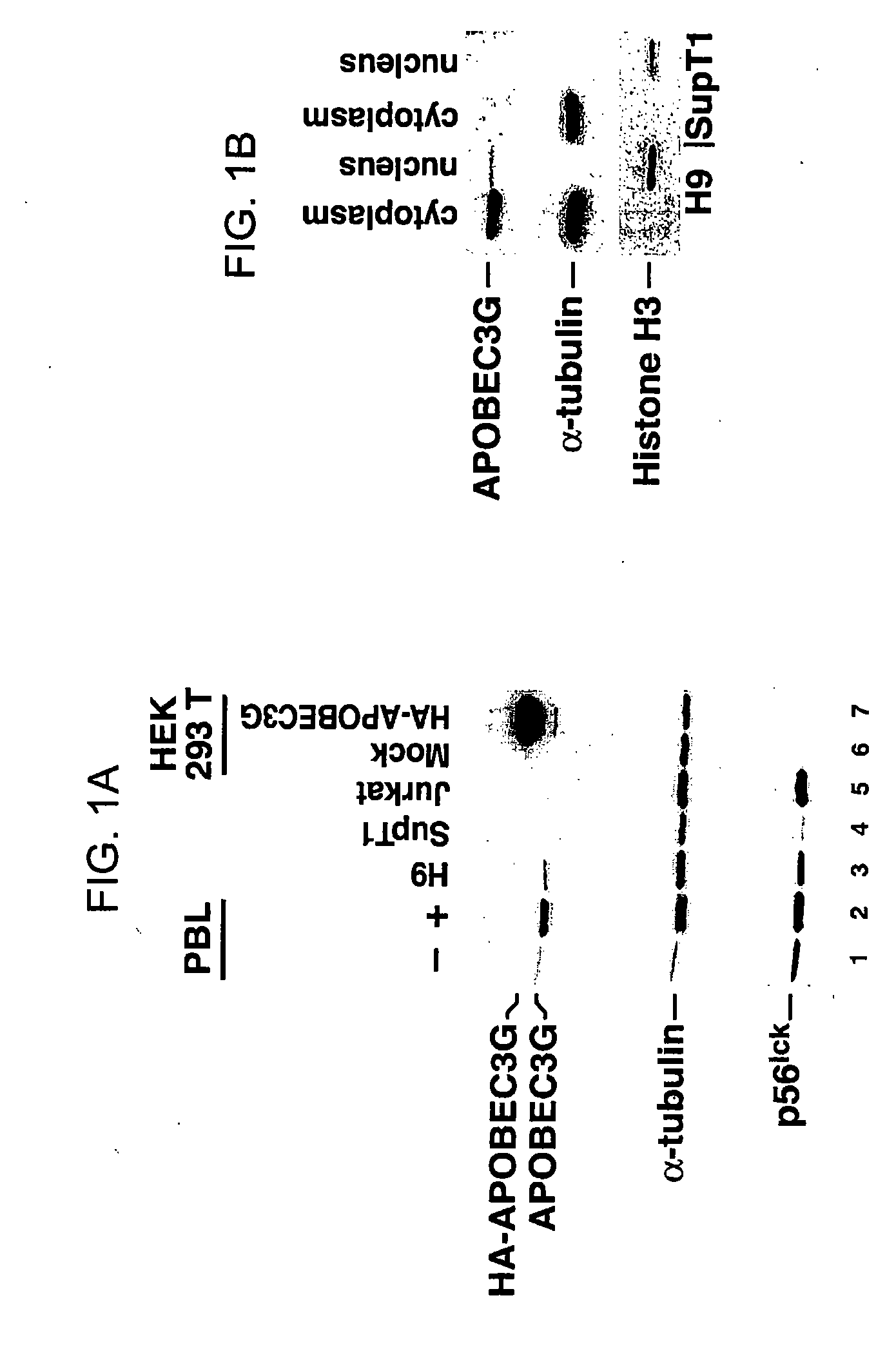
[0257] Human peripheral blood lymphocytes (PBLs) were isolated from blood and then incubated with anti-CD3 antibody (α-CD3), anti-CD28 antibody (α-CD28), IL-9, IL-12, IL-13, IL-15, or IFN-γ for 48 hours. Cells were lysed, the lysates clarified, and the protein concentration of the lysates was quantitated. Equal amounts of proteins in the lysates were separated by SDS-PAGE. After separation of the proteins by SDS-PAGE, the proteins were transferred to a membrane, and antibody to APOBEC3G was used to detect APOBEC3G protein on the membrane. The results are depicted in FIGS. 1-13.
[0258]FIG. 11 depicts the effect of PHA, anti-CD3 antibody, anti-CD28 antibody, and IL-15 on the level of APOBEC3G in peripheral blood lymphocytes. PBLs treated with PHA, anti-CD3 antibody, anti-CD28 antibody, or IL15 display increased expression of the APOBECG enzyme. Western blot of cell lysates deriving from stimulated and unstimulated p...
example 3
The Effect of an N-Terminal Fragment of APOBEC3G on Vif-Induced Degradation of APOBEC3G
[0274] HEK 293 cells were co-transfected with an HA-APOBEC3G expression vector or with increasing amounts (2 μg or 4 μg) of an HA-(1-104)APOBEC3G expression vector; and either an HIV-1 Vif or a control expression vector. The HA-APOBEC3G expression vector encodes wild-type (e.g., full-length) APOBEC3G fused in-frame to the hemagglutinin-epitope tag at the N-terminus. The HA-(1-104)APOBEC3G expression vector encodes amino acids 1-104 of APOBEC3G fused in-frame to the hemagglutinin-epitope tag. Cell lysates were clarified by centrifugation, and equal amounts of protein separated by SDS-PAGE. The separated proteins were transferred to nitrocellulose membranes, and both HA-APOBEC3G and HA-(1-104)APOBEC3G on the membranes were detected by anti-HA antibody. In cells transfected with the HA-APOBEC3G expression vector (and not co-transfected with the Vif expression vector), HA-APOBEC3G was produced that m...
PUM
| Property | Measurement | Unit |
|---|---|---|
| molecular weight | aaaaa | aaaaa |
| molecular weight | aaaaa | aaaaa |
| temperature | aaaaa | aaaaa |
Abstract
Description
Claims
Application Information
 - R&D
- Intellectual Property
- Life Sciences
- Materials
- Tech Scout
- Unparalleled Data Quality
- Higher Quality Content
- 60% Fewer Hallucinations
Browse by: Latest US Patents, China's latest patents, Technical Efficacy Thesaurus, Application Domain, Technology Topic, Popular Technical Reports.
© 2025 PatSnap. All rights reserved.Legal|Privacy policy|Modern Slavery Act Transparency Statement|Sitemap|About US| Contact US: help@patsnap.com