Hyperspectral Imaging in Diabetes and Peripheral Vascular Disease

a hyperspectral imaging and peripheral vascular disease technology, applied in the field of medical tissues hyperspectral and multispectral imaging, can solve the problems of unsatisfactory relying on medial history and physical examination alone, poor pharmacologic treatment of pad, and about 10% of pad patients experiencing classic symptoms of intermittent clarification, etc., to reduce the resolution of hu

Active Publication Date: 2007-10-04
HYPERMED IMAGING
View PDF41 Cites 46 Cited by
  • Summary
  • Abstract
  • Description
  • Claims
  • Application Information

AI Technical Summary

Benefits of technology

[0020] One embodiment of the invention is directed to a medical instrument comprising a first stage optic responsive to illumination of tissue, a spectral separator, one or more polarizers, an imaging sensor, a diagnostic processor, a filter control interface, a general purpose operating module to assess the state of tissue in diabetic subjects following a set of instructions, and a calibrator. Preferably, the instrument further comprises a second stage optic responsive to illumination of tissue. Preferably, the set of instructions comprises preprocessing hyperspectral information, building a visual image, defining a region of interest in tissue, converting the visual image into units of optical density by taking a negative logarithm of each decimal base, decomposing a spectra for each pixel into several independent components, determining three planes for an RGB pseudo-color image, determining a sharpness factor plane, converting the RGB pseudo-color image to a hue-saturation-value / intensity image having a plane, adjusting the hue-saturation-value / intensity image plane with the sharpness factor plane, converting the hue-saturation-value / intensity image back to the RGB pseudo-color image, removing outliners beyond a standard deviation and stretching image between 0 and 1, displaying the region of interest in pseudo-colors; and characterizing a metabolic state of the tissue of interest.
[0021] Preferably, the region of interest is one of a pixel, a specified region or an entire field of view. Preferably, determining three planes for an RGB pseudo-color image comprises one or more characteristic features of the spectra, determining a sharpness factor plane comprises a combination of the images at different wavelengths, removing outliners beyond a standard deviation comprises three standard deviations, displaying the region of interest in pseudo-colors comprises one of performing one in combination with a color photoimage of a subject, in addition to a color photo image of a subject, and projecting onto the tissue of interest.
[0022] Preferably, defining the color intensity plane as apparent concentration of one or a mathematical combination of oxygenated Hb, deoxygenated Hb, and total Hb, oxygen saturation, defining the color intensity plane as reflectance in blue-green-orange region, adjusting the hue saturation comprises adjusting a color resolution of the pseudo-color image according to quality of apparent concentration of one or a mathematical combination of oxygenated Hb, deoxygenated Hb, and total Hb, oxygen saturation, adjusting the hue saturation further comprises one or a combination of reducing resolution of hue and saturation color planes by binning the image, resizing the image, and smoothing the image through filtering higher frequency components out, and further interpolating the smoothed color planes on a grid of higher resolution intensity plane.

Problems solved by technology

Problems with die foot that affect the ambulatory nature of the patient are not ony important from the standpoint of physical risk, but also convey an emotional risk as well, as these problems disrupt the fundamental independence of the patient by limiting his or her ability to walk.
Although pharmacologic treatments for PAD have traditionally been poor, 2.1 million nevertheless receive pharmacologic treatment for the symptoms of PAD, and current diagnostic tests are not considered to be very sensitive indicators of disease progression or response to therapy.
One difficulty in diagnosing PAD is that in the general population, only about 10% of persons with PAD experience classic symptoms of intermittent clarification.
Relying on medial history and physical examination alone is unsatisfactory.
As in the case of diabetic foot disease, current technologies have fallen short.
Another problem face by physicians is disease of the peripheral veins.
Venous occlusive disease due to incompetent valves in veins designed to prevent backflow and deep vein thrombosis, results in venous congestion and eventually stasis ulcers.
This, in turn, can lead to increased pressure on the dermis, resulting in tissue ischemia and eventual death, and ultimately result in an ulcer.4 There are a number of factors that weigh heavily in the process of ulcerations5—affecting different aspects of the foot—that lead to a combination of effects that greatly increase the risk of ulceration:6 Neuropathy—Results in a loss of protective sensation in the foot, exposing patients to undue, sudden or repetitive stress.
Can cause a lack of awareness of damage to the foot as it be occurs and physical defects and deformities7 which lead to even greater physical stresses on the foot.
It can also lead to increased risk of cracking and the development of fissures in calluses, creating a potential entry for bacteria and increased risk of infection.8 Microcirculatory Changes—Often seen in association with hyperglycemic damage.9 Functional abnormalities occur at several levels, including hyaline basement membrane thickening and capillary leakage.
On a histologic level, it is well known that diabetes causes a thickening of the endothelial basement membrane which in turn may lead to impaired endothelial cell function.
Musculoskeletal Abnormalities—Include altered foot mechanics, limited joint mobility, and bony deformities, and can lead to harmful changes in biomechanics and gait.
This increases pressures associated with various regions of the foot.
Alteration or atrophy of fat pads from increased pressure can lead to skin loss or callus, both of which increase the risk of ulceration by two orders of magnitude.
Foot pathology is major source of morbidity among diabetics and is a leading cause of hospitalization.
None of these methods are discriminatory for feet at risk, and none of them provide any information about the spatial variability across the foot.
Currently there is no method to accurately assess the predisposition to serious foot complications, to define the real extent of disease or to track the efficacy of therapeutics over time.

Method used


Image

  • Hyperspectral Imaging in Diabetes and Peripheral Vascular Disease
  • Hyperspectral Imaging in Diabetes and Peripheral Vascular Disease
  • Hyperspectral Imaging in Diabetes and Peripheral Vascular Disease

Examples


Embodiment Construction

Background of Hyperspectral Imaging

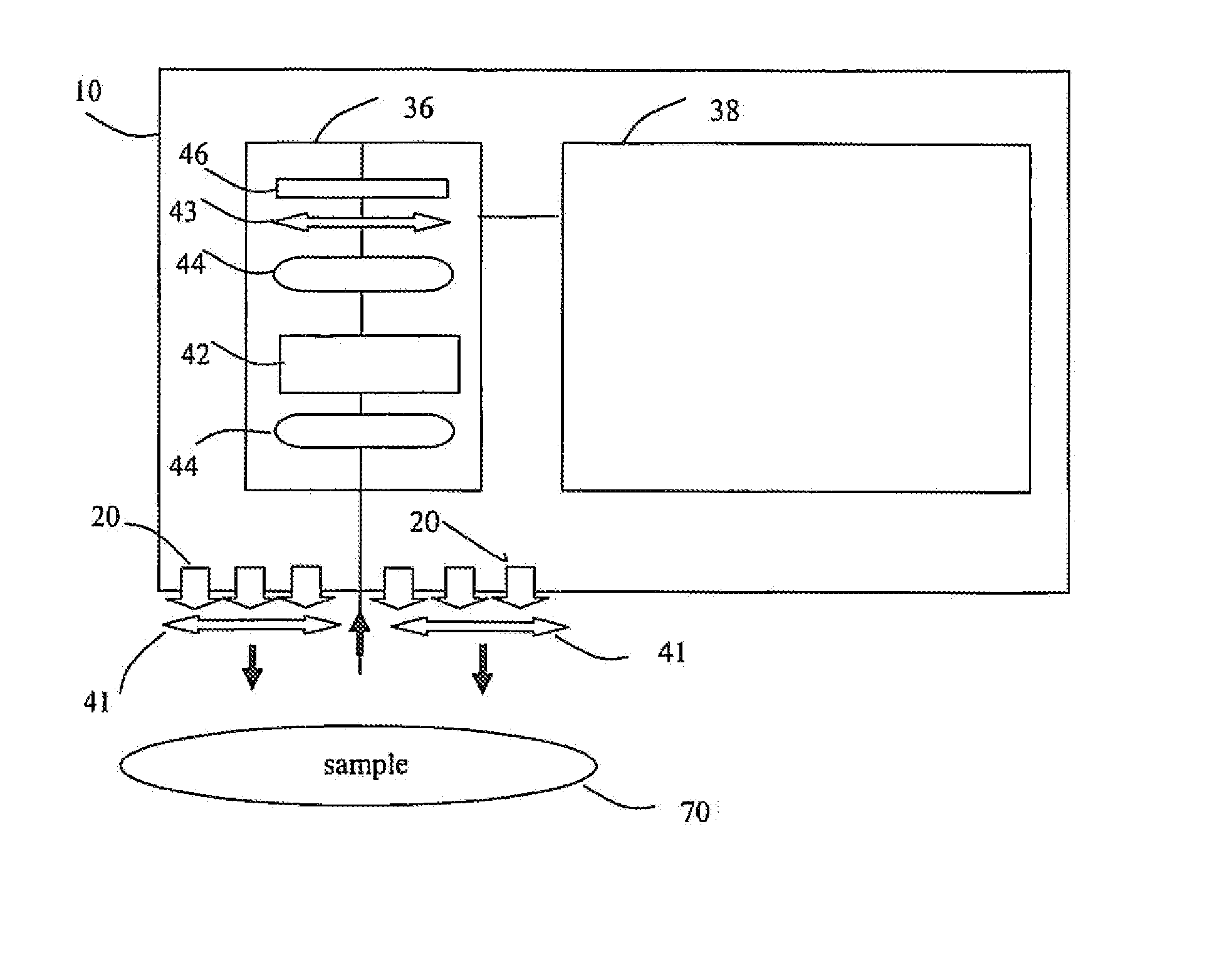
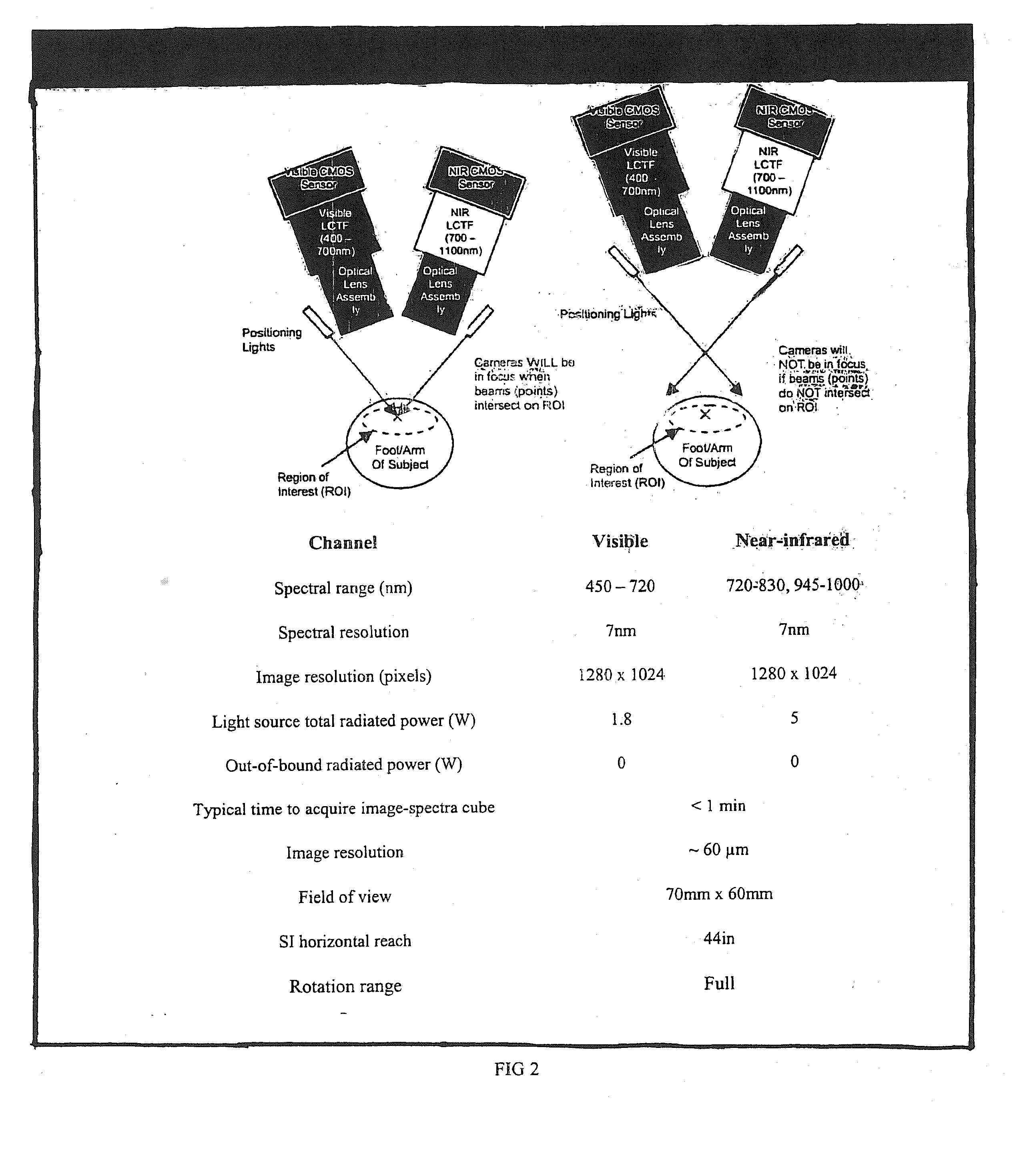
[0037] HSI or hyperspectral imaging is a novel method of “imaging spectroscopy” that generates a “gradient map” of a region of interest based on local chemical composition. HSI has been used in satellite investigation of suspected chemical weapons production areas22, geological features23, and the condition of agricultural fields24 and has recently been applied to the investigation of physiologic and pathologic changes in living tissue in animal and human studies to provide information as to the health or disease of tissue that is otherwise unavailable.25 MHSI for medical applications (MHSI) has been shown to accurately predict viability and survival of tissue deprived of adequate perfusion, and to differentiate diseased (e.g. tumor) and isehemic tissue from normal tissue.27

[0038] Spectroscopy is used in medicine to monitor metabolic status in a variety of tissues. One of the most common spectroscopic applications is in pulse oximetry, which uti...


PUM


Abstract

The invention is directed to methods and systems of hyperspectral and multispectral imaging of medical tissues. In particular, the invention is directed to new devices, tools and processes for the detection and evaluation of diseases and disorders such as, but not limited to diabetes and peripheral vascular disease, that incorporate hyperspectral or multispectral imaging.

Description

REFERENCE TO RELATED APPLICATIONS [0001] The present application claims priority to U.S. Provisional Patent Application Ser. No. 60 / 784,476 filed Mar. 22, 2006 entitled Combiniatons of Hyperspectrial Imaging Methods with Other Evaluation Methods and is a continuation in part of U.S. patent application Ser. No. 11 / 396,941 filed Apr. 4, 2006 entitied Hyperspectrial Imaging in Diabetes and Peripheral Vascular Disease, which claims priority to U.S. Provisional Patent Application Ser. No. 60 / 667,677 entitled Hyperspectral Imaging in Diabetes, filed Apr. 4, 2005, and U.S. Provisional Patent Application Ser. No. 60 / 785,977 entitled Hyperspectral Imaging of Angiogenesis, filed Mar. 27, 2006, the contents of which are hereby incorporated by reference.BACKGROUND [0002] 1. Field of the Invention [0003] The invention is directed to methods and systems of hyperspectral and multispectral imaging of medical tissues. In particular, the invention is directed to new devices, tools and processes for t...

Claims


Application Information

Patent Timeline
Patent Type & Authority Applications(United States)
IPC IPC(8): A61B5/00
CPCA61B5/0059A61B5/14551A61B2560/0228A61B5/445A61B5/412
Inventor FREEMAN, JENNYPANASYUK, SVETLANAHOPMEIER, MICHAELSCHOMACKER, KEVINBRAND, DEREK
Owner HYPERMED IMAGING
Why Eureka
Try Eureka