Esophageal dilation and stent delivery system and method of use

a technology which is applied in the field of esophageal dilation and stent delivery system and method of use, can solve the problems of swollen, scarring, and stiff lining of the esophagus, and achieve the effect of effectively dilatation at the predetermined poin

Inactive Publication Date: 2007-10-04
BOARD OF RGT THE UNIV OF TEXAS SYST
View PDF17 Cites 28 Cited by
  • Summary
  • Abstract
  • Description
  • Claims
  • Application Information

AI Technical Summary

Benefits of technology

[0012] In another embodiment, the disclosure further provides a method for delivering a prosthesis into a body vessel or cavity of a mammal using assemblies disclosed herein, wherein the method comprises inserting a guide wire within a body vessel or cavity, coupling a first dilator cap to the distal end of an endoscope to form a endoscope-dilator assembly, extending the endoscope-dilator assembly through the interior of the vessel or cavity to a predetermined point so as to effectively dilate the predetermined point, and withdrawing the endoscope-dilator assembly. In accordance with certain aspects of this embodiment, this method further comprises selecting a second dilator cap from a kit comprising a plurality of dilator caps, the second dilator cap having proximal end diameter different from the proximal end diameter of the first dilator cap; coupling the second dilator cap to the distal end of the endoscope to form a second endoscope-dilator assembly; and, extending the second endoscope-dilator assembly through the interior of the vessel or cavity to the predetermined point.
[0013] In another embodiment, the present disclosure provides an assembly for the delivery and fixation of a stent in a body vessel or cavity, the assembly comprising a guide wire; an endoscope having a distal end and a proximal end; a dilator cap having a distal end, a proximal end, and a length no more than one quarter of the length of the endoscope; a stent; and a flexible stent delivery device, the dilator cap being substantially transparent or translucent and coupled to the distal end of the endoscope. In accordance with this embodiment, the dilator cap can be selected from a kit comprising a plurality of dilator caps, each of which have different proximal end and / or distal end diameters. Additionally, the stent can further comprise one or more apertures or rings circumferentially located on a flange of the stent.
[0014] In a further embodiment, the disclosure further provides a method for delivering a prosthesis or stent into a body vessel or cavity, comprising: providing a stent delivery assembly; inserting a guide wire having a distal tip and a proximal end within a body vessel or cavity until the guide wire distal tip reaches a desired location in the body vessel or cavity; extending an endoscope having a dilator cap attached to the distal end thereof through the interior of the vessel or cavity to a predetermined point; inserting a stent through the vessel or cavity to the predetermined point with a stent delivery device; withdrawing the endoscope-dilator assembly from the body vessel or cavity; and withdrawing the stent delivery device from the body vessel or cavity, wherein the method is performed independent of an expanding device, such as a balloon expander. In accordance with this embodiment, the method can further comprise attaching the stent to a wall of the vessel or cavity using an attachment means, such as a surgical suture, that is capable of passing through one or more apertures or rings in a flange of the stent and into a wall of the vessel or cavity.
[0015] The disclosure further provides a method for delivering a stent into the esophagus of a mammal for treating esophageal strictures, comprising: extending a guide wire into the esophagus and extending the guide wire past a stricture in the esophagus; inserting a endoscope having a dilator cap coupled thereto (an endoscope-dilator assembly) into the esophagus, extending through the esophageal stricture; inserting a stent into the esophagus, over the endoscope; inserting the stent into the esophageal stricture using a pusher catheter; and withdrawing the endoscope having the dilator cap affixed thereto from the stricture, while simultaneously maintaining the stent in position with the pusher catheter. This method can be performed independent of an expanding device associated with the dilator or the stent. In further accordance with this embodiment of the present disclosure, the pusher catheter, guide wire, and endoscope-dilator cap assembly can be withdrawn separately, or in combination, or simultaneously. Additionally, the method can further comprise attaching the stent to a wall of the vessel or cavity using an attachment means that is capable of passing through one or more apertures or rings in a flange of the stent and into a wall of the vessel or cavity.
[0016] In yet another embodiment, the present disclosure provides a kit for use in endoscopic surgery and similar surgical procedures. Such a kit comprises a plurality of conically-shaped dilator caps having a distal end, a proximal end, and a tapered edge between the distal end and the proximal end. These dilator caps are translucent or transparent, have a proximal end with a diameter ranging from about 5 mm to about 15 mm, and have a distal end with a diameter ranging from about 2 mm to about 8 mm. In accordance with this embodiment, one or more of the dilator caps in the kit can comprise at least one radio-opaque element. Further, such dilator caps can optionally have one or more graduations on or within a surface.
[0017] In a further embodiment, the current disclosure provides a stent for use in therapeutic applications, the stent comprising a distal flange, a proximal flange, a medial sleeve portion intermediate between the distal flange and the proximal flange, and a support structure within the medial sleeve, wherein the proximal flange comprises one or more apertures or rings for the attachment of the stent to a biological body using any number of attachment means.

Problems solved by technology

An esophageal stricture is a gradual narrowing of the esophagus, which can lead to swallowing difficulties.
When the lining of the esophagus is damaged, scarring develops, and the lining of the esophagus becomes stiff.
In time, as this scar tissue continues to build up, the esophagus begins to narrow in that area, resulting in swallowing difficulties (dysphagia).
This scarring will produce damaged tissue in the form of a ring that narrows the opening of the esophagus.
Occurring anywhere in the esophagus, the cancer can exist as a stricture, mass, or plaque, and often results in painful dysphagia (difficulty swallowing and the sensation that food is sticking on the way down the esophagus).
While surgical removal or treatment of strictures, especially carcinomas in the esophagus, can sometimes be effective, the majority of patients exhibiting esophageal tumors are not candidates for surgery.
Such techniques, while effective, also have notable disadvantages.
For example, photodynamic laser therapy is expensive, typically requiring several treatment sessions before results are evident.
Further, certain submucosal tumors, and certain pulmonary tumors causing dysphagia by esophageal compression, cannot be treated by laser therapy techniques.
Rigid plastic prosthesis insertion is also not without problems.
Placement of such rigid, non-elastic plastic stents can be traumatic due to their large size, and can often result in perforation of the esophageal wall.
However, while dilation of esophageal strictures is a useful approach to the alleviation of the symptoms typically associated with this complication, such as dysphagia, it is not without associated risks and complications.
The problem with this technique is that it requires the use of a long dilator (on the order of 90 mm or more), and the expandable stents used are subject to recoil, necessitating several procedures to insert different, larger stents so as to effectively open the stricture to a point that dysphagia is alleviated.
However, using this technique, no dilator is used, and so the width of the stent is constrained, typically to about 4-8 mm.
In order to widen the stricture to a point that relieves at least some of the dysphagia associated with it, several procedures must typically be performed in order to gradually widen the gap in the stricture by inserting consecutively larger stents, a process that can be both costly and painful.
Resultant and subsequent sludge formation and / or ingrowth or overgrowth of the stent may cause obstruction of the metal endoprosthesis, necessitating surgical removal and re-stenting.
Plastic stents, such as those plastic prostheses made of Teflon®, polyethylene, and polyurethane, have also been used in the dilation of esophageal strictures, but with less success than that shown with metal stents.
However, the use of these stents typically result in incrustation and sludge (obstruction) formation of varying degrees, which can eventually lead to premature occlusion of the stented stricture.
Additionally, these stents have been shown to be less cost-effective and have higher reintervention costs than other dilation prostheses [Lammer, J., et al., Radiology, 201: pp.

Method used


Image

  • Esophageal dilation and stent delivery system and method of use
  • Esophageal dilation and stent delivery system and method of use
  • Esophageal dilation and stent delivery system and method of use

Examples


Embodiment Construction

[0030] One or more illustrative embodiments are presented below. Not all features of an actual implementation are described or shown in this application for the sake of clarity. It is understood that in the development of an actual embodiment incorporating the present invention, numerous implementation-specific decisions must be made to achieve the developer's goals, such as compliance with system-related, business-related, government-related and other constraints, which vary by implementation and from time to time. While a developer's efforts might be complex and time-consuming, such efforts would be, nevertheless, a routine undertaking for those of ordinary skill the art having benefit of this disclosure.

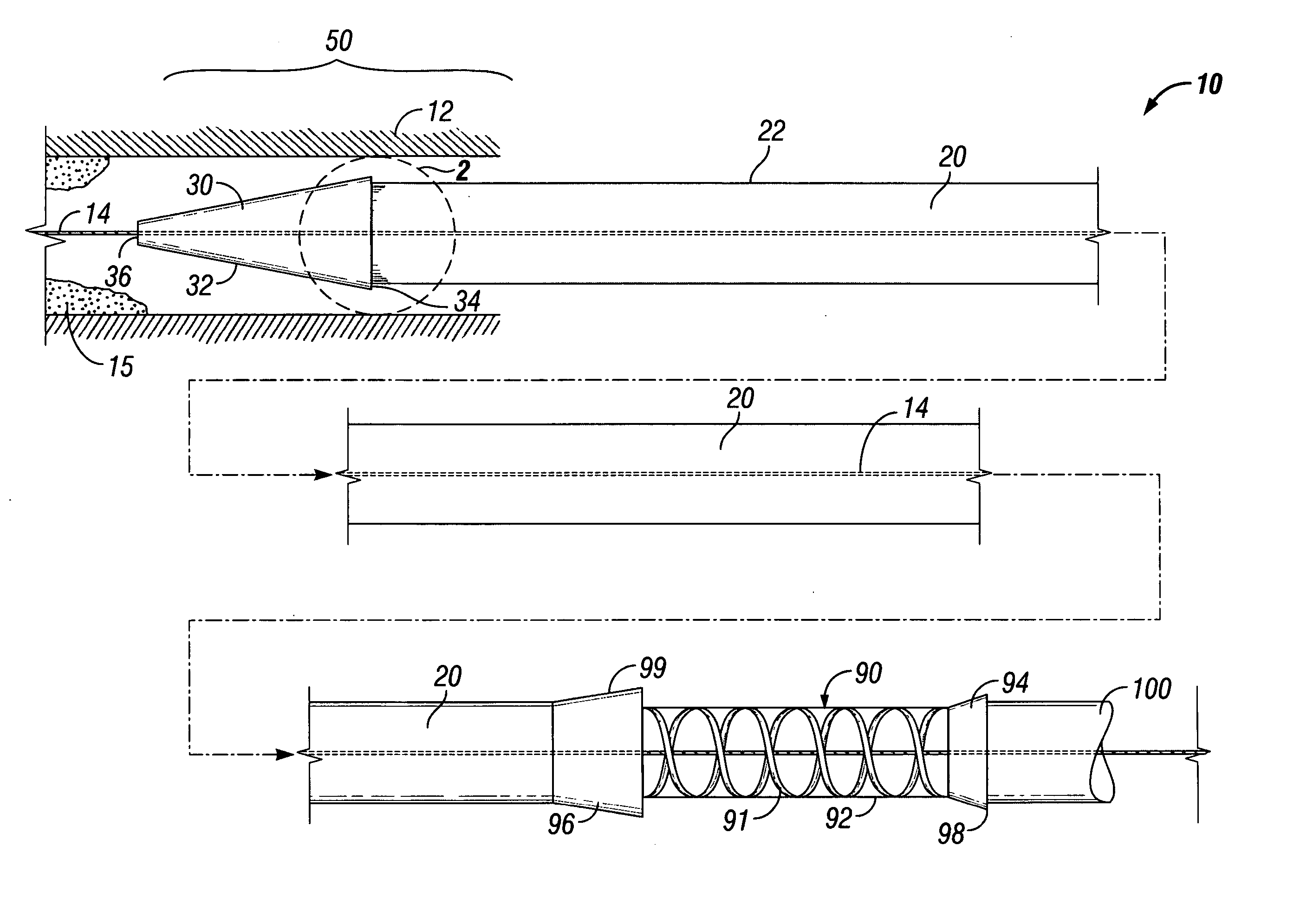
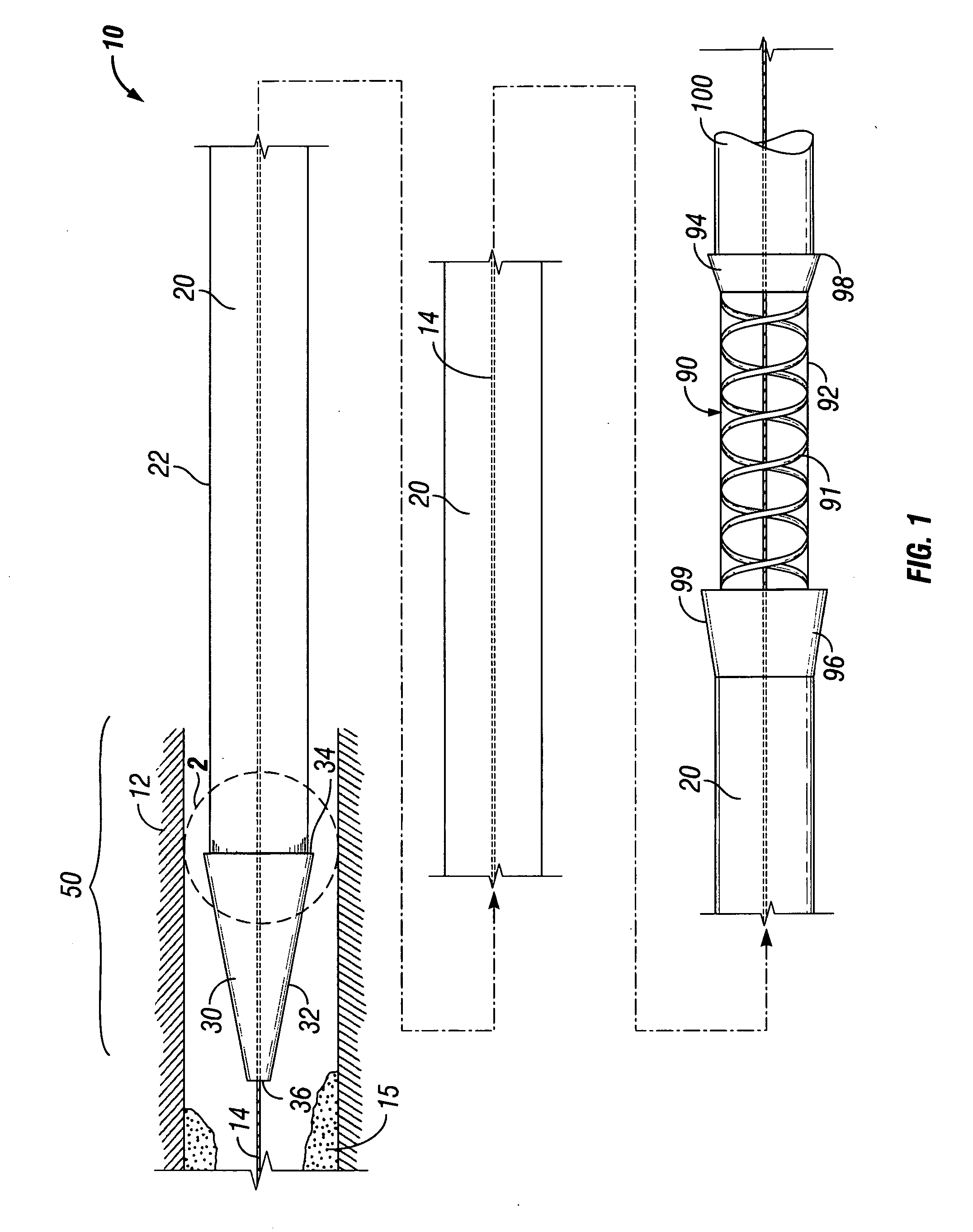
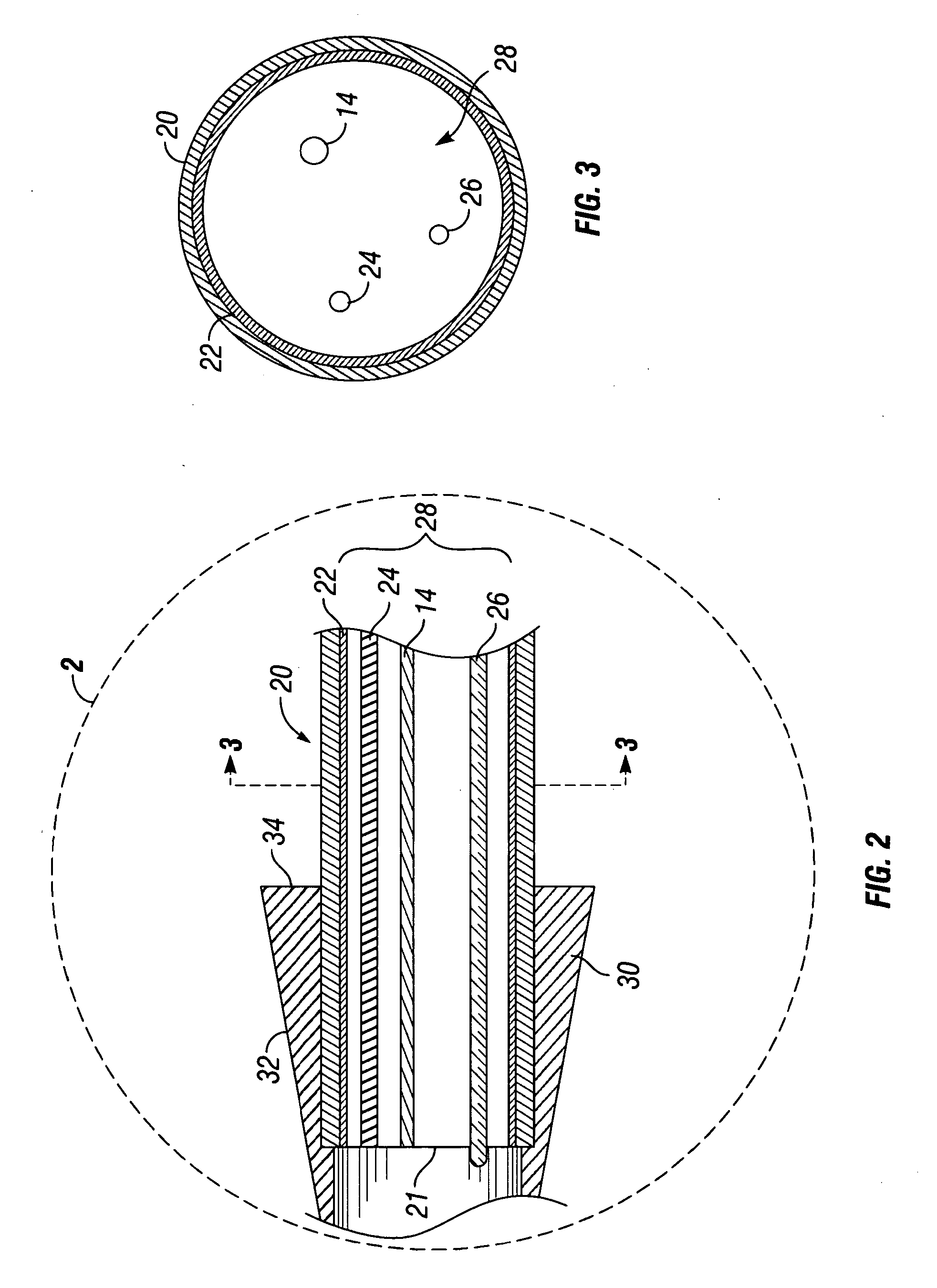
[0031] In general terms, the applicant has created an assembly and method for dilating and introducing a stent or similar prosthesis into a vessel or cavity, such as through a stricture within the esophagus of a mammal, which is easier and quicker than previous dilation methods. ...


PUM


Abstract

Methods and assemblies for dilating and/or delivering a stent into a body cavity or vessel is described. The assembly is particularly suited for the dilation, delivery and fixation of a stent in a body cavity, such as in a stricture in the esophagus, and includes a guidewire, an endoscope having a dilator cap affixed to its distal end, a stent, and a flexible stent delivery device. The dilator cap can be customized to various sizes, depending upon the interior diameter of the stricture to be dilated, and can be interchanged with the other dilator caps on the distal end of the endoscope. The endoscope-dilator assembly can be slid distally down the guidewire to the stricture to be dilated, and the stent can then be delivered distally along the guidewire using a delivery device. The stent delivery device and the endoscope-dilator assembly can be retracted proximally along the guidewire through the interior of the stent, leaving the stent in place in the stricture.

Description

FIELD OF THE INVENTION [0001] This disclosure relates generally to dilation of strictures and a body implantable treatment device, and more particularly to a dilation and stent delivery system and other prostheses intended for fixation in body lumens, including the esophagus, as well as methods of use thereof. DESCRIPTION OF RELATED ART [0002] An esophageal stricture is a gradual narrowing of the esophagus, which can lead to swallowing difficulties. The strictures can be caused by scar tissue that builds up in the esophagus. When the lining of the esophagus is damaged, scarring develops, and the lining of the esophagus becomes stiff. In time, as this scar tissue continues to build up, the esophagus begins to narrow in that area, resulting in swallowing difficulties (dysphagia). One of the conditions that can lead to esophageal strictures is gastroesophageal reflux disease, wherein excessive acid is refluxed from the stomach up and into the esophagus. This causes an inflammation in t...

Claims


Application Information

Patent Timeline
Patent Type & Authority Applications(United States)
IPC IPC(8): A61F2/06A61F2/04A61F2/82A61F2/94
CPCA61F2/82A61F2/848A61F2/88A61F2/94A61F2/95A61F2230/0067A61F2250/0091A61F2220/0008A61F2220/005A61F2220/0066A61F2220/0075A61F2002/044
Inventor RAJU, GOTTUMUKKALA S.
Owner BOARD OF RGT THE UNIV OF TEXAS SYST
Why Eureka
Try Eureka