Method to confirm immunosuppression in human patients by measuring lymphocyte activation

a technology lymphocyte activation, which is applied in the field of human immunodeficiency virus detection in human patients by measuring lymphocyte activation, can solve the problems of difficult to measure the function of t lymphocytes (t cells) or their response to specific antigens, and the current methods of measuring immune function are tedious, time-consuming, and poorly adapted to the clinical laboratory setting. , to achieve the effect of convenient, reliable and rapid measuremen

Inactive Publication Date: 2007-10-18
SOTTONG PETER +1
View PDF12 Cites 0 Cited by
  • Summary
  • Abstract
  • Description
  • Claims
  • Application Information

AI Technical Summary

Benefits of technology

[0019] The subject invention provides a convenient, reliable, and rapid method for analyzing the function of various sets or subsets of lymphocytes. The method of the invention involves exposing a population of cells to at least one inducing agent (e.g. a mitogen or an antigen, with or without a co-stimulatory agent); separating a desired subset of cells by means of the interaction of a specific b...

Problems solved by technology

The function of T lymphocytes (T cells) or their response to specific antigens is more difficult to measure.
Second, T cells respond to antigens only when they are presented by other cells in the context of major histocompatibility antigens on the surface of the presenting cell.
Current methods for measuring immune function are tedious, time consuming, and poorly adapted to the clinical laboratory setting.
Major disadvantages of flow cytometry include the requirement for complex and expensive equipment, the requirement for significant “hands on” time since each sample must be run using many manual steps, and the necessity of utilizing highly trained individuals to analyze results.
These disadvantages are particularly acute in a clinical laboratory which must process multiple patient specimens daily and where the need for consistent and reliable results is extremely important.
The major difficulty with all cell counting techniques is that they do not measure the function of specific cells or their responses to specific or non-specific stimuli, such as specific antigens or mitogens.
In addition, these methods are tedious and subject to poor reproducibility.
Because they evaluate the div...

Method used


Image

  • Method to confirm immunosuppression in human patients by measuring lymphocyte activation
  • Method to confirm immunosuppression in human patients by measuring lymphocyte activation
  • Method to confirm immunosuppression in human patients by measuring lymphocyte activation

Examples


example 1

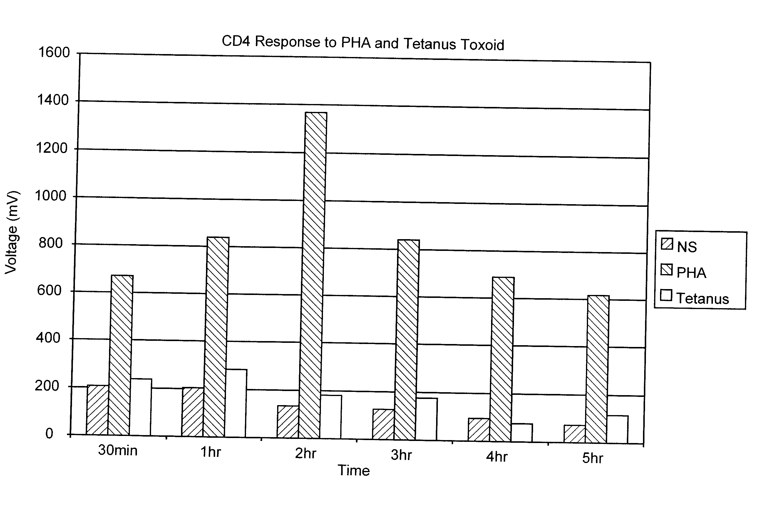
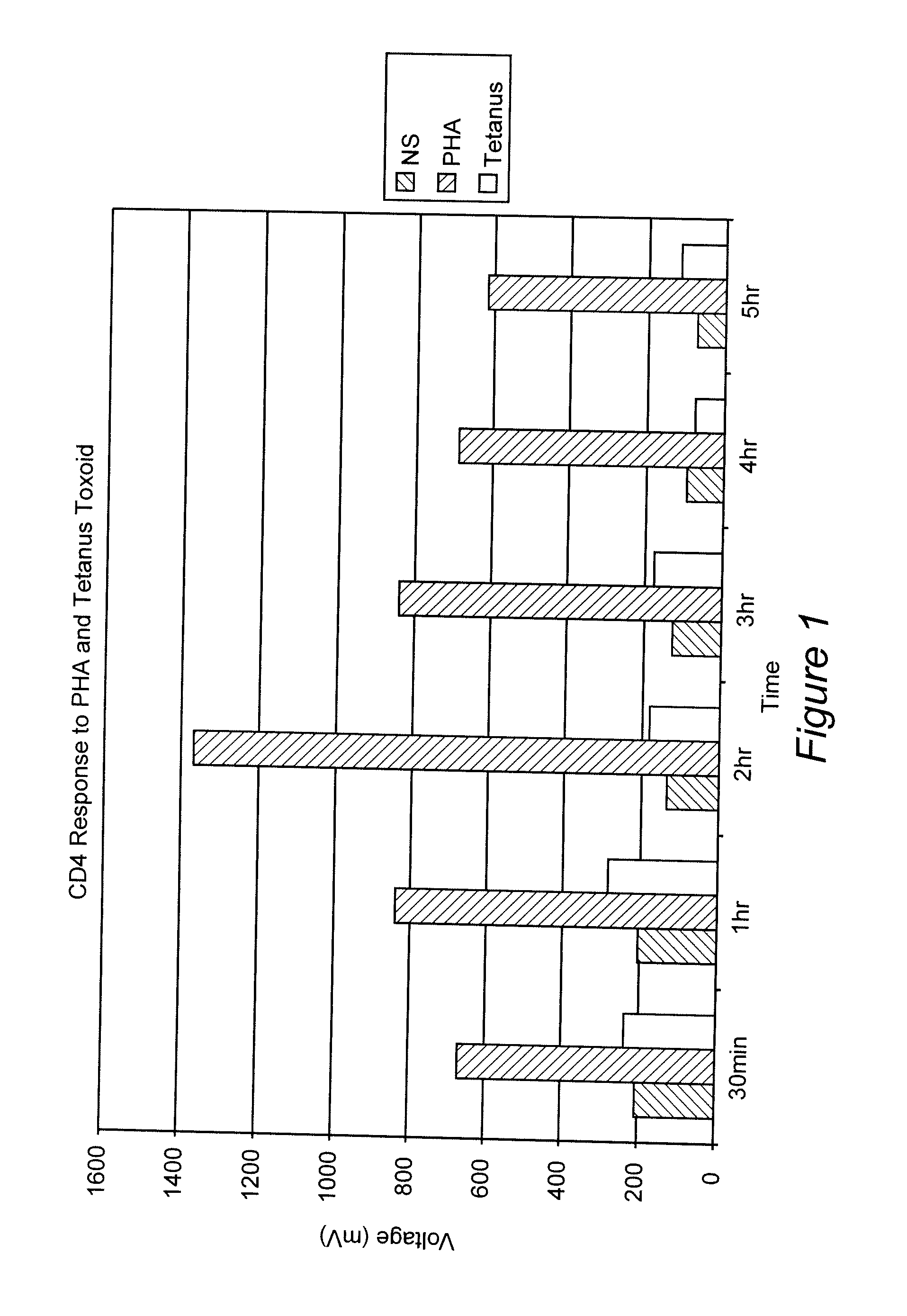
Measurement of the Response of T-Lymphocytes to Phytohemagglutinin (PHA) and Tetanus Toxoid Using Whole Blood as the Sample

[0064] A blood sample was obtained from a normal donor and collected in heparin as an anti-coagulant. Aliquots of the blood were diluted 1:10 in RPMI 1640 cell culture media (Biowhittaker, Walkersville, Md.). Replicates received either no addition, the mitogen PHA (Sigma-Aldrich, St. Louis, MO) at 1%, or the antigen Tetanus Toxoid (University of Massachusetts Medical Center, Jamaica Plains, Mass.) at 1 ug / mL final concentration. Samples were incubated at 37° C. At various time points (30 min, 1 hr, 2 hr, 3 hr, 4 hr and 5 hr), 100 uL of the samples were removed, incubated with paramagnetic beads coated with the binding substance anti-CD4 (Dynal, Oslo, Norway) (20 uL) for 30 minutes at room temperature. The cells and beads were placed next to a permanent magnet that was positioned such that the magnet field was in a direction perpendicular to gravity. Bead:cell c...

example 2

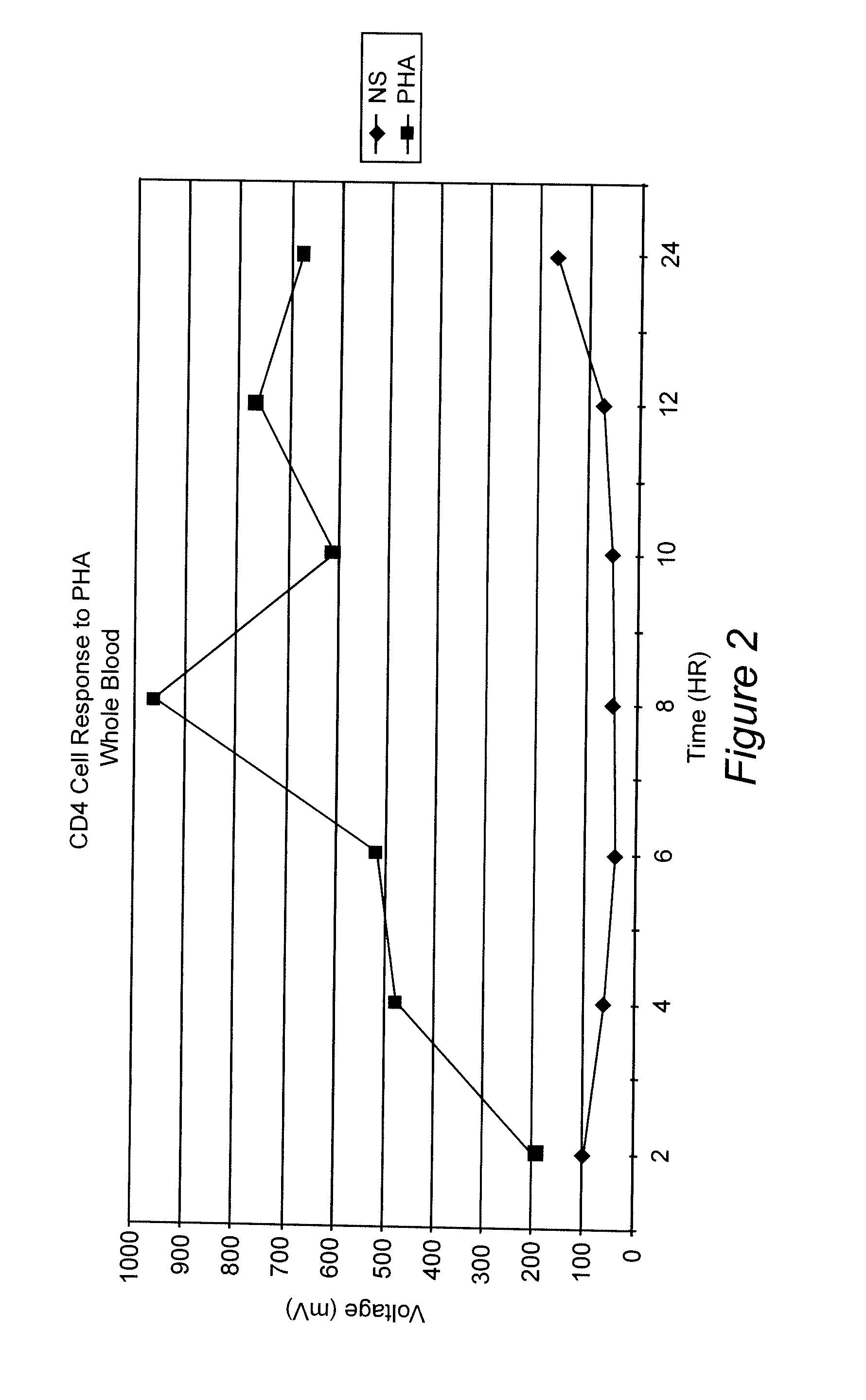
Whole Blood Response After Stimulation With Phytohemmaglutinin (PHA)

[0067] A blood sample was obtained from a normal donor and collected in heparin as an anti-coagulant. Aliquots of the blood were diluted 1:10 in RPMI 1640. Replicates received either no addition or the mitogen PHA at 1% final concentration. Samples were incubated at 37° C. At various time points (2 hr, 4 hr, 6 hr, 8 hr and 24 hr), 100 uL of the samples were removed, incubated with anti-CD4 coated paramagnetic beads (20 uL) for 30 minutes at room temperature. The cells and beads were placed next to a permanent magnet positioned such that the magnetic field was in a direction perpendicular to gravity. Bead:cell complexes were washed (3 times in RPMI 1640) and lysed in a detergent solution. Assay Reagent (luciferin / luciferase) was added to the samples ATP production was determined as described above. FIG. 2 shows the results of this assay. An increase in ATP over background can be seen at all time points. The response...

example 3

T-lymphocyte Response: Co-stimulation with Antigen and Antibody Coated Paramagnetic Particles

[0069] A blood sample was obtained from a normal donor and collected in heparin as an anti-coagulant. Aliquots of the blood (50 uL) were diluted 1:2 in RPMI 1640 and placed into wells of a 96-well microtiter plate. Replicates received either no addition, or the antigen Tetanus Toxoid at 1 μg / mL final concentration plus anti-CD3 / CD28 coated paramagnetic beads (0.5 uL) (Dynal, Oslo, Norway). Samples were incubated for various times (30 min, 1 hr, 2 hr, and 3 hr), at 37° C. After incubation, the 96-well strips were removed and placed in a magnet tray. Bead:cell complexes were washed (3 times in a wash buffer containing 1% BSA) and lysed in a hypotonic solution. An aliquot of the sample (50 uL) was removed to an opaque plate and Assay Reagent (luciferin / luciferase) was added. The samples were read and Relative Light Units were determined. The ATP production was then calculated from an ATP calib...


PUM


Abstract

Methods for measuring the function of lymphocytes and their responses to mitogens or specific antigens, with or without co-stimulatory agents, is provided. The methods are suitable for measurement of the responses of lymphoid cells when they are a subpopulation of cells, and also for measuring the function of specific subsets of lymphoid cells, each subpopulation or subset of a subpopulation having characteristic determinants on their cell surface. The invention also relates to test kits used in performing such methods. The methods of the invention facilitate screening of complex biological fluids, such as whole blood, by means of incubating a sample of the fluid with a mitogen or antigen, with or without co-stimulatory elements, separating the selected subset of interest, e.g., via affinity separation, and detecting the presence of an internal cellular component, advantageously ATP, that is increased as a result of the response.

Description

[0001] The present application claims benefit of U.S. provisional patent application 60 / 283,935, filed Apr. 17, 2001. The present application is a divisional application of U.S. patent application Ser. No. 10 / 123,163 filed Apr. 17, 2002.BACKGROUND OF THE INVENTION [0002] 1. Field of the Invention [0003] The invention relates to rapid methods for measuring the function of lymphocytes and their responses to mitogens or specific antigens. In particular, the methods rapidly detect the presence of an internal cellular component, advantageously adenosine triphosphate ATP, that is increased as a result of an immune response. [0004] 2. Background of the Invention [0005] The immune system is central to control of infectious diseases and cancer. Lymphocytes, a class of white blood cells, are critical cell types that are responsible for the activities of the immune system. Lymphocytes are divided into two major categories: T lymphocytes and B lymphocytes. Overall assessment of the function of ...

Claims


Application Information

Patent Timeline
IPC IPC(8): C12Q1/02G01N33/569
CPCG01N33/56972
Inventor SOTTONG, PETERKOWALSKI, RICHARD J.
Owner SOTTONG PETER
Why Eureka
Try Eureka