Nucleic acid amplification methods

a technology of nucleic acid and amplification method, applied in the field of nucleic acid amplification method, can solve the problems of preventing their general applicability in clinical practice, time and laborious process, and achieving rapid and sensitive detection, sensitive and standardized detection, and rapid and sensitive detection

Inactive Publication Date: 2007-11-22
MT SINAI SCHOOL OF MEDICINE
View PDF49 Cites 14 Cited by
  • Summary
  • Abstract
  • Description
  • Claims
  • Application Information

AI Technical Summary

Benefits of technology

[0012] An improved method, which allows for rapid, sensitive and standardized detection and quantitation of nucleic acids from pathogenic microorganisms from samples from patients with infectious diseases has now been developed. The improved methodology also allows for rapid and sensitive detection and quantitation of genetic variations in nucleic acids in samples from patients with genetic diseases or neoplasia.

Problems solved by technology

While all of these techniques are powerful tools for the detection and identification of minute amounts of a target nucleic acid in a sample, they all suffer from various problems, which have prevented their general applicability in the clinical laboratory setting for use in routine diagnostic techniques.
One of the most difficult problems is preparation of the target nucleic acid prior to carrying out its amplification and detection.
This process is time and labor intensive and, thus, generally unsuitable for a clinical setting, where rapid and accurate results are required.
Another problem, especially for PCR and SDA, is that conditions for amplifying the target nucleic acid for subsequent detection and optional quantitation vary with each test, i.e., there are no constant conditions favoring test standardization.
Such microorganisms cause infectious diseases that represent a major threat to human health.
The precise extent of the problem is not clear, however, since current cultural methods for detecting mycobacteria are cumbersome, slow and of questionable sensitivity.
The methods referred to above are relatively complex procedures that, as noted, suffer from drawbacks making them difficult to use in the clinical diagnostic laboratory for routine diagnosis and epidemiological studies of infectious diseases and genetic abnormalities.
The extensive time and labor required for target nucleic acid preparation, as well as variability in amplification templates ( e.g., the specific target nucleic acid whose detection is being measured) and conditions, render such procedures unsuitable for standardization and automation required in a clinical laboratory setting.

Method used


Image

  • Nucleic acid amplification methods
  • Nucleic acid amplification methods
  • Nucleic acid amplification methods

Examples


example 1

Detection of HIV-1 RNA in A Sample

Preparation of Oligonucleotide Probes

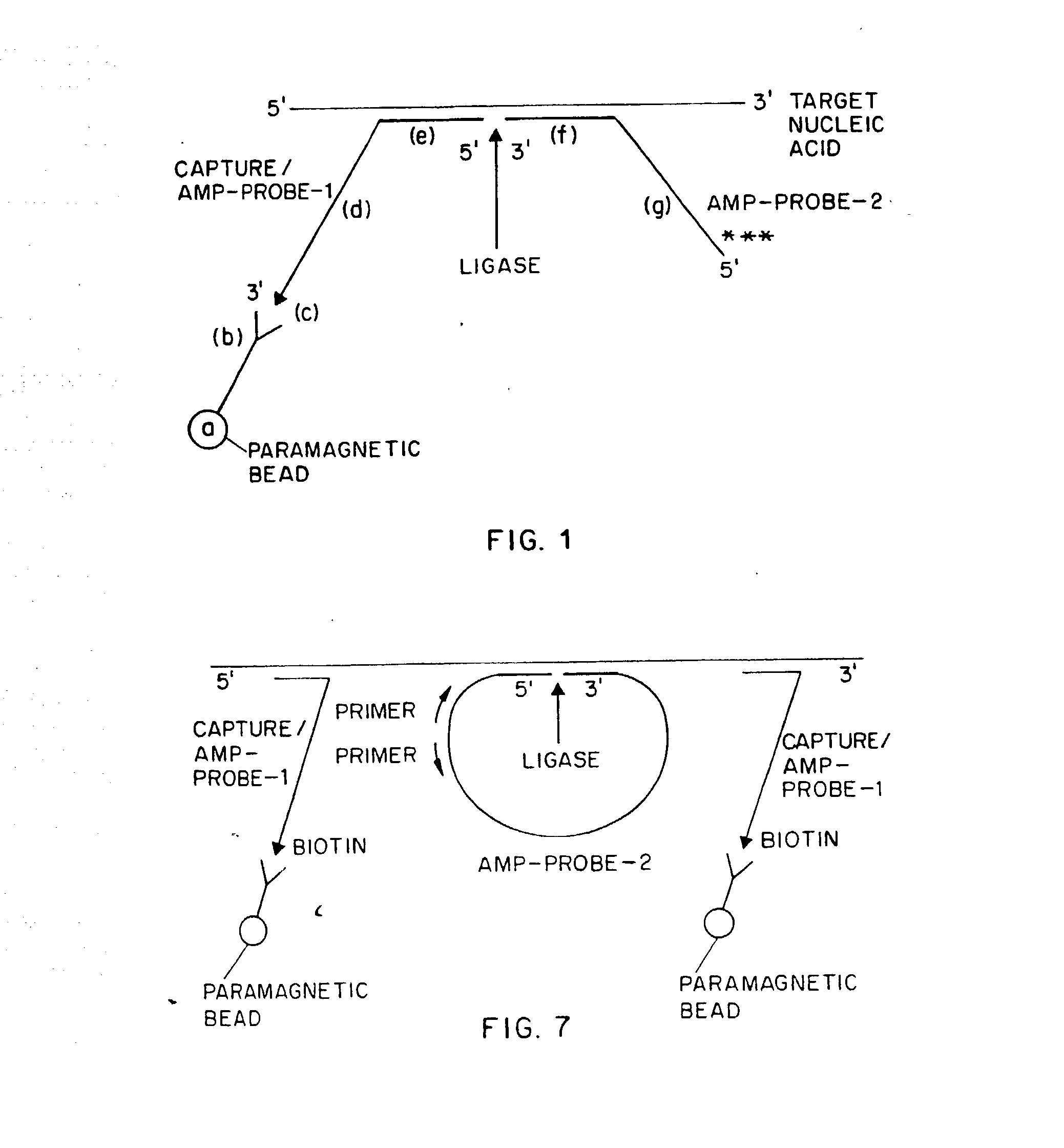
[0198] A pair of oligodeoxyribonucleotide probes, designated Capture / Amp-probe-1 (HIV) and Amp-probe-2 (HIV), respectively for detecting the gag region of HIV-1 RNA were prepared by automated synthesis via an automated DNA synthesizer (Applied Biosystems, Inc.) using known oligonucleotide synthetic techniques. Capture / Amp-probe-1 (HIV) is an oligodeoxyribonucleotide comprising 59 nucleotides and a 3′ biotin moiety, which is added by using a 3′-biotinylated nucleoside triphosphate as the last step in the synthesis. The Capture / Amp-probe-1 (HIV) used in this Example has the following nucleotide sequence (also listed below as SEQ ID NO. 1):

 1              11          215′- CCATCTTCCT GCTAATTTTA AGACCTGGTA31             41          51     ACAGGATTTC CCCGGGAATT CAAGCTTGG - 3′

[0199] The nucleotides at positions 24-59 comprise the generic 3′ end of the probe. Within this region are recognition sequences for SmaI (CC...

example 2

Direct Detection of HIV-1 RNA in A Sample

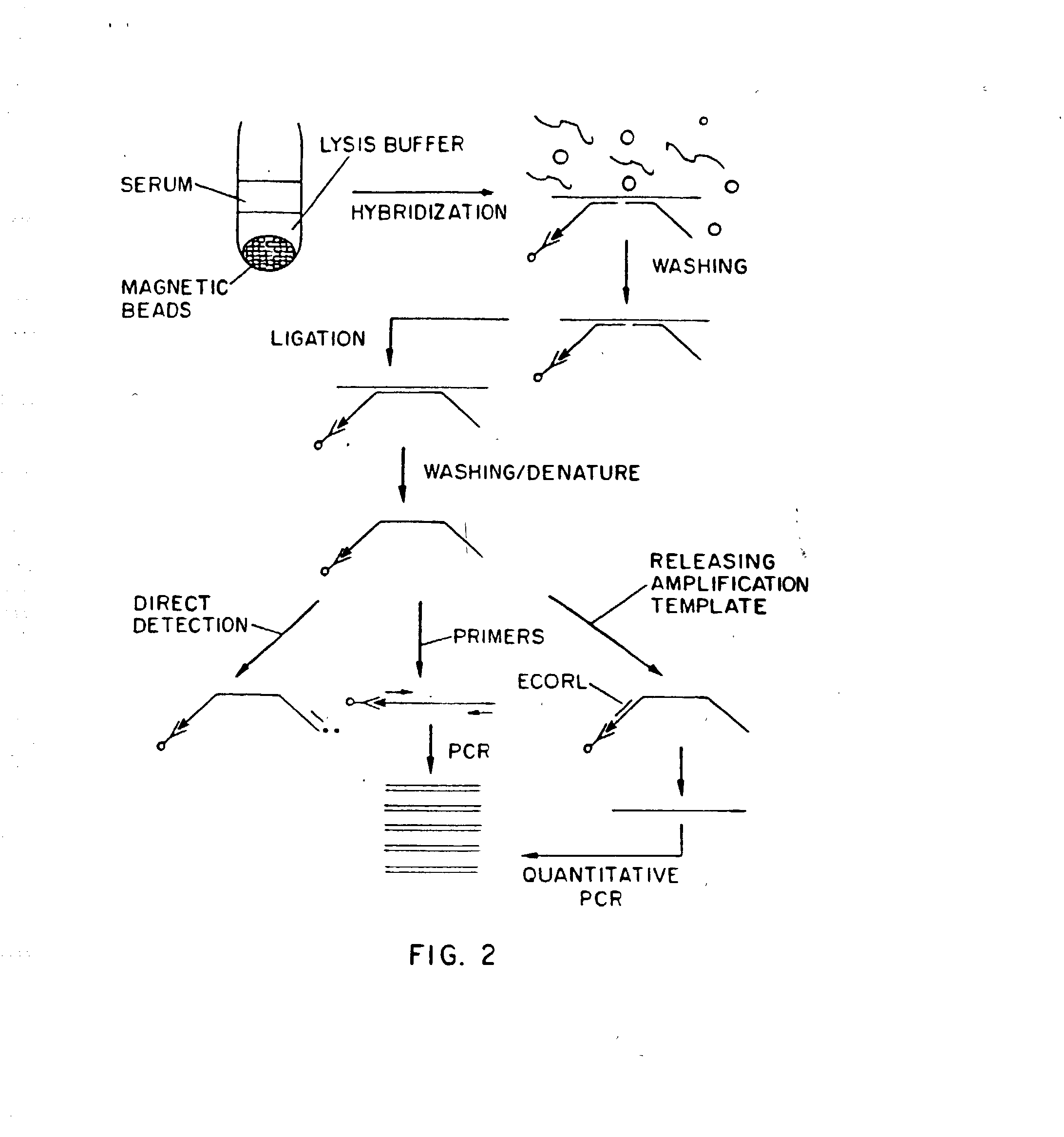
[0212] The ability of the present method to directly detect the presence of HIV-1 RNA in a sample was also determined. The probes used in this Example are the same as in Example 1 (SEQ ID NOS. 1 and 2). For direct detection, Amp-probe-2 (HIV) (SEQ ID NO. 2) was labeled at its 5′ end with 32P by the T4 phosphokinase reaction using 32P-γ-ATP. The various reaction mixtures were as follows: [0213] 1. Streptavidin-coated paramagnetic beads, 3′-biotinylated Capture / Amp-probe-1 (HIV) (SEQ ID NO. 1), Amp-probe-2 (HIV) (SEQ ID NO. 2) 5 (32P), HIV-1 RNA transcript. [0214] 2. Streptavidin-coated paramagnetic beads, 3′-biotinylated Capture / Amp-probe-1 (HIV), Amp-probe-2 (HIV) 5′(32P). [0215] 3. Streptavidin-coated paramagnetic beads, Amp-probe-2 (HIV) 5′(32P), HIV-1 RNA transcript.

[0216] Hybridizations, using each of the above three reaction mixtures, were carried out in 20 μl of a 1M GnSCN buffer comprising 1M GnSCN, 0.5% NP-40 (Nonidet P-40, N-lauroy...

example 3

Detection of Mycobacterium Avium-Intracellulaire (MAI) in Patient Samples

[0218] A recent paper (Boddinghaus et al., J. Clin. Microbiol. 28:1751, 1990) has reported successful identification of Mycobacteria species and differentiation among the species by amplification of 16S ribosomal RNAs (rRNAs). The advantages of using bacterial 16S rRNAs as targets for amplification and identification were provided by Rogall et al., J. Gen. Microbiol., 136:1915, 1990 as follows: 1) rRNA is an essential constituent of bacterial ribosomes; 2) comparative analysis of rRNA sequences reveals some stretches of highly conserved sequences and other stretches having a considerable amount of variability; 3) rRNA is present in large copy numbers, i.e. 103 to 104 molecules per cell, thus facilitating the development of sensitive detection assays; 4) the nucleotide sequence of 16S rRNA can be rapidly determined without any cloning procedures and the sequence of most Mycobacterial 16S rRNAs are known.

[0219]...


PUM

PropertyMeasurementUnit
pHaaaaaaaaaa
diameteraaaaaaaaaa
temperatureaaaaaaaaaa

Abstract

The present invention relates to assays and kits for carrying out said assays for the rapid, automated detection of infectious pathogenic agents and normal and abnormal genes. The present invention further relates to methods for general amplification of genomic DNA and total mRNAs and for analyzing differential mRNA expression using the amplification methods disclosed herein.

Description

INTRODUCTION [0001] The present invention relates to assays and kits for carrying out said assays for the rapid, automated detection of infectious pathogenic agents and normal and abnormal genes. The present invention further relates to methods for general amplification of genomic DNA and total mRNAs and for analyzing differential mRNA expression using the amplification methods disclosed herein. BACKGROUND OF THE INVENTION [0002] A number of techniques have been developed recently to meet the demands for rapid and accurate detection of infectious agents, such as viruses, bacteria and fungi, and detection of normal and abnormal genes. Such techniques, which generally involve the amplification and detection (and subsequent measurement) of minute amounts of target nucleic acids (either DNA or RNA) in a test sample, include inter alia the polymerase chain reaction (PCR) (Saiki, et al., Science 230:1350, 1985; Saiki et al., Science 239:487, 1988; PCR Technology, Henry A. Erlich, ed., Sto...

Claims


Application Information

Patent Timeline
Patent Type & Authority Applications(United States)
IPC IPC(8): C12P19/34C12Q1/68G01N33/53C12N15/09G01N21/78G01N33/566G01N33/58
CPCC12Q1/682C12Q1/6827C12Q1/6834C12Q1/6858C12Q2600/158C12Q2561/125C12Q2525/197C12Q2525/161C12Q2537/143C12Q2525/307C12Q2565/519C12Q2563/143C12Q2531/125C12Q2531/119C12Q2531/113
Inventor ZHANG, DAVID Y.
Owner MT SINAI SCHOOL OF MEDICINE
Why Eureka
Try Eureka