Use of Leptin in Wound Healing
a technology of leptin and wound healing, applied in the direction of peptide/protein ingredients, instruments, drug compositions, etc., can solve the problems of severely impaired wound healing phenotype, deprived affected tissue of oxygen and nutrients, and disrupted normal continuity of structures, etc., to repair, promote and/or accelerate wound contraction, promote and/or accelerate wound healing
- Summary
- Abstract
- Description
- Claims
- Application Information
AI Technical Summary
Benefits of technology
Problems solved by technology
Method used
Image
Examples
example 1
Preparation of Animals
[0116] Protocols involving mice experiments were first reviewed and approved by the Yale and Cedars-Sinai Animal Care and Use Committees, observing all appropriate institutional guidelines. Female C57BL / 6J mice (Jackson Laboratories, Bar Harbor, Me.) were used between 6-8 weeks of age. After wounding procedures, the mice were singly housed in microisolator cages.
example 2
Conducting of Wounding Procedure, Treatment and Collection of Wound Tissue
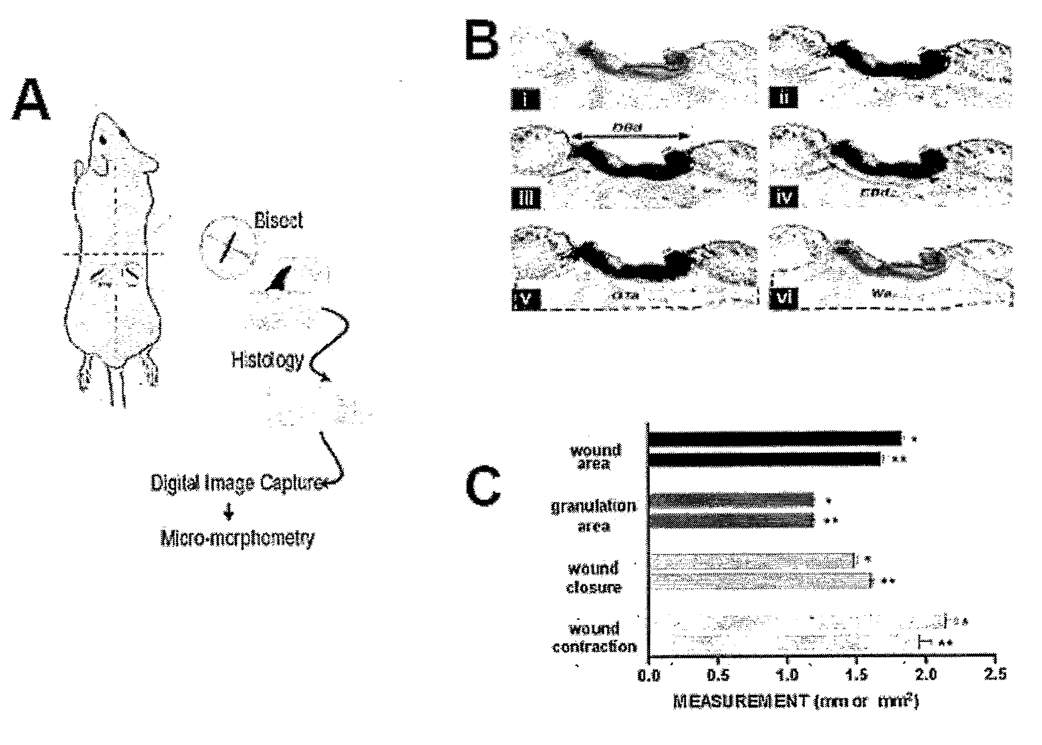
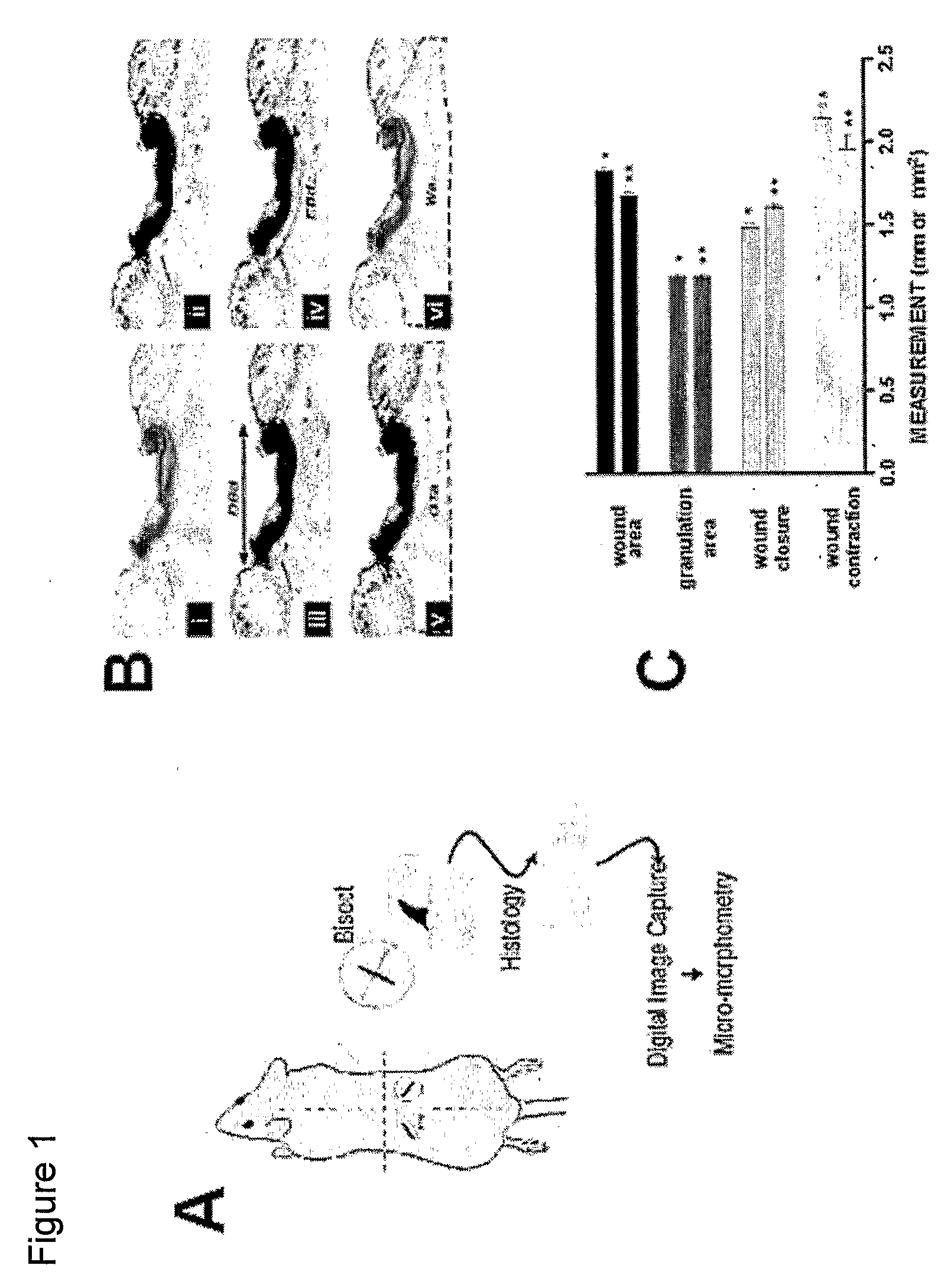
[0117] The animals were anesthetized with ketamine (10 mg / kg, i.m.) and Xylazine (40 mg / kg, i.p.). After shaving and disinfecting the skin with 70% ethanol, an 8-mm line was traced on each side on the mid-dorsal region with a surgical skin marker (see FIG. 1A). The skin was firmly retracted and bilateral full thickness dermal wounds were created using fine surgical scissors. The panniculus carnosum was always cut but care was taken not to damage the abdominal wall. Preliminary leptin dose-response experiments were performed using a dose range of 0.1-50 μg leptin / wound (Calbiochem, La Jolla, Calif.). In subsequent experiments, wounds of each mouse received a topical treatment with a pre-established optimal dose of leptin (10 μg / wound) or saline in a volume of 15 μl (n=22). Time points of 24-96 hours were evaluated by morphometric analysis. Wound borders were not mechanically juxtaposed and no dressing was appl...
example 3
Macroscopic, Micromorphometric and Histopathological Analysis
[0118] Macroscopic images of wounds were captured using an Olympus Camedia Digital Camera C-3040ZOOM with an Olympus Super Bright Zoom Lens (7.1-21.3 mm Lens) (Olympus Corporation, Japan). For micromorphometric analysis, H&E slides were randomly coded and digital images were acquired for analysis with an IPLab Spectrum v. 3.2.4 digital microscopy software program (Scanalytics Fairfax, Va.; see FIG. 1A). An image obtained from a graduated stage micrometer was used to calibrate the imaging software for automatic conversion of pixel units to millimeters. To evaluate wound contraction, the distance between dermis borders was measured by tracing a straight-line between the normal dermis tissues on each side of the wound (DBd; FIG. 1B, iii). To assess wound closure, re-epithelialization was measured as the length between the migrating epithelial tongues along the surface of the unhealed wound (EBd; FIG. 1B, iv). Granulation tis...
PUM
| Property | Measurement | Unit |
|---|---|---|
| Pharmaceutically acceptable | aaaaa | aaaaa |
| Contraction enthalpy | aaaaa | aaaaa |
Abstract
Description
Claims
Application Information
 - R&D
- Intellectual Property
- Life Sciences
- Materials
- Tech Scout
- Unparalleled Data Quality
- Higher Quality Content
- 60% Fewer Hallucinations
Browse by: Latest US Patents, China's latest patents, Technical Efficacy Thesaurus, Application Domain, Technology Topic, Popular Technical Reports.
© 2025 PatSnap. All rights reserved.Legal|Privacy policy|Modern Slavery Act Transparency Statement|Sitemap|About US| Contact US: help@patsnap.com


