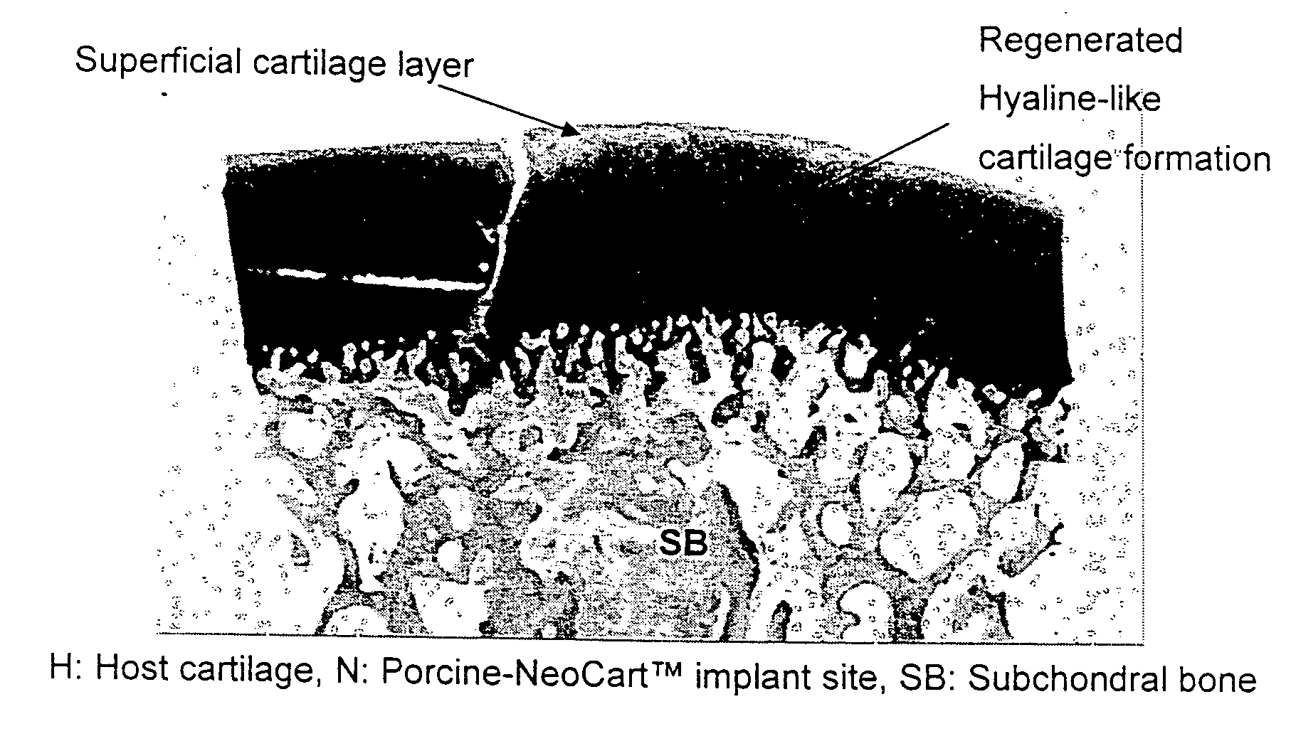
Method for in situ repair of injured, damaged, diseased or aged articular cartilage
a technology of articular cartilage and in situ repair, which is applied in the field of treatment of injured, damaged, diseased or aged articular cartilage, and can solve the problems of debilitating disability, affecting mobility, and damage to the articular cartilage of active individuals and older generation adults
- Summary
- Abstract
- Description
- Claims
- Application Information
AI Technical Summary
Benefits of technology
Problems solved by technology
Method used
Image
Examples
example 1
Isolation of Chondrocytes from Source Tissue
[0362] This example describes the procedure used for isolation of chondrocytes from swine cartilage.
[0363] Chondrocytes were enzymatically isolated from cartilage harvested under sterile conditions from the hind limbs of 6-month old swine. The femur was detached from the tibia and the trachea head exposed. Strips of cartilage were removed from the trachea using a surgical blade.
[0364] The cartilage was minced, digested in a 0.15% collagenase type I solution in DMEM / Nutrient Mixture F-12 (DMEM / F-12) 1:1 mixture with 1% penicillin-streptomycin (P / S) and gently rotated for 18 hours at 37° C. Chondrocytes were collected and rinsed twice by centrifugation at 1500 rpm for 5 min. Chondrocytes were re-suspended in DMEM / F-12 containing 1% penicillin-streptomysin and 10% FBS.
[0365] Chondrocytes were expanded for about 5 days at 37° C.
example 2
The Production of Human Neo-Cartilage Construct
[0366] This example describes conditions for production of neo-cartilage for human use.
[0367] The patient undergoes arthroscopic biopsy of a small (200-500 mg) piece of healthy cartilage from the ipsilateral knee. The biopsy is taken from the non-weight bearing portion of the femoral condyle or from the femoral notch as deemed most appropriate for the patient. The biopsy sample is placed into a sterile, non-cytotoxic, non-pyrogenic specimen container which is packaged and shipped to the laboratory.
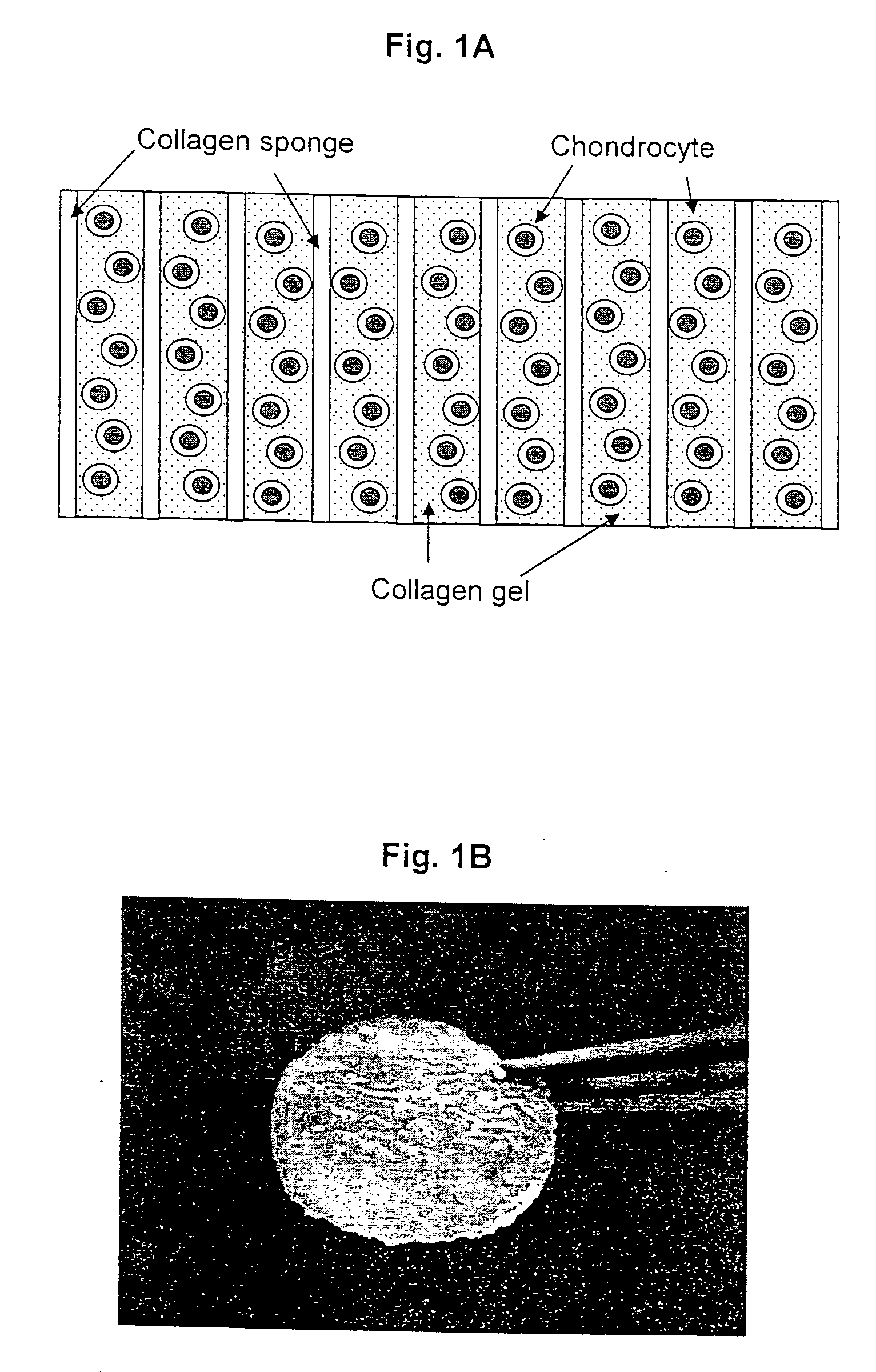
[0368] At the laboratory the biopsy sample is examined against acceptance criteria and then transferred to the chondrocyte isolation and expansion area. Samples from the biopsy specimen transport buffer are tested for sterility and for mycoplasma. The expanded chondrocytes are suspended in VITROGEN® gellable collagen solution, commercially available from Cohesion Corp., Palo Alto, Calif. A pre-formed collagen sponge (22×22 mm square and 2-4...
example 3
Preparation of Support Matrices
[0371] This example illustrates preparation of the cellular support matrix, also called the TESS matrix.
[0372] 300 grams of a 1% aqueous atelocollagen solution (VITROGEN®), maintained at pH 3.0, is poured into a 10×20 cm tray. This tray is then placed in a 5 liter container. A 50 ml open container containing 30 ml of a 3% aqueous ammonia solution is then placed next to the tray, in the 5 liter chamber, containing 300 grams of said 1% aqueous solution of atelocollagen. The 5 liter container containing the open trays of atelocollagen and ammonia is then sealed and left to stand at room temperature for 12 hours. During this period the ammonia gas, released from the open container of aqueous ammonia and confined within the sealed 5 liter container, is reacted with the aqueous atelocollagen resulting in gelling said aqueous solution of atelocollagen.
[0373] The collagenous gel is then washed with water overnight and, subsequently, freeze-dried to yield a ...
PUM
| Property | Measurement | Unit |
|---|---|---|
| frequency | aaaaa | aaaaa |
| pressure | aaaaa | aaaaa |
| temperature | aaaaa | aaaaa |
Abstract
Description
Claims
Application Information
 - R&D
- Intellectual Property
- Life Sciences
- Materials
- Tech Scout
- Unparalleled Data Quality
- Higher Quality Content
- 60% Fewer Hallucinations
Browse by: Latest US Patents, China's latest patents, Technical Efficacy Thesaurus, Application Domain, Technology Topic, Popular Technical Reports.
© 2025 PatSnap. All rights reserved.Legal|Privacy policy|Modern Slavery Act Transparency Statement|Sitemap|About US| Contact US: help@patsnap.com


