Covalent tethering of functional groups to proteins

a functional group and functional group technology, applied in the field of biochemical assays and reagents, can solve the problems of limited labeling limited fluorescence of intrinsically labeled proteins such as gfp, and poor functional sfv expression in reducing environments, so as to reduce imaging efficiency and efficient fluorescence of intrinsically labeled proteins

Active Publication Date: 2008-01-31
PROMEGA CORP
View PDF41 Cites 40 Cited by
  • Summary
  • Abstract
  • Description
  • Claims
  • Application Information

AI Technical Summary

Benefits of technology

[0012] In one embodiment, substrates of the invention are permeable to the plasma membranes of cells. For instance, as described herein the plasma membranes of prokaryotic (E. coli) and eukaryotic (CHO—K1) cells were permeable to TAMRA-C14H24O4—Cl and biotin-C18H32O4—Cl and, these substrates were rapidly and efficiently loaded into and washed out of cells in the absence of a mutant hydrolase. In the presence of a mutant hydrolase, at least a portion of the substrate was prevented from being washed out of the cells. Thus, the bound portion of the substrate can serve as a marker or as a means to capture the mutant hydrolase or a fusion thereof.
[0035] When performing image analysis on a cell, it may be desirable to fix the cell with a preservative (fixative) such as paraformaldehyde, acetone or methanol which generally maintains most features of cellular structure. Such fixed cells are then often analyzed by adding fluorescent stains or fluorescently labeled antibodies to reveal specific structures within the cells. Another method to fluorescently label cells is to express a fluorescent protein, e.g., GFP, in cells prior to fixation. Unfortunately, the efficient fluorescence of these proteins is dependent on protein structure, which can be disrupted by preservatives, thus decreasing the efficiency of imaging in those cells.

Problems solved by technology

However, while GFP-based indicators are currently employed in a variety of assays, e.g., measuring pH (Kneen et al., 1998; Llopis et al., 1998; Miesenböck et al., 1998), Ca2+ (Miyawaki et al., 1997; Rosomer et al., 1997), and membrane potential (Siegel et al., 1997), the fluorescence of intrinsically labeled proteins such as GFP is limited by the properties of protein structure, e.g., a limited range of fluorescent colors and relatively low intrinsic brightness (Cubitt et al., 1995; Ormö et al., 1996), and To address the deficiencies of GFP labeling in situ, Griffen et al.
Furthermore, labeling proteins by FLASH is limited by the range of fluorophores that may be used.
Nevertheless, functional sFv expression may be relatively poor in reducing environments.

Method used


Image

  • Covalent tethering of functional groups to proteins
  • Covalent tethering of functional groups to proteins
  • Covalent tethering of functional groups to proteins

Examples


example i

General Methodologies

[0190] Unless defined otherwise, all technical and scientific terms used herein have the same meaning as commonly understood by one of ordinary skill in the field of molecular biology and cellular signaling and modeling. Generally, the nomenclature used herein and the laboratory procedures in spectroscopy, drug discovery, cell culture, molecular genetics, plastic manufacture, polymer chemistry, diagnostics, amino acid and nucleic acid chemistry, and alkane chemistry described below are those well known and commonly employed in the art. Standard techniques are typically used for preparation of plastics, signal detection, recombinant nucleic acid methods, polynucleotide synthesis, and microbial culture and transformation (e.g., electroporation, lipofection).

[0191] The techniques and procedures are generally performed according to conventional methods in the art and various general references (see generally, Sambrook et. al. Molecular Cloning: A laboratory manual...

example ii

A DhaA-Based Tethering System

A. Wild-Type and Mutant DhaA Proteins and Fusions Thereof

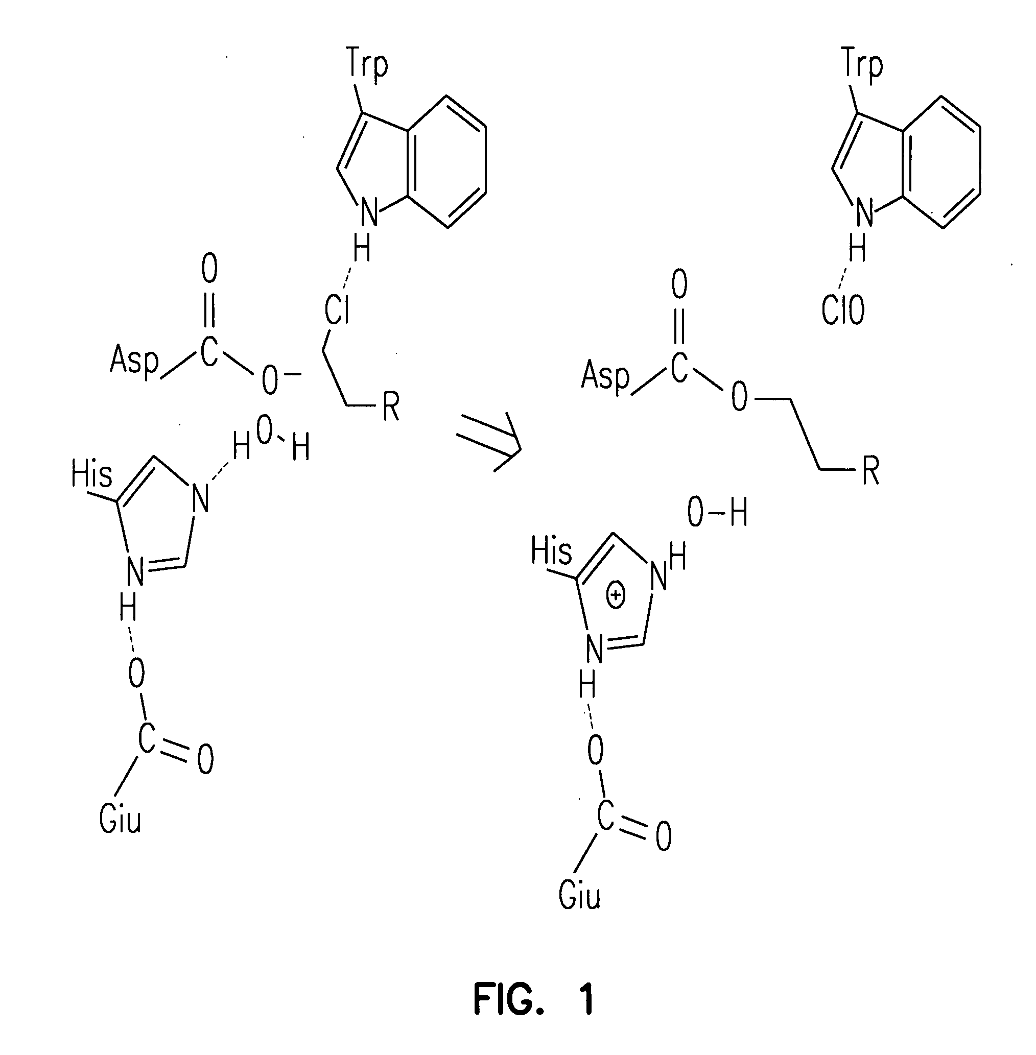
[0209] A halo-alkane dehydrogenase from Rhodococcus rhodochrous is a product of the DhaA gene (MW about 33 kDa). This enzyme cleaves carbon-halogen bonds in aliphatic and aromatic halogenated compounds, e.g., HaloC3-HaloC10. The catalytic center of DhaA is a typical “catalytic triad”, comprising a nucleophile, an acid and a histidine residue. It is likely that substrate binds to DhaA to form an E·S complex, after which nucleophilic attack by Asp106 forms an ester intermediate, His272 then activates H2O that hydrolyzes the intermediate, releasing product from the catalytic center. To determine whether a point mutation of the catalytic His272 residue impairs enzymatic activity of the enzyme so as to enable covalent tethering of a functional group (FG) to this protein, mutant DhaAs were prepared.

Materials and Methods

[0210] To prepare mutant DhaA vectors, Promega's in vitro mutagenesis kit which ...

example iii

Tethering of Luciferase to a Solid Support via a Mutant DhaA and a Substrate of the Invention

Materials and Methods

[0235] phRLuc-linker-DhaA.WT-Flag and phRLuc-linker-DhaA.H272F-Flag fusion cassettes were constructed by cloning the phRLuc coding region into the NheI / SalI sites of the pCIneo vector which contains a myristic acid attachment peptide coding sequence (MAS). Two primers (5′-GCTTCACTTGTCGTCATCGTCCTTGTAGTCA-3′; SEQ ID NO:11) and (5′-GCTTCACTTGTCGTCATCGTCCTTGTAGTCA-3′; SEQ ID NO:12) were designed to add NheI and SalI sites to the 5′ and 3′ coding regions, respectively, of phRLuc and to amplify a 900 bp fragment from a phRLuc template (pGL3 vector, Promega). Then, a myristic acid attachment peptide coding sequence was excised with NheI and SalI restriction enzymes and the amplified fragment containing phRLuc was inserted into the NheI / SalI restriction sites of pCIneo.DhaA.(WT or H272F)-Flag vector. The sequence of each construct was confirmed by DNA sequencing. Promega's TN...


PUM

PropertyMeasurementUnit
lengthaaaaaaaaaa
w/waaaaaaaaaa
pHaaaaaaaaaa

Abstract

A mutant hydrolase optionally fused to a protein of interest is provided. The mutant hydrolase is capable of forming a bond with a substrate for the corresponding nonmutant (wild-type) hydrolase which is more stable than the bond formed between the wild-type hydrolase and the substrate. Substrates for hydrolases comprising one or more functional groups are also provided, as well as methods of using the mutant hydrolase and the substrates of the invention. Also provided is a fusion protein capable of forming a stable bond with a substrate and cells which express the fusion protein.

Description

CROSS-REFERENCE TO RELATED APPLICATIONS [0001] This application is a divisional of U.S. patent application Ser. No. 10 / 768,976, filed Jan. 30, 2004, which claims the benefit of the filing date of U.S. application Ser. No. 60 / 444,094, filed Jan. 31, 2003, and U.S. application Ser. No. 60 / 474,659, filed May 30, 2003, under 35 U.S.C. §119(e), and incorporates those applications by reference herein.FIELD OF THE INVENTION [0002] This invention relates to the field of biochemical assays and reagents. More specifically, this invention relates to mutant proteins covalently linked (tethered) to one or more functional groups and to methods for their use. BACKGROUND OF THE INVENTION [0003] The specific detection of molecules is a keystone in understanding the role of that molecule in the cell. Labels, e.g., those that are covalently linked to a molecule of interest, permit the ready detection of that molecule in a complex mixture. The label may be one that is added by chemical synthesis in vit...

Claims


Application Information

Patent Timeline
Patent Type & Authority Applications(United States)
IPC IPC(8): C12Q1/02C07C233/00C07C271/06C07D209/00C07D209/34G01N33/573C07D221/00C07D311/02C07D311/82C07D403/00C12NC12N9/14C12N9/86C12Q1/26C12Q1/34G01N33/50G01N33/58
CPCC12N9/14C12N9/86Y10S436/80G01N33/5005G01N33/58C12Q1/34C07D209/48C07D233/64C07D311/78C07D311/82C07D405/06C07D493/10C07D495/04C12Y308/01005C07C217/08C07C271/16C07F5/022
Inventor WOOD, KEITH V.KLAUBERT, DIETERLOS, GEORGYI V.BULLEIT, ROBERT F.MCDOUGALL, MARKZIMPRICH, CHAD
Owner PROMEGA CORP
Why Eureka
Try Eureka