Conventionally,
cell injection has been conducted manually; however, long training, low throughput, and low success rates from poor reproducibility in manual operations call for the
elimination of direct human involvement and
fully automated injection systems.
The laborious manual injection task easily causes fatigue in injection technicians and hinders performance consistency and success rates.
The current manual technology is not capable of meeting the needs of such high-throughput applications.
Currently, no automated, high-throughput
zebrafish embryo injection systems are available.
Although the microrobots can to a certain extent facilitate
cell injection by a
human operator without long training, the human involvement still exists in the process of cell injection, resulting in a low throughput and reproducibility.
However, switching from one embryo to another was conducted manually, and thus, injection was
time consuming.
One drawback of this
system is that manual alignment of the two stages was required before injection.
The large alignment error would greatly influence the injection performance.
More importantly, the low stiffness of the MEMS
injector requires that the hard embryo chorion be removed in order to facilitate the injection, which may affect subsequent embryonic development, making the system unsuitable for zebrafish or mouse embryo injection.
Additionally, randomly dispersing embryos slows down the injection speed due to the embryo searching process.
In this system, introducing oocytes into regular patterned wells is conducted manually, which is tedious and
time consuming.
The
silicon substrate is not optically transparent, making the observation, monitoring, and control of the injection process difficult.
Thus, the detection methods proposed are not suitable to use for
zebrafish embryo injection.
First, precisely aligning microneedles with regularly positioned cells is difficult.
Manual alignment (in-plane or x-y alignment) through
microscopic observation from an off-optical-axis angle cannot guarantee a high accuracy.
Second, determining the
vertical distance (out-of-plane or z) between microneedle tips and cells is difficult.
Size differences from one cell to another (e.g., zebrafish embryos can differ by 200-300 cm) make
vertical alignment / positioning impossible.
These requirements for microneedles make the selection of MEMS-based solutions inappropriate.
In summary, parallel injection with MEMS microneedle arrays is not applicable to
zebrafish embryo injection.
It should be understood that despite their relatively
large size (˜600 μm and ˜1.2 mm including chorion), zebrafish embryos have a delicate structure and can be easily damaged.
They are also highly deformable, making the automatic manipulation task difficult.
Therefore specific difficulties in achieving automated
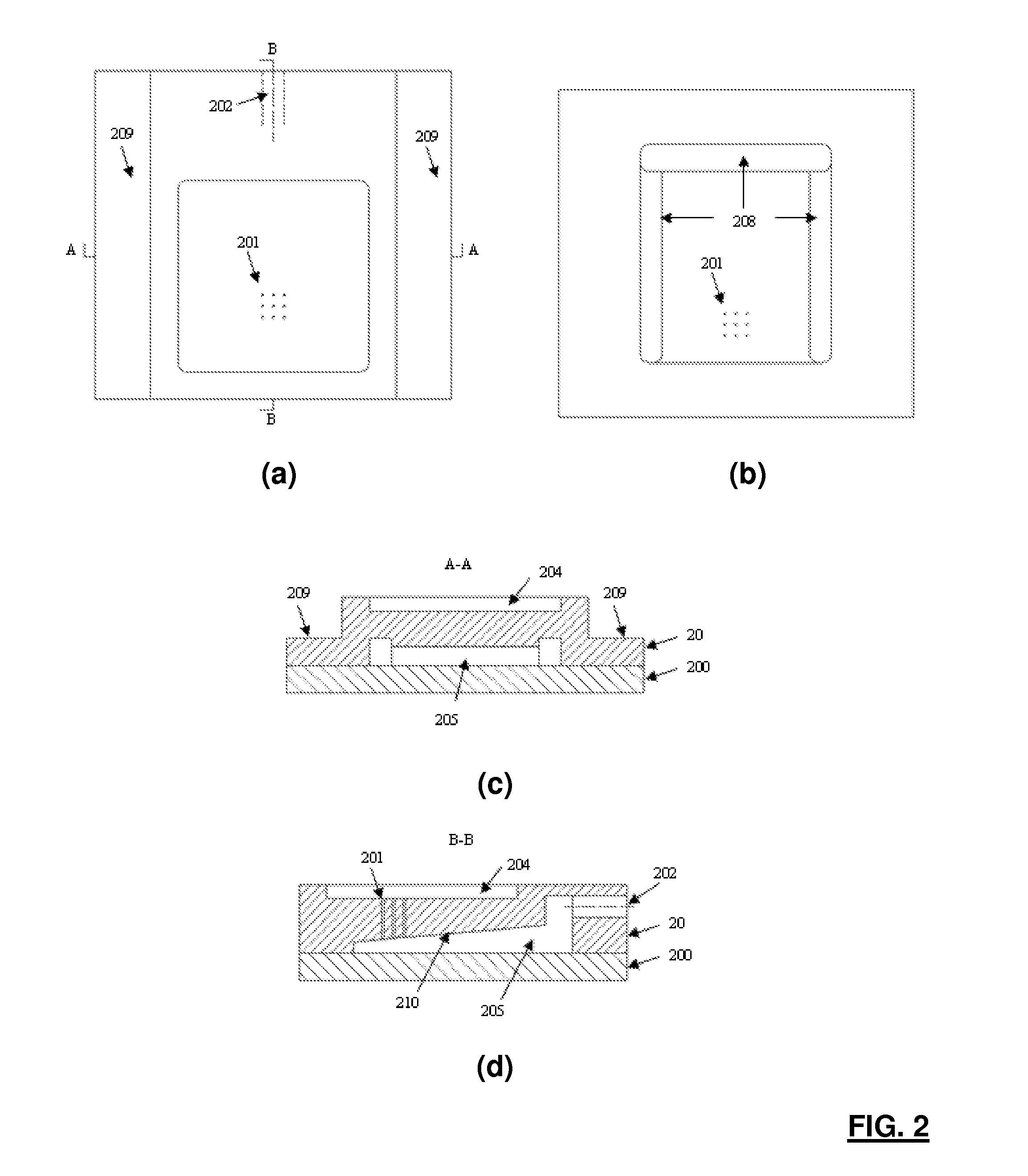
zebrafish embryo injection include: (i) the ability to quickly (i.e. seconds) immobilize a large number of zebrafish embryos into a
regular pattern; (ii) the ability to automatically and robustly identify cell structures for vision-based
position control (i.e.
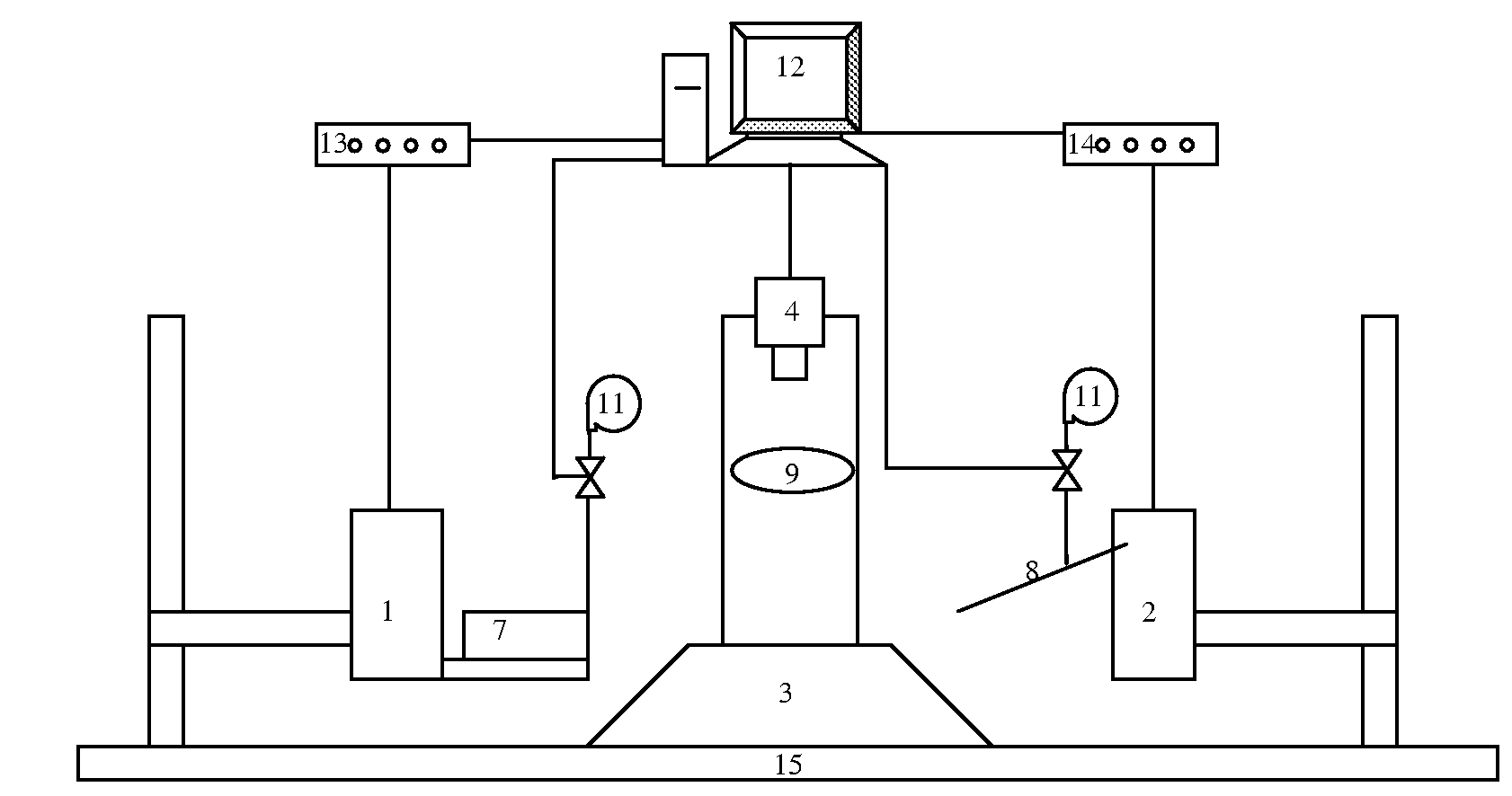
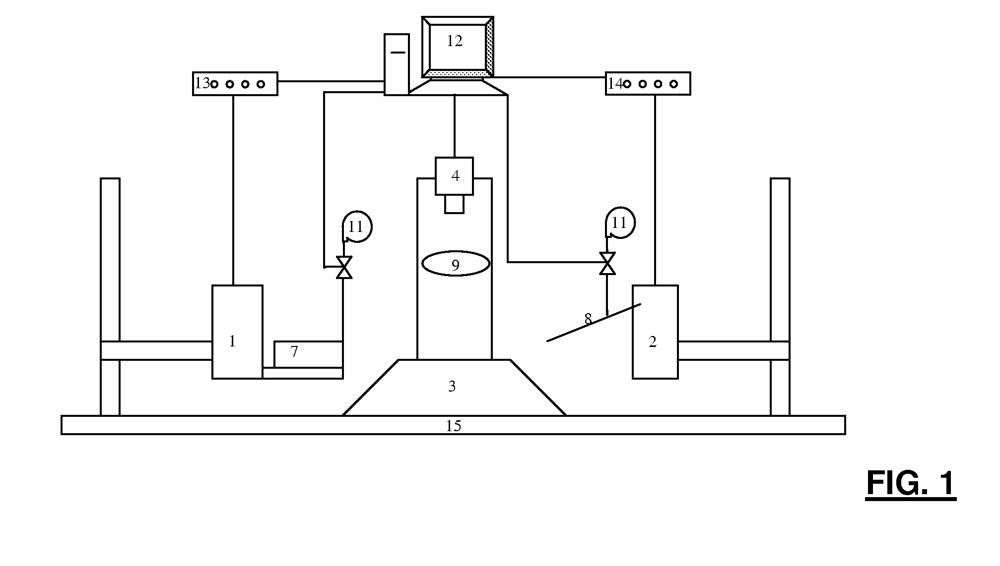
visual servoing) and account for size differences across embryos; and (iii) the ability to co-ordinately control two motorized positioning devices to achieve robust, high-speed zebrafish embryo injection.

