

Device for detection of molecules in biological fluids
a biological fluid and detection device technology, applied in the field of assays, can solve the problems of inability to administer treatment therapies within a certain time, inability to detect molecules in biological fluids,
- Summary
- Abstract
- Description
- Claims
- Application Information
AI Technical Summary
Problems solved by technology
Method used
Image

Examples
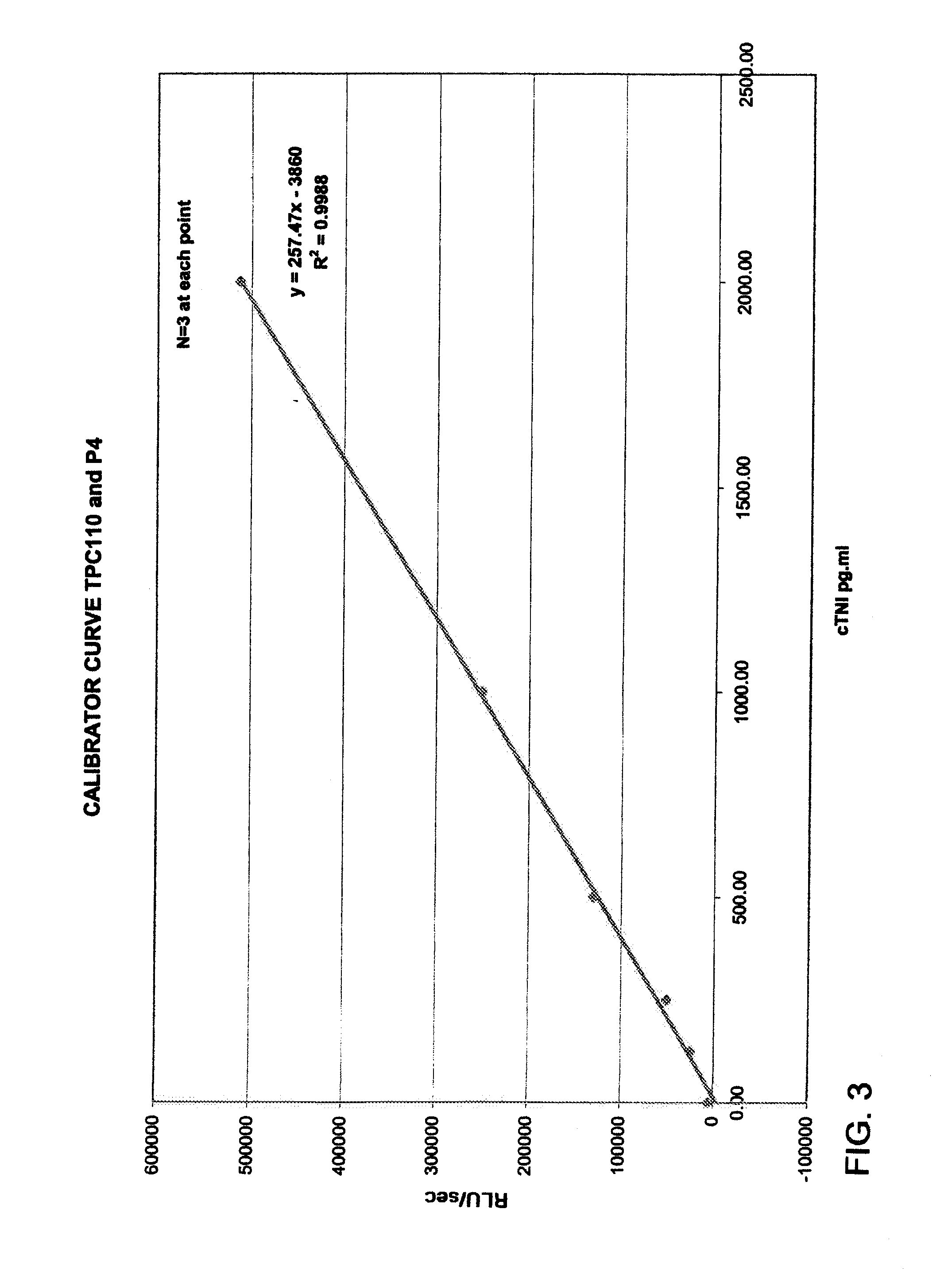
example 1
[0112]Calibration levels of 0, 125, 250, 500, 1000, and 2000 pg / ml cTNI in serum were run through the device using the procedures described above. The results were graphed and are seen in FIG. 3. A straight-line correlation (r2=0.9988) between the concentration and the fluorescent output is indicated.
example 2
[0113]A serum sample containing 500 pg / ml cTNI was placed in the device and the stir cycle run for two minutes. Three minutes after the cycle ended (i.e. 5 minutes total assay run time), the trapping zone was continuously scanned. Data was recorded every second. A sample containing no cTNI (i.e. a negative sample) was similarly processed. FIG. 4 is a graph of the results. The graph shows the fluorescent output (RLU / sec) versus the read interval (sec), where 0 corresponds to 5 minutes total assay run time. The 500 pg / ml sample began rising above the negative sample at a read interval of about 35 sec and peaked at about 41 sec, or about 5 minutes 41 seconds total assay run time. The graph showed that cTNI can be detected at a relatively low abundance within a minute of completing the stir and incubation cycles. A baseline was visible at both concentrations (0 and 500 pg / ml). Thus, cTNI was detectable at low levels and within an early time frame.
example 3
[0114]A series of serum samples were obtained from infected and non-infected individuals. The assay was used to determine the level of anti-HCV antibodies in each sample. The measurements were performed using a Night Owl instrument. The configuration of the strip and cassette remained the same, with the exception that a cocktail of peptides, from known epitopes of the HCV genome, were utilized as the trapping entities (i.e. second antibody). The trapping entities were biotinylated at the N-terminal residue and then overlaid on a neutravidin zone. The fluorescent signal entity used in the conjugate was goat anti-human phycoerythrin.
[0115]The procedure used was identical to that described above, with the exception that the sample was stirred using a standard magnetic stir plate. The images were captured on the Night Owl using a 20 msec exposure at 5 or 10 minute total assay run times. The number of DNA copies for each sample was provided by the sample supplier (Promeddx Inc.); a level...
PUM
 Abstract
Description
Claims
Application Information