Emission ratiometric indicators of phosphoinositides

Active Publication Date: 2008-04-24
THE JOHN HOPKINS UNIV SCHOOL OF MEDICINE
View PDF5 Cites 3 Cited by
  • Summary
  • Abstract
  • Description
  • Claims
  • Application Information

AI Technical Summary

Problems solved by technology

Due to the involvement of these PIs in such diverse functions, a cell must precisely control their spatial and temporal dynamics to avoid abnormalities, yet there are no reliable methods available for measuring PIP3, PI(3,4)P2 dynamics within subcellular compartments with high spatiotemporal resolution.
Previously employed indicators cause artifacts due to cell movements and additionally cannot be targeted to subcellular locations.
For dynamic monitoring of the lipid molecules, live-cell imaging using PH domains fused with fluorescent proteins (10-13, 32, 33) has been widely used, however their dependency on translocation limits their abilities to examine different pools of PIs.

Method used


Image

  • Emission ratiometric indicators of phosphoinositides
  • Emission ratiometric indicators of phosphoinositides
  • Emission ratiometric indicators of phosphoinositides

Examples


example 1

Methods

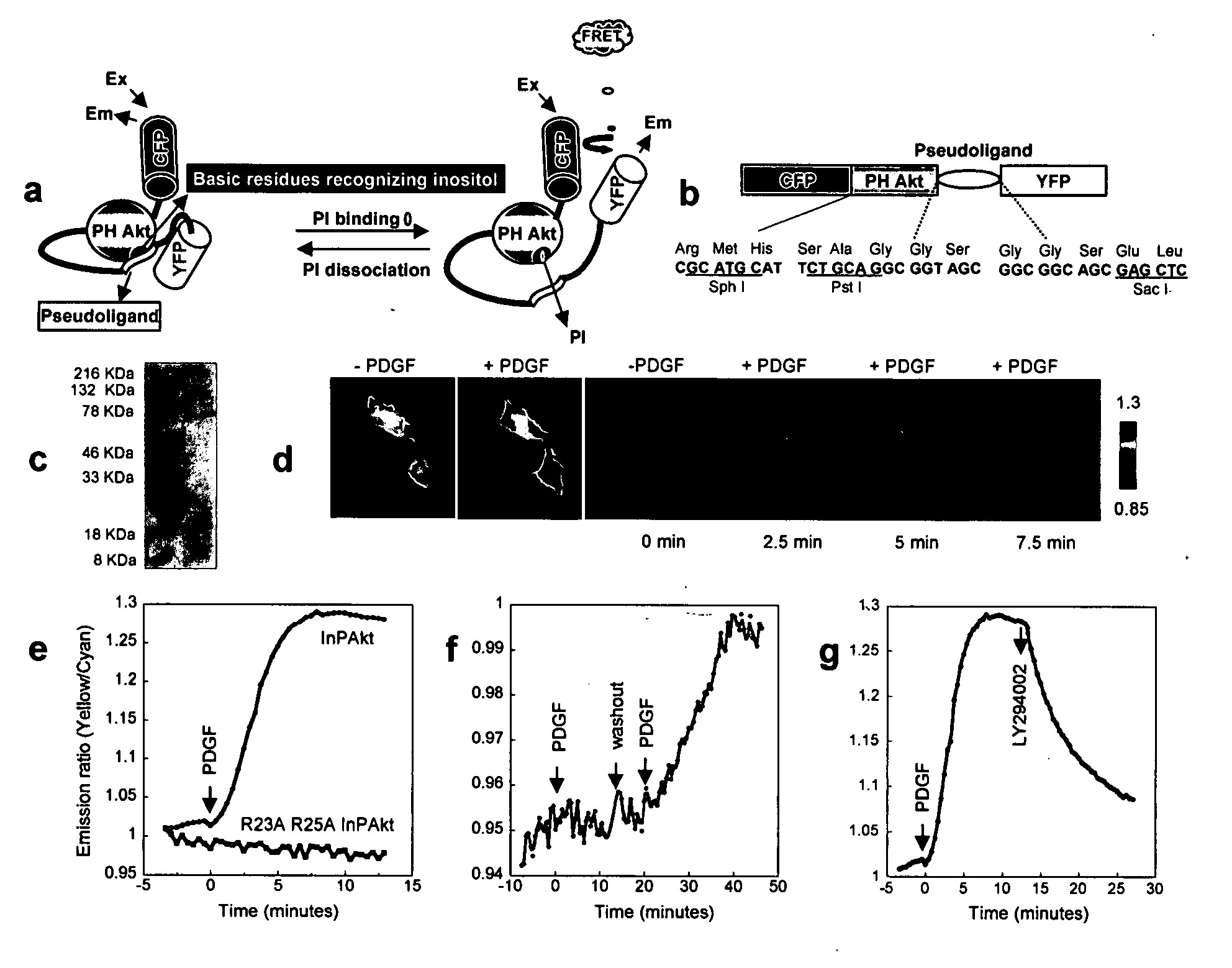
[0057] Reporter construction. The Akt / PKB PH domain (1-164) was created by PCR using full length human Akt (SEQ ID NO:1) as the template. The pseudoligand peptide sequence VAEEEDDEEEDEDD (SEQ ID NO:3) was inserted between the C-terminus of PH domain and N-terminus of improved versions of YFP, Citrine (16) or Venus (17). Double mutation R23A / R25A was incorporated by the QUICKCHANGE™ method (Stratagene) (FIG. 1). The constructs were first generated in pRSET B (Invitrogen) and subcloned into modified pcDNA3 (Invitrogen) behind a Kozako sequence for mammalian expression.

[0058] For plasma membrane targeting of InPAkt, the sequence KKKKKSKTKCVIM (SEQ ID NO:4) was used. For nuclear targeting, the nuclear localization signal (NLS) PKKKRKVEDA (SEQ ID NO:5) was attached to the C terminus of the Venus-containing construct.

[0059] Cell Culture. HEK293 and NIH3T3 cells were plated onto sterilized glass coverslips in 35-mm dishes and grown to 50-90% confluency in DMEM (10% FBS) at 37° C....

example 2

Development of a FRET Based Phosphoinositide Indicator

[0061] Design of the reporter. To measure the spatiotemporal dynamics of PIs in living cells, we employed a general molecular design in which binding of PIs in the “sensing unit” of the indicator can be translated into a change in the “reporting unit.” A pair of fluorescent proteins that can undergo fluorescence resonance energy transfer (FRET), enhanced cyan fluorescent protein (ECFP) and improved versions of yellow fluorescent proteins (YFP) namely Citrine (16) or Venus (17), was employed as the reporting unit (FIGS. 1A, 1B). These two fluorophores can be genetically fused to a conformationally responsive element, the conformational change of which alters the relative distance and / or orientation of the two fluorophores and generates a change in the emission ratio. Recently many such reporters have been developed for measuring signaling molecules like Ca2+, cGMP, cAMP, or signaling events such as protein phosphorylation (14, 15...

example 3

Differential Dynamics Stimulated by Different Growth Factors

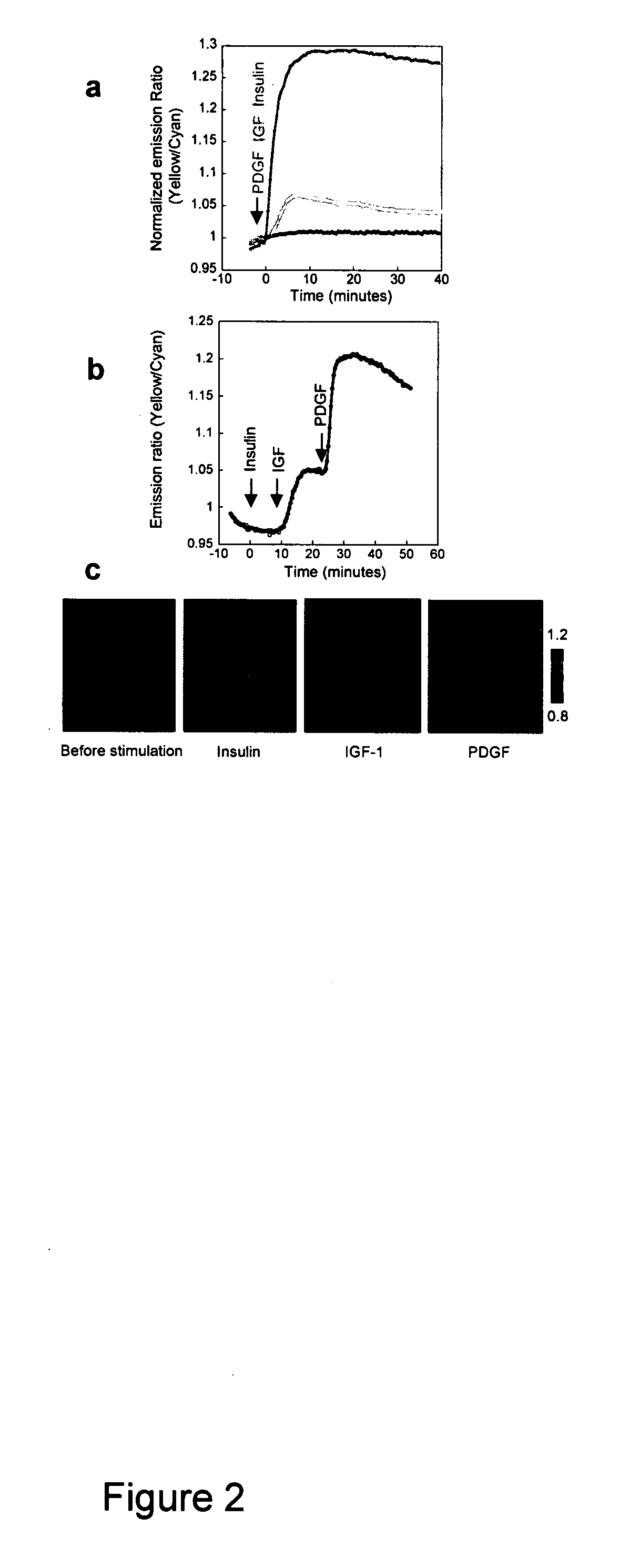
[0068] Although PI3K is a crucial regulatory component shared by various growth factor pathways, their differential coupling to PI3K isoforms (23), and distinct modes of negative regulation (24), could lead to significant difference in PI dynamics, thereby differentiating downstream signaling. To compare the accumulation of PIP3 and PI(3,4)P2 induced by the activation of different tyrosine kinase receptor pathways, we applied three different ligands, namely insulin, insulin-like growth factor 1 (IGF-1) and PDGF, to serum-starved NIH3T3 cells expressing InPAkt. As shown above, PDGF stimulation produced an acute response that reached a plateau of 25.4±3.7% ratio increase in 5-9 min (t1 / 2=3.45±0.65 min) (FIG. 2A). In contrast, addition of 50 nM IGF-1 to activate insulin like growth factor receptor (IGF-IR) produced a more gradual response of 6.5±0.8% (n=5) in 10-12 min (t1 / 2=5.4±0.4 min) (FIG. 2A). Finally, stimulation with 1...


PUM

PropertyMeasurementUnit
Angleaaaaaaaaaa
Resonance energyaaaaaaaaaa
Fluorescenceaaaaaaaaaa

Abstract

Emission ratiometric indicators of phosphoinositides comprise a fusion protein comprising a pleckstrin homology (PH) domain of Akt (also known as protein kinase B) and a “pseudoligand” containing acidic amino acid residues, which is sandwiched between resonance energy transfer (RET) pairs, such as cyan and yellow mutants of GFP (a FRET pair). Such indicators can be used, inter alia, to monitor spatiotemporal dynamics of phosphoinositides and in high throughput assays for inhibitors of P13K, including drug screening assays.

Description

[0001] This application claims the benefit of co-pending application Ser. No. 60 / 830,811 filed Jul. 14, 2006, which is incorporated herein by reference in its entirety. [0002] The patent or application file contains at least one drawing executed in color. Copies of this patent or patent application publication with color drawing(s) will be provided by the Office upon request and payment of the necessary fee.FIELD OF THE INVENTION [0003] The invention relates to the monitoring of phosphoinositides in living mammalian cells. BACKGROUND OF THE INVENTION [0004] Ligand activation of the receptors in the plasma membrane initiates cell signaling which propagates into various intracellular compartments through multi-step processes that depend on cellular second messengers. It has been shown that these short lived small molecules not only amplify the signal in the relay but also confer specificity by their temporal and spatial distribution in the cell. [0005] Although a relatively minor comp...

Claims


Application Information

Patent Timeline
IPC IPC(8): C07H21/04C12N5/00G01N33/53G01N33/566
CPCG01N33/542G01N2500/02G01N2333/9121G01N33/92
Inventor ZHANG, JINANANTHANARAYANAN, BHARATH
Owner THE JOHN HOPKINS UNIV SCHOOL OF MEDICINE
Why Eureka
Try Eureka