Protein Microscope

a protein microscope and microscope technology, applied in the field of protein microscopes, can solve the problems of inability to molecular imaging at the source of conventional esi, the inability to identify biochemical species in a single way, and the specificity of these methods

Inactive Publication Date: 2008-06-26
GEORGE WASHINGTON UNIVERSITY
View PDF16 Cites 92 Cited by
  • Summary
  • Abstract
  • Description
  • Claims
  • Application Information

AI Technical Summary

Benefits of technology

[0040]Another advantage of the present invention is that it provides spatially resolved chemical analysis of the sample correlated with the surface topography. Topographical analysis is achieved by scanning a sharp SNOM probe across the sample at constant distance from the surface. Chemical analysis by means of the laser-induced ionization mass spectrometry is achieved by delivering pulsed laser radiation to the sample surface through the same sharp SNOM probe, and consequent collection and analysis of mass spectra from the plume generated on the sample by the laser radiation.
is that it provides spatially resolved chemical analysis of the sample correlated with the surface topography. Topographical analysis is achieved by scanning a sharp SNOM probe across the sample at constant distance from the surface. Chemical analysis by means of the laser-induced ionization mass spectrometry is achieved by delivering pulsed laser radiation to the sample surface through the same sharp SNOM probe, and consequent collection and analysis of mass spectra from the plume generated on the sample by the laser radiation.

Problems solved by technology

Although several analytical methods offer high sensitivity and spatial resolution (fluorescence measurement, voltammetric microelectrodes, etc.), the selectivity and specificity of these methods seldom allows for the unambiguous identification of biochemical species.
In the mass-spectral analysis of biological materials, however, most spatial distribution information is lost during the sample preparation step, wherein cells are ground and thoroughly mixed to form a homogenized slurry which is then placed in a mass spectrometer for analysis.
The conventional ESI source is not amenable to molecular imaging, as it requires a liquid sample.
However, three obstacles exist with the MALDI technique.
First, the mixing and co-crystallization of the sample with the light-absorbing matrix largely obscures the original spatial distribution of analytes (e.g., through lateral mixing).
Second, the need to transfer the sample into a vacuum environment for mass analysis considerably restricts the choice of samples.
Significantly, both of these requirements for successful MALDI analysis exclude the possibility of in vivo measurements.
The third obstacle associated with the MALDI technique is that the laws of physics and practical considerations limit the focusable size of the laser spot so that it is larger than the wavelength of the laser light, resulting in a laser spot larger than most cells of interest and, thereby, diminishing the value of MALDI in view of the need for sampling from smaller regions.

Method used


Image

  • Protein Microscope
  • Protein Microscope
  • Protein Microscope

Examples


Embodiment Construction

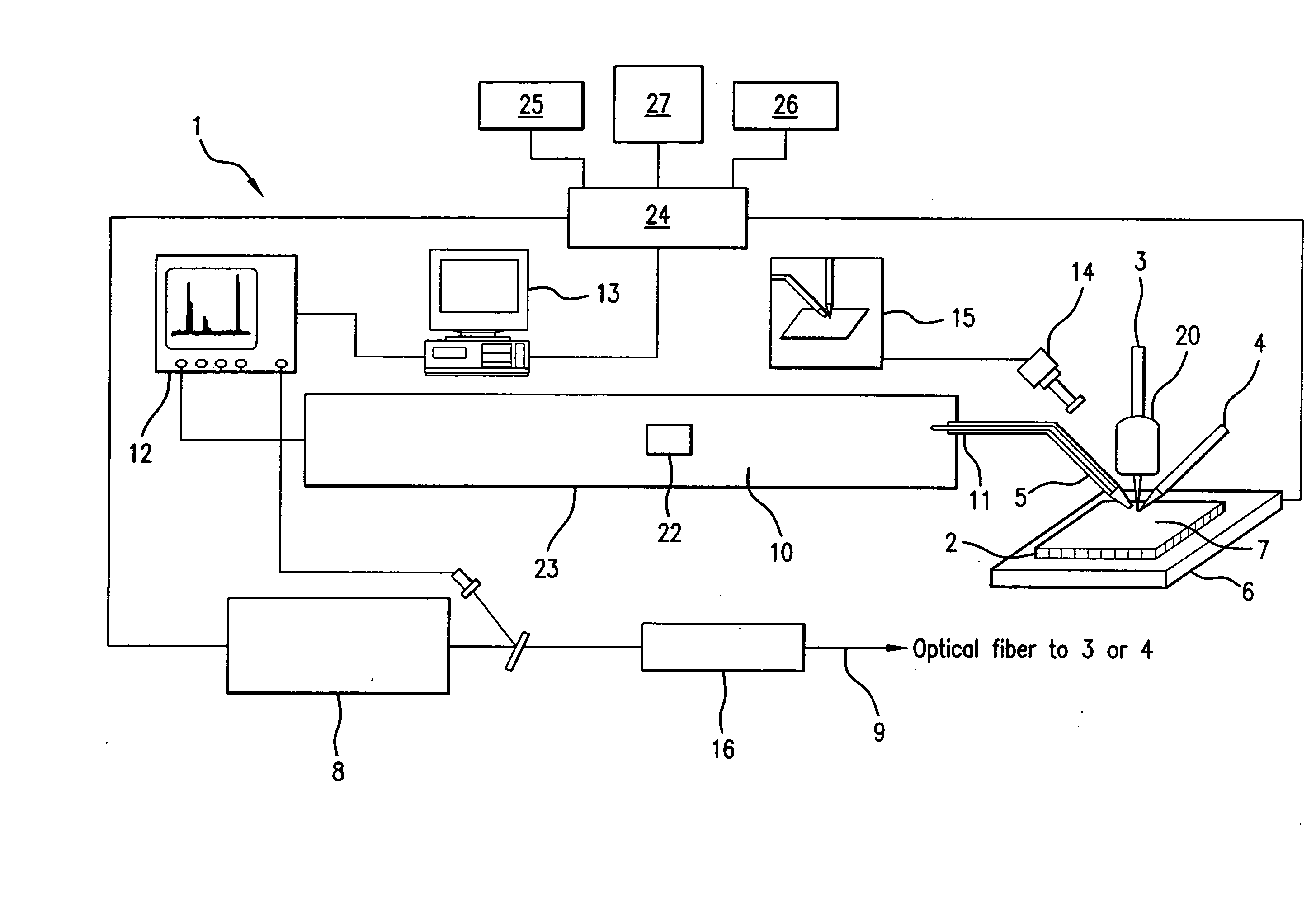
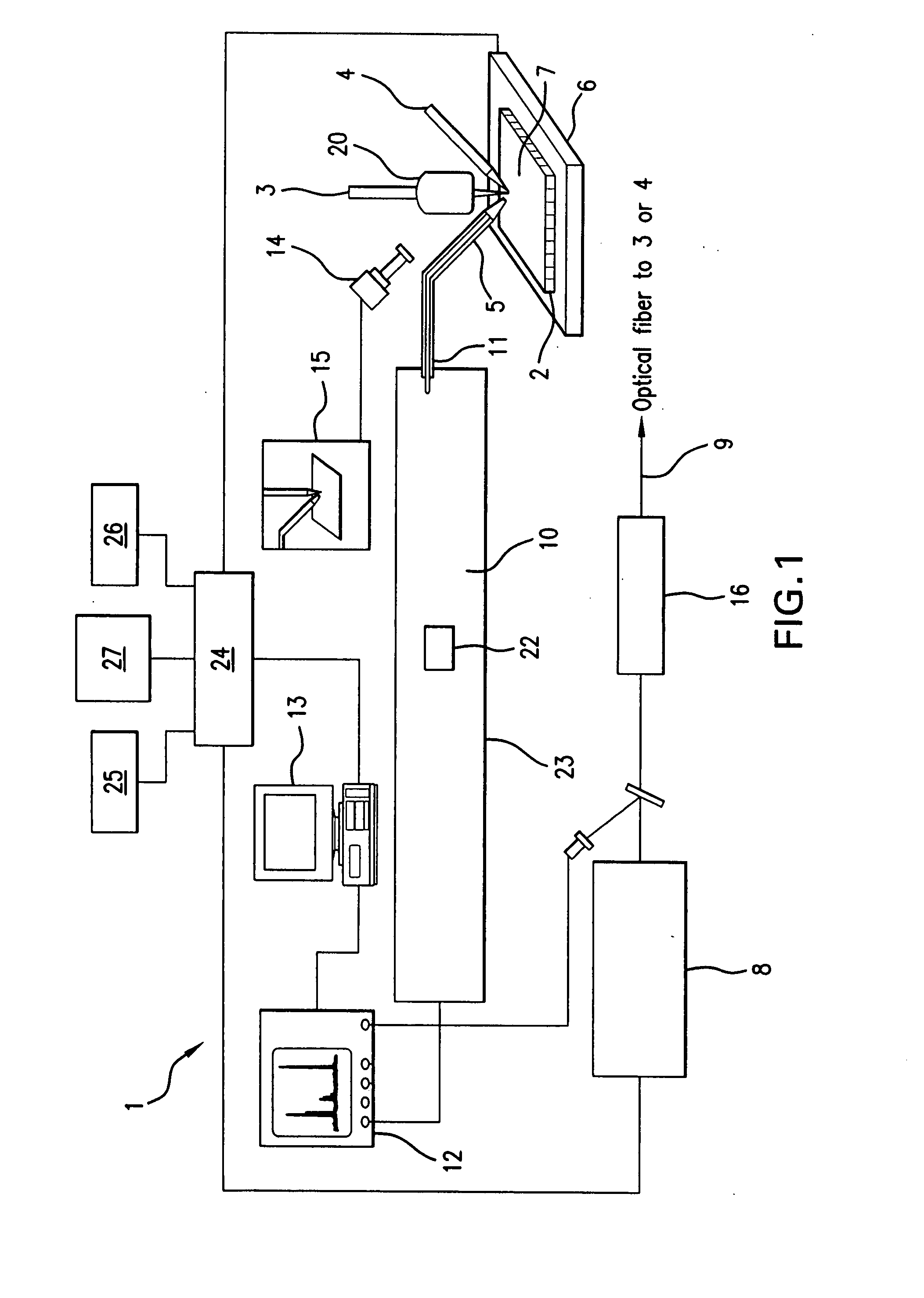
[0046]The present invention provides a novel system and method for analyzing and imaging samples of interest. The sample, also referred to as the target material, normally comprises a mixture of analyte materials and light-absorbing matrix substances. The sample can be solid or liquid and is composed of one or more materials selected from the groups consisting of peptides, proteins, lipids, carbohydrates, organic compounds and inorganic compounds. A particularly preferred material comprises tissue samples such as, for example, those disclosed in U.S. Pat. No. 5,808,300, which is hereby incorporated by reference herein in its entirety.

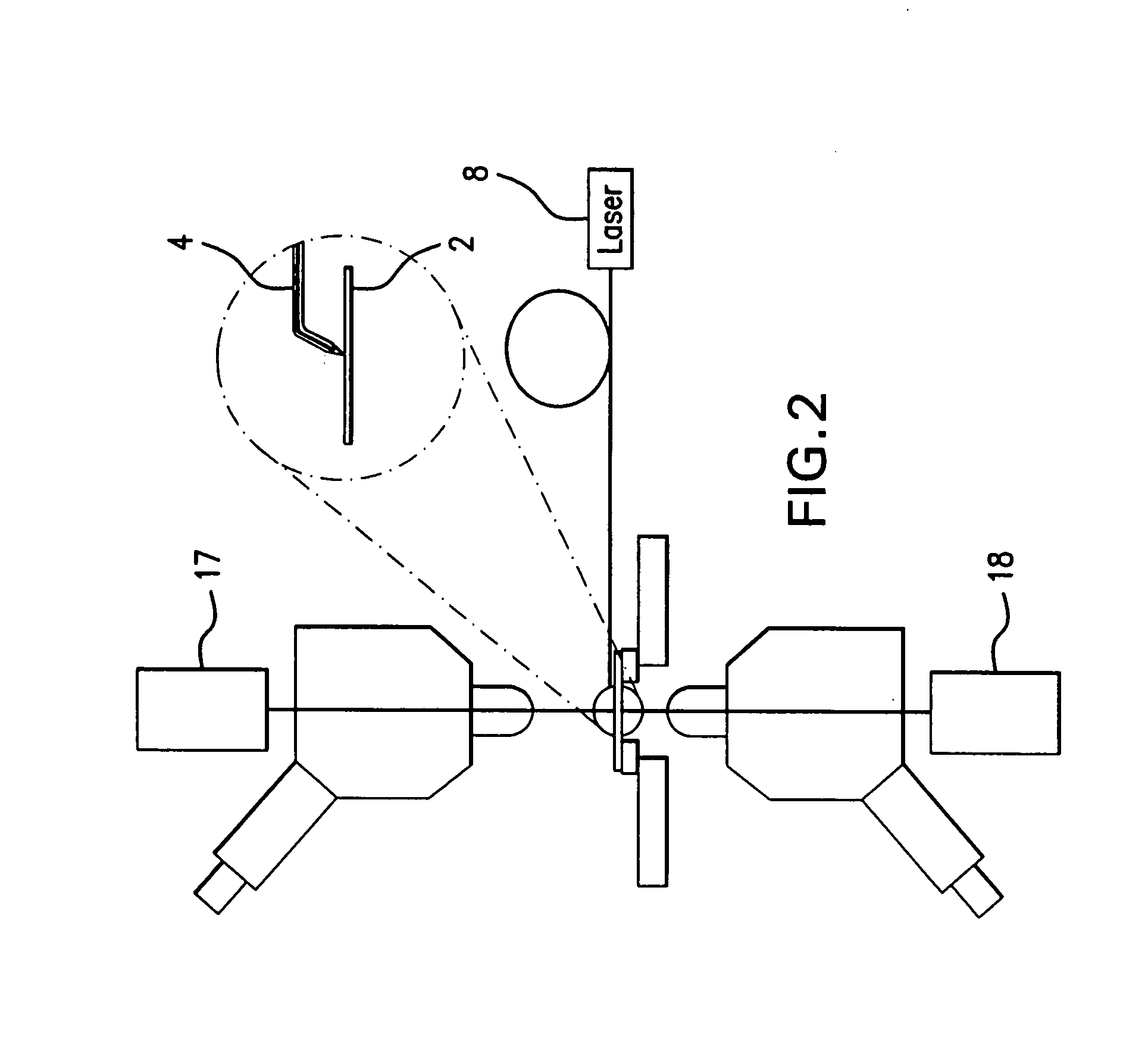
[0047]The sample is deposited in a matrix on a target surface of a sample support. When illuminated with the laser beam, the matrix molecules are ionized and evaporated. The ionized matrix molecules subsequently ionize the analyte molecules through charge transfer process. At the same time, the analyte molecules, analyte ions and fragmented analyte ions...


PUM


Abstract

A system and method for analyzing and imaging a sample containing molecules of interest combines modified MALDI mass spectrometer and SNOM devices and techniques, and includes: (A) an atmospheric-pressure or near-atmospheric-pressure ionization region; (B) a sample holder for holding the sample; (C) a laser for illuminating said sample; (D) a mass spectrometer having at least one evacuated vacuum chamber; (E) an atmospheric pressure interface connecting said ionization region and said mass spectrometer; (F) a scanning near-field optical microscopy instrument comprising a near-field probe for scanning the sample; a vacuum capillary nozzle for sucking in particles which are desorbed by said laser, the nozzle being connected to an inlet orifice of said atmospheric pressure interface; a scanner platform connected to the sample holder, the platform being movable to a distance within a near-field distance of the probe; and a controller for maintaining distance information about a current distance between said probe and said sample; (G) a recording device for recording topography and mass spectrum measurements made during scanning of the sample with the near-field probe; (H) a plotting device for plotting said topography and mass spectrum measurements as separate x-y mappings; and (I) an imaging device for providing images of the x-y mappings.

Description

FIELD OF THE INVENTION[0001]The present invention is directed to an instrument for identifying peptides, proteins and other biomolecules within a tissue or cell and providing information on their spatial and temporal distribution. More particularly, this invention relates to an instrument for identifying peptides, proteins and other biomolecules within a tissue or cell, which provides information on their spatial and temporal distribution down to submicron resolution, and which analyzes their activity in vivo.BACKGROUND OF THE INVENTION[0002]Living organisms function through the interactions of biomolecules intricately distributed in space and varying in time. Often, spatial variations within a tissue or cell hold the key to understanding the function of molecular components. The abundance of biomolecules can span a wide range. For example, protein concentrations range from millimolar (10−3 M) to attomolar (10−18 M), and perhaps less. Copy numbers as low as 101 to 102 protein molecu...

Claims


Application Information

Patent Timeline
Patent Type & Authority Applications(United States)
IPC IPC(8): B01D59/44G01Q10/00G01Q30/02G01Q60/18
CPCB82Y20/00B82Y35/00H01J49/0004G01Q60/22G01Q30/02
Inventor VERTES, AKOSREEVES, MARK E.KASHANCHI, FATAH
Owner GEORGE WASHINGTON UNIVERSITY
Why Eureka
Try Eureka