A method for determining glycosylation and terminal modification of samples during protein purification process

a protein purification and terminal modification technology, applied in the biotechnology field, can solve the problems of large amount of sample, uneven glycosylation, and complicated sample handling, and achieve the effects of improving the resolution and accuracy of test results, simple sample handling, and rapid determination of glycosylation

Active Publication Date: 2015-12-17
LIVZON MABPHARM
View PDF1 Cites 4 Cited by
  • Summary
  • Abstract
  • Description
  • Claims
  • Application Information

AI Technical Summary

Benefits of technology

[0036]In contrast, the method of the present invention uses ESI-MS to measure antibody multiply-charged ions, and then perform deconvolution calculation, thus greatly improving the resolution and accuracy of the test results (<30 ppm). In addition, the present invention is characterized in simple sample handling, requiring only a reducing agent to conduct reaction (45 min), small amount of sample (5 μg), and UPLC-MS detection of samples just completed within 16 min, meanwhile the data for antibody glycosylation, N-terminal pyroglutamination and C-terminal de-lysination can be obtained. The present invention is particularly applicable to experiments with a small amount of test sample, such as clones screening, and rapid batch detection during process development. Meanwhile, the present invention is also applicable to sample detection in conventional dosage (100 μg). The method of the present invention can detect glycosylation and terminal modification of different antibodies, such as IgG1, and IgG2, and also can be used to test the sample in antibody process development. Furthermore, in combination with carboxypeptidase B and cation-exchange chromatography (CEX-HPLC), the method of the present invention can also be applied to characterize and identify the structures of antibody charge isomers.
[0037]Immuneglobulin can be divided into five categories, IgG, IgA, IgM, Ig

Problems solved by technology

IgG glycosylation is uneven because of different glycoform and content.
Enzymatic quantitative fluorescence labeling method is a classic quantitative assay for determining IgG1 glycosylation, but the sample handling is quite complicated and time-consuming, and requires a large amount of sample.
However, these methods have some disadvantages, such as weak enzyme cleavage site selectivity, or high cost, or complicated sample handling, and hence they are inappropriate for batch testing of conventional or process development samples.
Application of LC-MS in peptide mapping analysis is theoretically possible to determine the glycosylation

Method used


Image

  • A method for determining glycosylation and terminal modification of samples during protein purification process
  • A method for determining glycosylation and terminal modification of samples during protein purification process
  • A method for determining glycosylation and terminal modification of samples during protein purification process

Examples


example 1

Determination of Conditions for Immunoglobulin Reducing Method

1.1 Determination of the Amount of Reducing Agent DTT

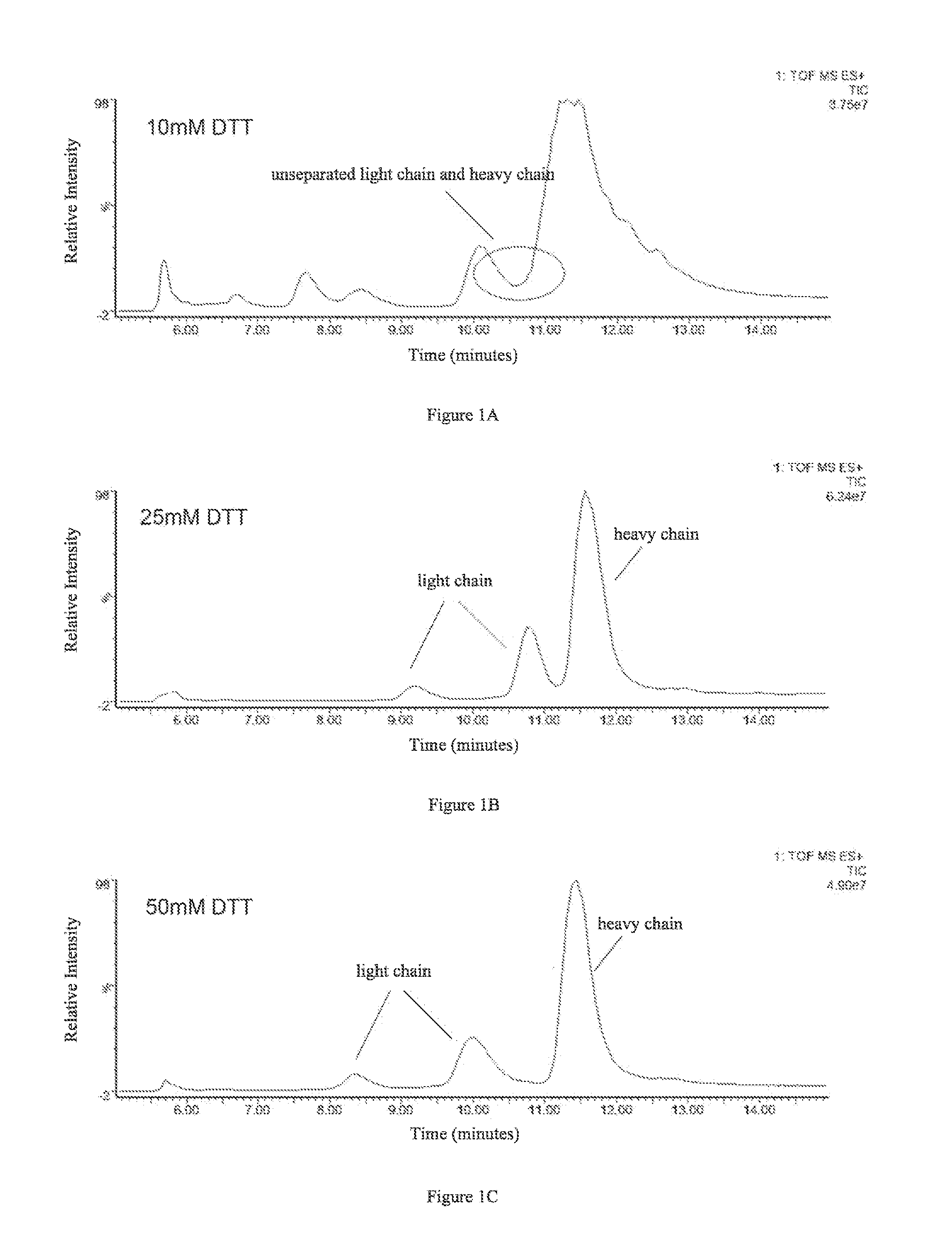
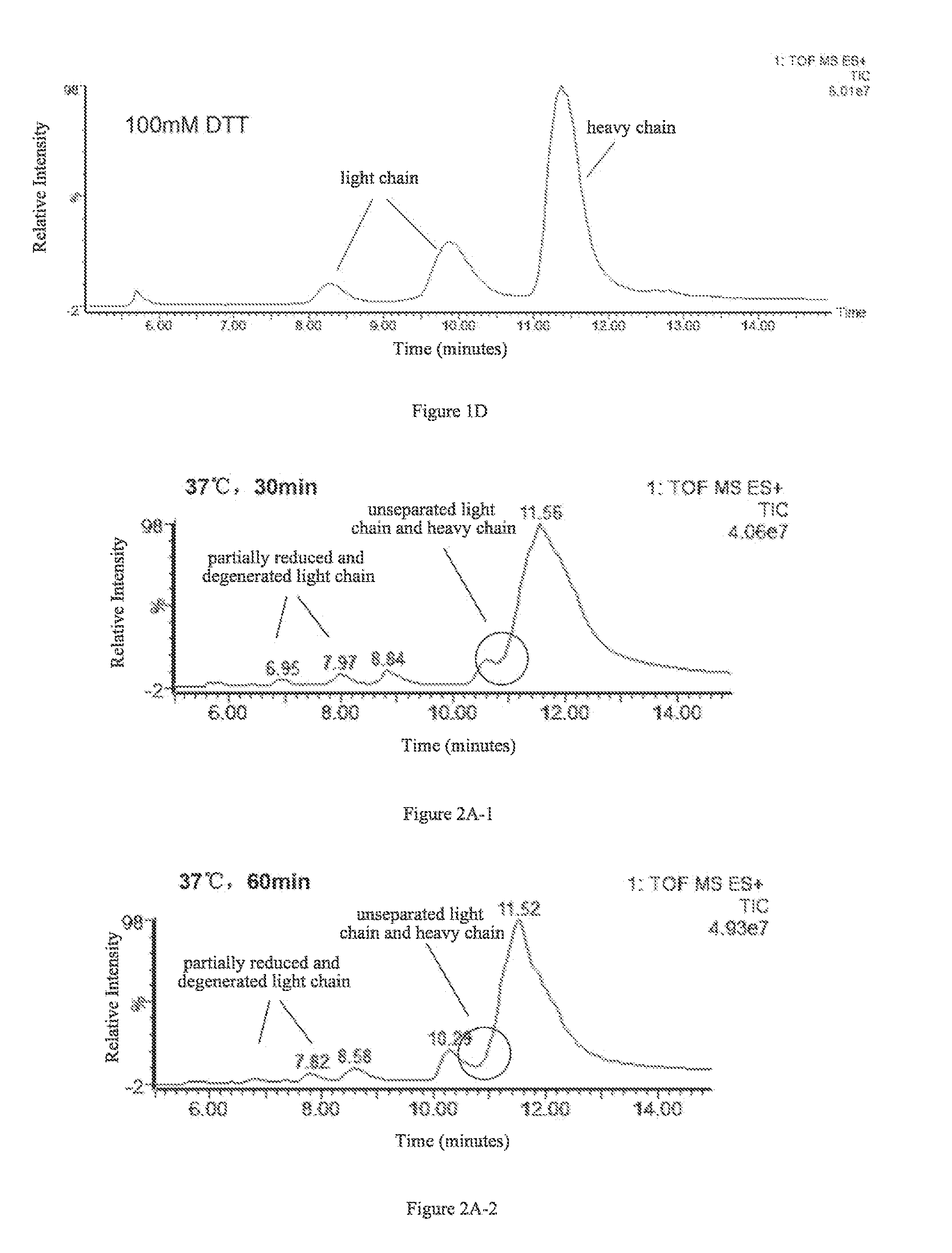
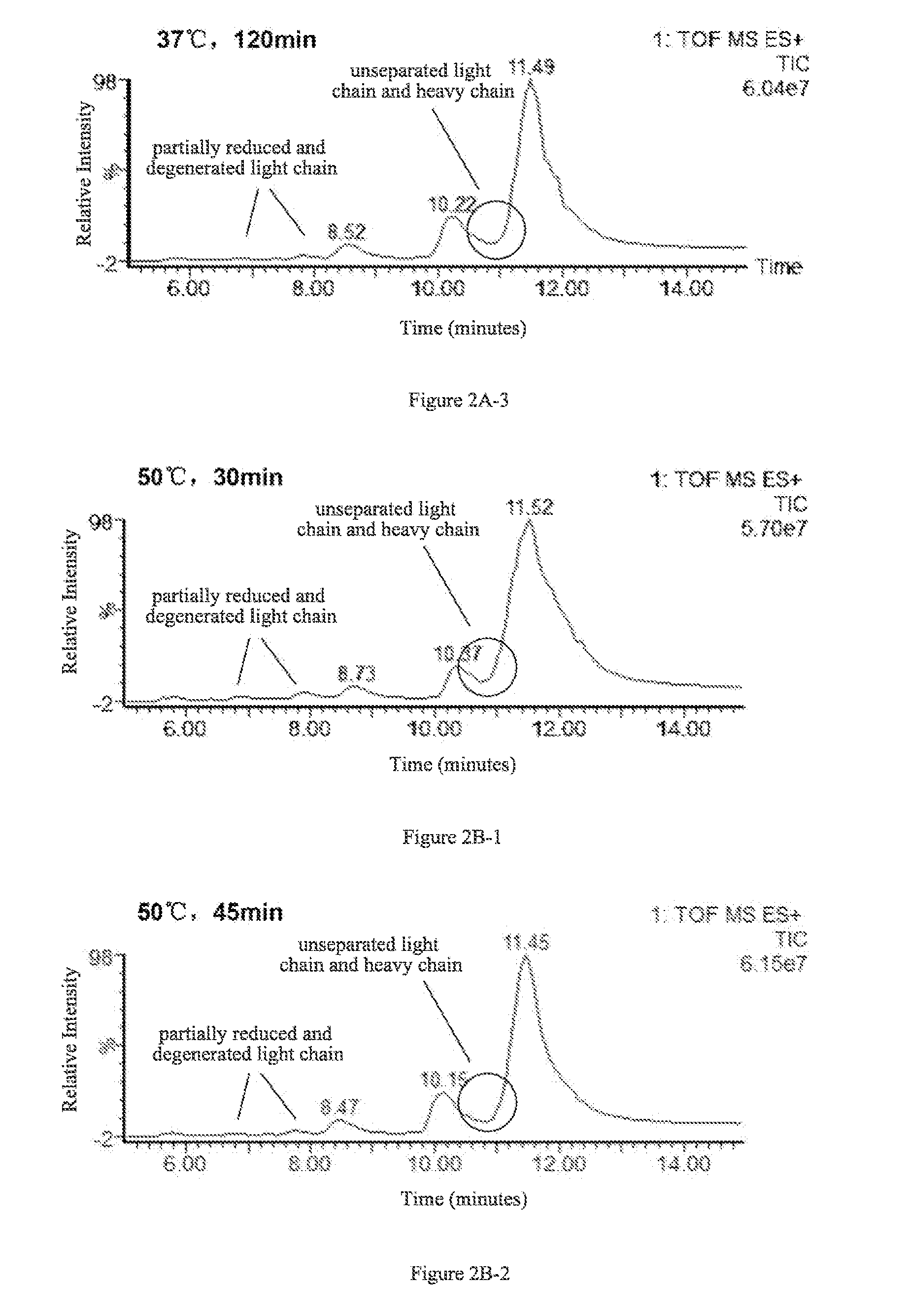
[0049]The effects of four different amounts of DTT on separation of the light and heavy chains were examined. 4 aliquots of 5 μg antibody protein A were respectively added to 10 μL 6M guanidine hydrochloride solution, followed by the addition of 0.1M DTT solution, 2 μL and 5 μL, and 0.5M DTT solution, 2 μL and 4 μL, as well as appropriate amounts of 6M guanidine hydrochloride solutions to make final DTT concentrations of 10 mM, 25 mM, 50 mM, and 100 mM, respectively. Then the resulting products were reacted with said IgG1 protein at 65° C. for 45 min.

[0050]C4 reverse-phase high pressure liquid chromatography was used to separate the light and heavy chains obtained in the reactions, and the liquid phase system used was UPLC (Waters, ACQUITY). Column: Waters, ACQUITYUPLC column, BEH C4, 1.7 μm (diameter), 300 Å (aperture), 2.1×50 mm. Chromatographic conditions were set as...

example 2

Determining the Glycosylation and Terminal Modification of Antibody a and Antibody B (IgG1) Using UPLC-MS Method of the Present Invention

[0069]The glycosylation and terminal modification of antibody A and antibody B were analyzed using the optimized reduction conditions (5 μg antibody A was added to 10 μL 6M guanidine hydrochloride solution, followed by the addition of 2 μL 0.5M DTT solution and finally an appropriate amount of 6M guanidine hydrochloride solution, to make a final DTT concentration of 50 mM; reacted for 45 min at 65° C.), UPLC separation (the same as Example 1.1), ESI-MS detection (the same as Example 1.1), and normalized data processing (the same as Example 1.2). The first amino acids at N-terminal of light chain and heavy chains of antibody A were both glutamine (Gin), for which pyroglutamic acid cyclization occurs readily. The first amino acids at N-terminal of light chain antibody B was glutamic acid (Glu), for which pyroglutamic acid cyclization does not occur r...

example 3

Determining the Glycosylation and Terminal Modification of Each Component of Antibody a after Purification Using UPLC-MS Method of the Present Invention

[0070]In purification process of antibody A, a conventional strong cation-exchange chromatography was used, with 20 mM sodium phosphate buffer as loading buffer, 20 mM sodium phosphate and 1M sodium chloride buffer (pH=6.0) as elution buffer, a flow rate of 200-400 cm / h. The eluting components were monitored by UV absorption at 280 nm. Then, the components of antibody A were collected in accordance with retention time: component 1 (4000-4300 min), component 2 (4300-4500 min), component 3 (4500-4650 min), component 4 (4650-4800 min), and component 5 (4800-5100 min). The glycosylation and terminal modifications of each component of the IgG1 were analyzed using the optimized reduction conditions (5 μg antibody A was added to 10 μL 6M guanidine hydrochloride solution, followed by the addition of 2 μL 0.5M DTT solution and finally an appr...


PUM


Abstract

The present invention provides a method for determining glycosylation and terminal modifications of immunoglobulin during immunoglobulin purification process, which can simultaneously and rapidly determine glycosylation, N-terminal pyroglutamination and C-terminal de-lysination of immunoglobulin. The method comprises: 1) separating immunoglobulin by using cation-exchange resin, and collecting different components in according to retention time; 2) denaturing the components of immunoglobulin obtained in step 1) with a denaturant, followed by reducing them with a reducing agent, to separate the light chain and heavy chain; 3) separating the light chain and heavy chain of immunoglobulin of step 2) by using reverse phase ultrahigh pressure liquid chromatography; 4) measuring the molecular weights of the light chain and heavy chain obtained in step 3) with mass spectrum; 5) analyzing the chromatographic data obtained in step 3) and the mass spectrometric data obtained in step 4) to determine glycosylation and terminal modifications of said immunoglobulin.

Description

TECHNICAL FIELD[0001]The present invention relates to biotechnology field. Specifically, the present invention provides a method for determining glycosylation and terminal modifications of samples during protein purification process. Meanwhile, the present invention also relates to a kit for determining protein glycosylation and terminal modification during immunoglobulin purification process.BACKGROUND[0002]Over the past decade, monoclonal antibodies achieved significant success and tremendous growth in bio-pharmaceutical industry, as well as the entire pharmaceutical industry. Compared with the traditional small molecule drugs, monoclonal antibodies possess many advantages including good specificity, significant therapeutic effect, less side effects, small administration amount, and so on. Regarding the characteristics of drug molecule, the antibodies have greater heterogeneity. This property of the antibodies is caused by a variety of factors, of which post-translational modifica...

Claims


Application Information

Patent Timeline
IPC IPC(8): G01N33/68G01N30/72G01N30/06
CPCG01N33/6848G01N30/06G01N30/7233G01N2030/027G01N2560/00G01N2030/065G01N2030/067G01N2440/38G01N2030/8831G01N33/6854Y10T436/24
Inventor ZHU, BAOGUOPENG, YUCAIYANG, JIAMING
Owner LIVZON MABPHARM
Why Eureka
Try Eureka