

Nanoparticle adjuvants for sub-unit vaccines
- Summary
- Abstract
- Description
- Claims
- Application Information
AI Technical Summary
Benefits of technology
Problems solved by technology
Method used
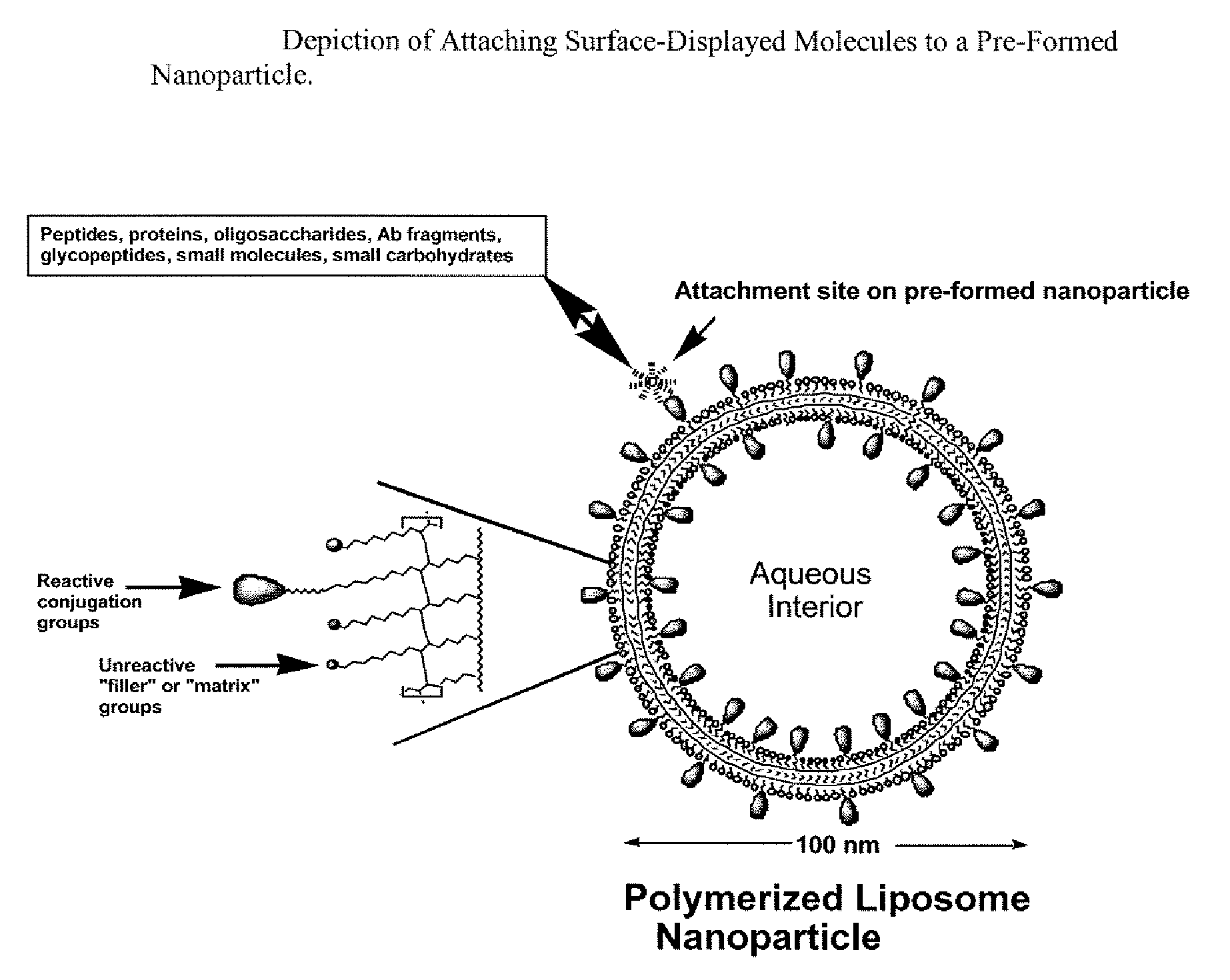
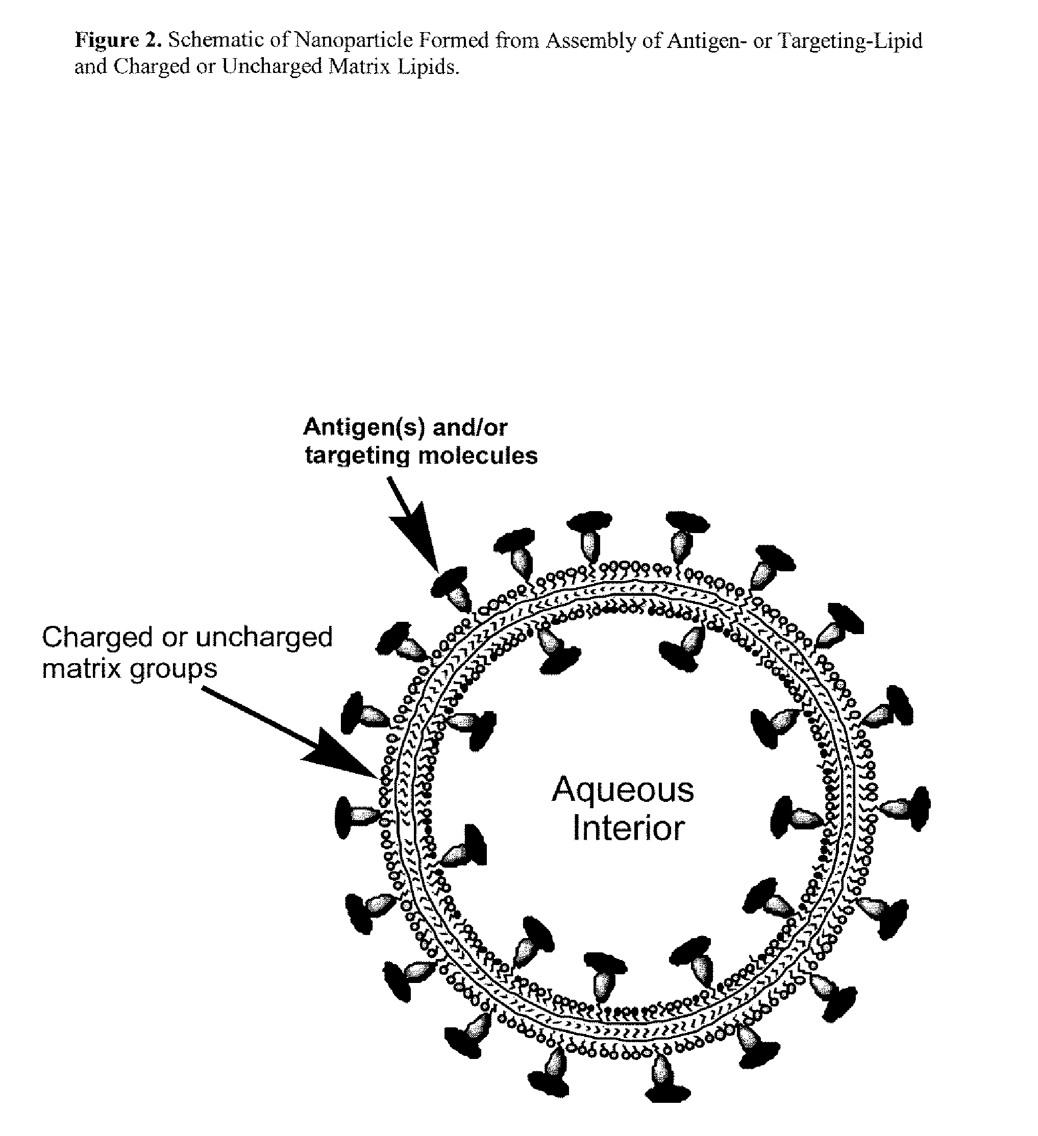
Image

Examples
example 1
PDA Liposome Preparation
[0124]Polymerized polydiacetylene (PDA) liposomes were prepared according to the method described by Spevak et al., “Carbohydrates in an Acidic Multivalent Assembly: Nanomolar P-Selectin Inhibitors,”J Med Chem 39(5):1018-1020 (1996), which is hereby incorporated by reference in its entirety. Briefly, polymerizable lipids and antigen- or targeting-lipids were mixed and evaporated to a film. Deionized water was added to the films to give a 1 mM (total lipid) suspension. The suspension was heated to between 70-80° C. and probe sonicated for 10 min. The resulting clear solution was then cooled to 5° C. for 20 min. and polymerized by UV light irradiation (254 nm). The deeply colored solutions were syringe filtered through 0.45 μm or 0.2 μm cellulose acetate filters in order to remove trace insoluble aggregates. Essentially all of the lipid material (>98%) is incorporated into the soluble liposomes. In the case of carbohydrate-displaying nanoparticles, Dionex Analy...
example 2
HPSO Liposome Preparation
[0125]HPSO liposomes were prepared according to the general method described by Wong et al., “A New Polymer-Lipid Hybrid Nanoparticle System Increases Cytotoxity of Doxorubicin Against Multidrug-Resistant Human Breast Cancer Cells,”Pharm Res 23(7):1574-1585 (2006), which is hereby incorporated by reference in its entirety. Briefly, lignoceric acid (Na+ salt form) lipid and antigen- or targeting-lipids (or antigen-chelating lipids) were mixed and evaporated to a film. HPSO dissolved in deionized water was added to the films to give a 6 mg / ml (total weight) suspension. The suspension was heated to between 70-80° C. and probe sonicated for 10 min. The resulting clear solution was then cooled. If an antigen-chelating lipid was used, the polymerized liposome was mixed with antigen and incubated for several minutes.
example 3
Candida albicans Glycoprotein Presentation on PDA Nanoparticles for use as a Vaccine.
[0126]Glycoproteins from the cell wall of Candida albicans are highly mannosylated, phosphomannoprotein complexes that are weak immunogens when administered alone or with adjuvants (CFA, Ribi RS-700, etc), but conjugation to a carrier protein like BSA or encapsulation in traditional liposomes elicits increased immune responses that protect mice against disseminated and mucocutaneous candidiasis (Han et al., “Antibody Response that Protects Against Disseminated Candidiasis,”Infect Immun 63:2714-2719(1995); Han et al., “A Vaccine and Monoclonal Antibodies that Enhance Mouse Resistance to Candida albicans Vaginal Infection,”Infect Immun 66:5771-5776 (1998); and Han et al., “Candida albicans Mannan Extract-Protein Conjugates Induce a Protective Immune Response Against Experimental Candidiasis,”J Infect Dis 179:1477-1484 (1999); which are hereby incorporated by reference in their entirety). Vaccine formu...
PUM
| Property | Measurement | Unit |
|---|---|---|
| Volume | aaaaa | aaaaa |
| Time | aaaaa | aaaaa |
| Equivalent mass | aaaaa | aaaaa |
Abstract
Description
Claims
Application Information