
Mri Guided Ultrasound Therapy Apparatus

a technology of ultrasound therapy and guided ultrasound, which is applied in the field of ultrasound therapy equipment, can solve the problems of b-mode ultrasound image, inability to completely display the relative tissue relationships and solid structure of the subject's region, and limited visible area, so as to reduce the interference of the magnetic field produced by the power used by ultrasound therapy equipment to the mri system, and facilitate reconstruction

Inactive Publication Date: 2008-11-06
RONGHAI SUPERSONIC MEDICINE EN
View PDF21 Cites 28 Cited by
  • Summary
  • Abstract
  • Description
  • Claims
  • Application Information

AI Technical Summary

Benefits of technology

[0034]Aiming at the conditions that the existing technologies mostly adopted the non-magnetic designs and operations within magnetic field, this invention is to adopt the existing open magnetic field and put the mechanical positioning means of the ultrasound transducer outside of the main magnetic field and reduce the space limitation for the mechanical positioning means of the ultrasound transducer. Meanwhile, the non-magnetic requirement on the mechanical positioning means become less greatly and particularly the problem of interference from magnetic field produced by working current of the transducer power cord to MRI system can be solved. Further, also the ultrasound transducer may be located outside of the main magnetic field. Thus, the interference of magnetic field produced by the power used by ultrasound therapy equipment to MRI system can be reduced further and the problem of interference from magnetic field produced by working current of the transducer power cord and the positioning means in US patents as above mentioned to MRI magnetic field has been be solved. Meanwhile, with this invention, the medical personnel may change and fix body position of a patient, monitor and operate a patient to some extent.
[0035]The existing high-intensity focused ultrasound therapy apparatus can be easily reconstructed and adopted by this invention.
[0036]With the development of MRI technology, more and more kinds of open magnetic fields can be selected. A lot of magnetic material is produced in north of China and the level of general techniques of permanent magnet open MRI system in China has reached the world advanced level. If the advantageous MRI technology in China is selected and used in conjunction with ultrasound therapy technology, the application cost can be decreased greatly and it has a good market expectation.
[0037]This invention may expand the application range of the conventional high-intensity focused ultrasound therapy. It is especially suitable for relatively complex tumor treatment and has good social benefits. Especially, the arrangement of MRI system in this invention is adaptive to the technical solutions disclosed in Chinese Patent No. 98100283.8 “High intensity focused ultrasound system for scanning and curing tumor” submitted before by the inventor of this invention and the ultrasound therapy system with an open flexible fluid container can be selected by this invention. Accordingly the safety and good effects of high-intensity focused ultrasound therapy can be ensured.
[0038]This invention may mostly or fully maintain the examining area in MRI (Magnetic Resonance Imaging) apparatus, but Chinese Patent No. 98805359.4 “MRI-guided therapeutic unit and methods” is very difficult to achieve this. The large examining area makes clinical observations more visual and convenient. MRI system may reserve many functions so as to promote its compatibility and availability ratio.

Problems solved by technology

But, B-mode ultrasound image is only a plane image with a certain angle and cannot completely display the relative tissue relationships and solid structure of the region of the subject to be treated, therapeutic acoustic path and the area behind the region of the subject.
Even though the 3-D ultrasound system is used, the visible area is still limited.
Besides, the ultrasound image is limited on the depth of observation and it almost cannot display the tissue behind the bone because the bone influences the image greatly.
The serious noises on the images exist during monitoring treatment.
Further, ultrasound images have poor capacity to identify the tissue boundary and particularly it is more difficult to identify small tumors and deep-bedded tumors.
At present, the major problem for a MRI guided ultrasound therapy apparatus focus is the interference between a MRI system and a focused ultrasound therapy system.
This kind of treatment needs repeated moving of patient and needs location for many times. The locating system is complex and a long time is needed to make locations.
Furthermore, the real-time monitoring and on-line monitoring during the treatment are difficult to be realized.
These technical solutions mainly have the following disadvantages: 1) Because the highly required non-magnetic designs and treatment on the therapy transducer and its positioning means, the technical complex and the cost are increased; 2) Because the positioning means are located within the magnetic field, the movement range of ultrasound transducer is limited strictly and furthermore the high-intensity focused ultrasound therapy needs a very precise locating system and the positioning means of the real therapy equipment in clinical applications are usually large, this technical solution increases the difficulties of equipment manufacturing and actual applications; 3) Because one part of the conductor used to supply power to the transducer is located within magnetic field, the magnetic field produced by the working current flow of the conductor will bring a big interference to MRI system, which is sensitive to magnetic signals; 4) It is difficult to perform assistant manual operations for an operator and also it is inconvenient for an operator to make clinical observations.
But, the technical solutions for designs of this magnetic field still have some unsatisfactory points: 1) Weak magnetic field intensity and uneven magnetic line distribution influences the image quality.
Under low field, the time for imaging is long and it is very difficult to realize the real-time imaging and the temperature measuring.
The effective magnetic resonance volume, i.e. magnetic field available for magnetic resonance imaging is only several centimeters; 2) Because the more complex superconductive technology is needed to be applied, the complexity of the equipment is high and it is very difficult to put it in practice and it is a long time to be a mature applicable technology for this equipment.

Method used


Image

  • Mri Guided Ultrasound Therapy Apparatus
  • Mri Guided Ultrasound Therapy Apparatus
  • Mri Guided Ultrasound Therapy Apparatus

Examples


embodiment 1

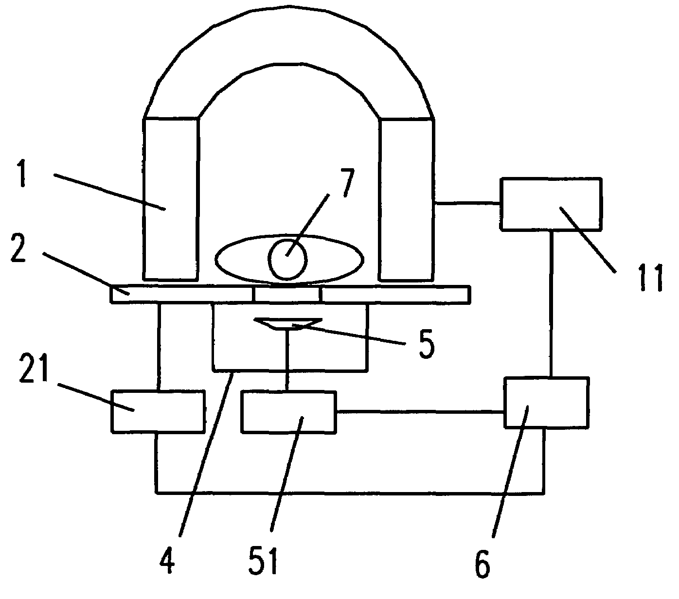
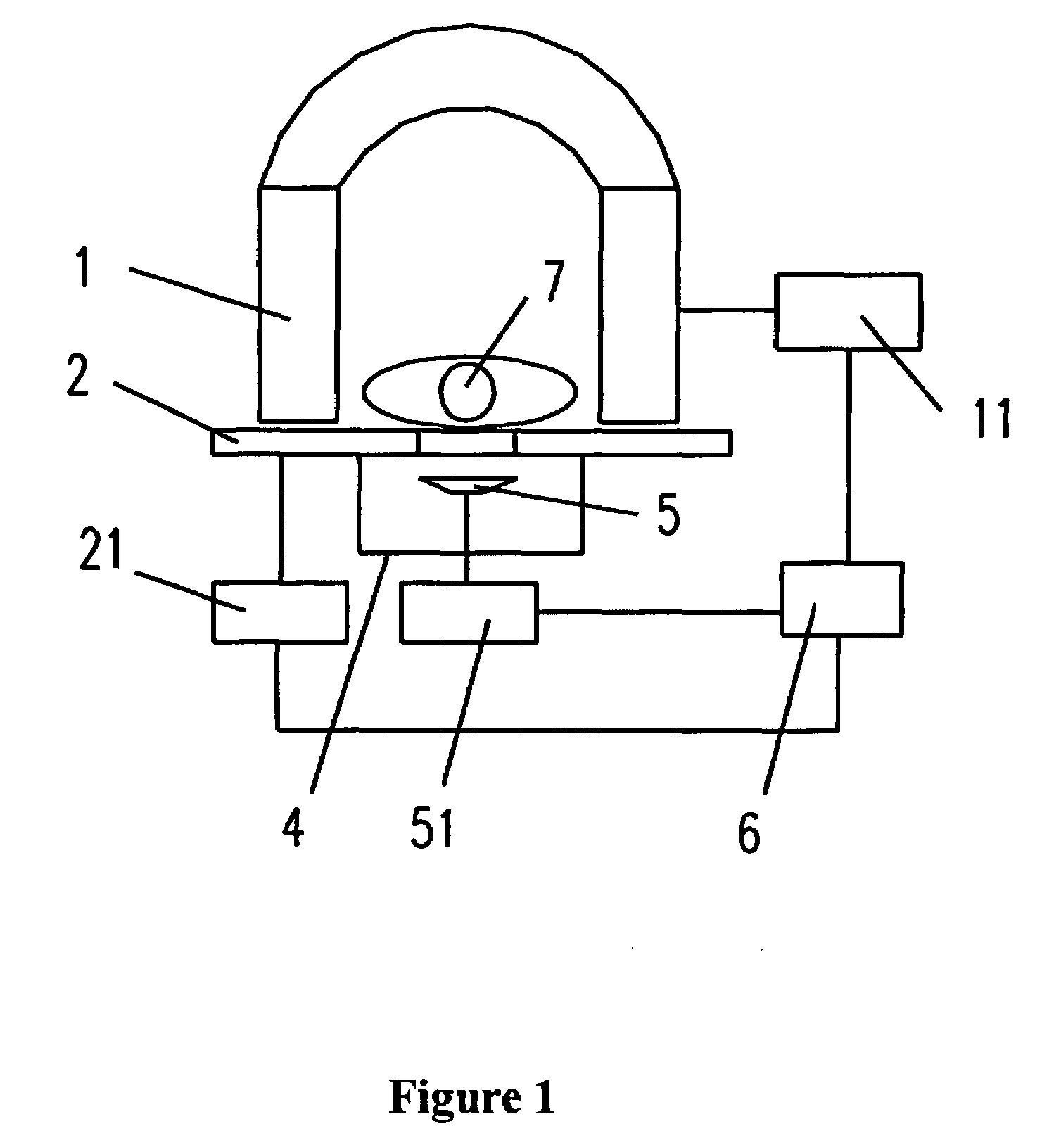
[0043]The system illustrated in FIG. 1 comprises magnet 1, treatment bed 2, water container 4, therapy transducer 5, control system 6, MRI image processing means 11, mechanical positioning means for treatment bed 21, mechanical positioning means for therapy transducer 51 and patient to be treated 7 located within the system. The magnet 1 is a 0.3 T permanent magnet (For example, 0.3 T NMR permanent magnet produced by Ningbo Heli Magnetech Co. Ltd.) with an downward open. The gradient field unit is used to code x\y\z three-dimensional space of the magnetic field. RF unit sends imaging sequence signals and receives the magnetic resonance response signals from the body and the MRI image processing means 11 rebuilds the tissue structural image and temperature image.

[0044]Therapy transducer 5 is a sphere-focusing piezoelectric transducer with a focal length of 100 mm-150 mm, a diameter of 120 mm-150 mm and a working frequency from 0.5 Mhz to 2 MHz. The therapy transducer 5 is connected t...

embodiment 2

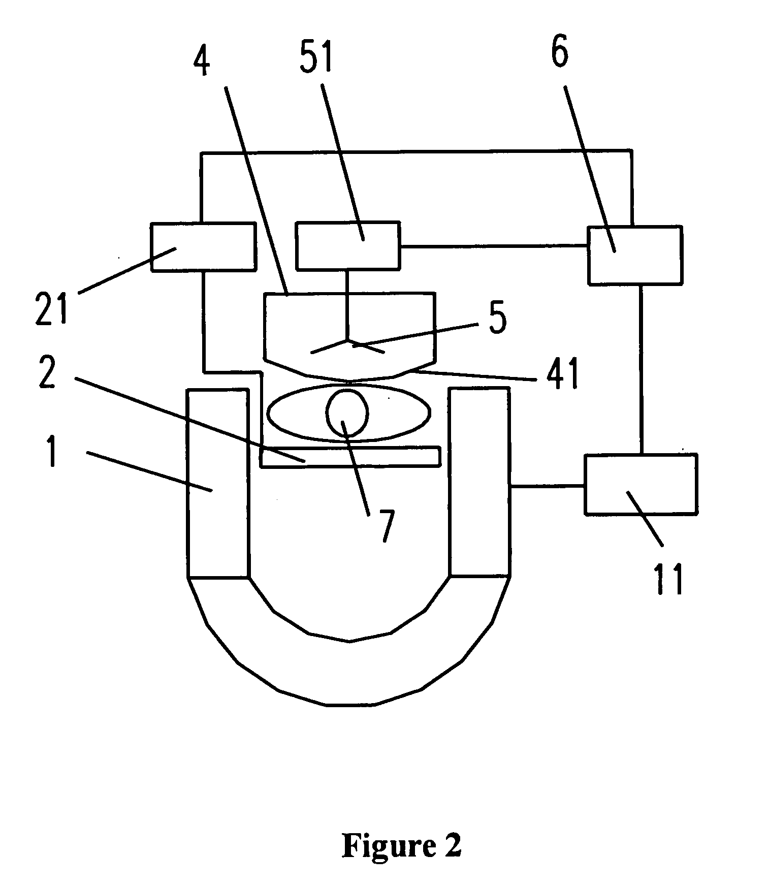
[0049]The system illustrated in FIG. 2 comprises magnet 1, treatment bed 2, water container 4, therapy transducer 5, control system 6, MRI image processing means 11, acoustic membrane 41, mechanical positioning means for treatment bed 21, mechanical positioning means for therapy transducer 51 and patient to be treated 7 located within the system. Magnet 1 of the system is a 0.3 T permanent magnet (For example, 0.3 T NMR permanent magnet produced by Ningbo Heli Magnetech Co. Ltd.) with an downward open.

[0050]The treatment bed 2 is located within the gap of the magnet 1 and supports the patient to be treated 7. The water container 4 and the therapy transducer 5 are mounted on the mechanical positioning means for therapy transducer 51. There is the acoustic membrane 41 at the surface of the water container 4. This membrane may prevent the medium water from overflowing.

[0051]In this embodiment, the ultrasound therapy is applied to a patient from up to down. Other components and their fu...


PUM


Abstract

This invention provides a MRI guided ultrasound therapy apparatus. It comprises a static field magnet adapted to apply a static magnetic field in an magnetic resonance volume at a predetermined disposition; at least one ultrasound energy applicator adapted to apply energy within an energy application zone at a predetermined disposition; and The mechanical positioning means for moving said ultrasound energy applicator to position the applicator so that the energy application zone intersects said magnetic resonance volume within said region of the subject. In that apparatus, the static field magnet is open at both ends or open at side. The sided open is upward or downward and the mechanical positioning means of this ultrasound energy applicator is close to and is located outside of this sided open. This invention reduces the space limitation for the mechanical positioning means of the ultrasound transducer. Meanwhile, the non-magnetic requirement on the mechanical positioning means become less greatly and particularly the problem of interference from magnetic field produced by working current of the transducer power cord to MRI system can be solved.

Description

FIELD OF THE INVENTION[0001]The present invention pertains to an ultrasound therapy apparatus and more specifically, to a MRI guided high-intensity focused ultrasound (HIFU) therapy apparatus.BACKGROUND OF THE INVENTION[0002]High-intensity focused ultrasound (HIFU) therapy is a non-invasive and non-traumatic approach. It is particularly applied to the patients with tumors. Comparing to the conventional surgeries or chemotherapy, HIFU therapy brings the patient less trauma and therefore its applications have been developed rapidly. Its indications include liver cancer, bone sarcoma, breast cancer, pancreas cancer, kidney cancer, soft tissue tumor and pelvic tumor.[0003]Most of the existing HIFU therapy use B-mode ultrasound imaging device to locate the region of the subject and to monitor the therapy. Adoption of B-mode ultrasound imaging device has the following advantages: low cost, real time imaging, having the same acoustic path as the therapeutic ultrasound, observing the tissue...

Claims


Application Information

Patent Timeline
IPC IPC(8): A61B5/055A61N7/00
CPCA61B19/22A61B2017/00084A61B2019/5236A61N7/02A61B34/70A61B2090/374A61N7/00
Inventor MU, MUCHEN, WENZHIWANG, HAIWANG, LONG
Owner RONGHAI SUPERSONIC MEDICINE EN
Learn More