

Fluorescent chlorotoxin conjugate and method for intra-operative visualization of cancer
a fluorescent chlorotoxin and cancer technology, applied in the field of fluorescent chlorotoxin conjugate and intraoperative visualization of cancer, can solve the problems of insufficient surgical judgment, more problematic, and inability to accurately identify tumor margins or small foci of cancer cells in the intraoperative area, and achieve the effect of little mmp-2 activity
- Summary
- Abstract
- Description
- Claims
- Application Information
AI Technical Summary
Benefits of technology
Problems solved by technology
Method used
Image

Examples
example 1
The Preparation and Characterization of a Representative Chlorotoxin Conjugate
CTX:Cy5.5
[0101]A representative chlorotoxin conjugate of the invention, CTX:Cy5.5 was prepared by conjugating Cy5.5 to chlorotoxin.
[0102]The conjugate was synthesized using a mixture of CTX (Alomone Labs, Israel, 250 ul of 2 mg / ml in 50 mM bicarbonate buffer, pH 8.5) and Cy5.5-NHS ester (Amersham Biosciences, Sweden, 43 ul of 10 mg / ml in anhydrous dimethylformamide). Conjugation was performed in dark at room temperature for 1 hour. Unconjugated dye was removed by dialysis against PBS (3 times) using Slide-A-Lyzer (Pierce Biotechnology, IL) membrane (Mr, cutoff, 3500) for 9 hours at 4° C. Samples were diluted with PBS to produce 1 and 10 uM of CTX solution and filtered with a 0.2 um syringe filter before use.
[0103]The concentration of CTX was quantified using bicinchoninic acid (BCA) assay (Pierce Biotechnology, Rockford, Ill.). The concentration of fluorophore in the same solution was quantified and evalua...
example 2
In Vitro Imaging with a Representative Chlorotoxin Conjugate
CTX:Cy5.5
[0104]9L / lacZ gliosarcoma cells (ATCC, VA) and primary human foreskin fibroblast (HFF) were maintained in DMEM and RPMI medium both supplemented with 1% sodium pyruvate, 1% streptomycin / penicillin and 10% FBS (Hyclone, UT), respectively. 2×10⁵ cells were seeded on sterile cover slips 36 hrs prior to labeling and confocal microscopy. Cells were cultured with 1 ml of CTX:CY5.5 conjugate (1 uM) for 2 hours in a 37° C. humidified incubator maintained at 5% CO2. Cover slips were washed 2 times in cell culture medium and 2 times in PBS buffer. Following this step, cell membranes were stained with 1 μM solution of FM 1-43FX (Molecular Probes, OR) for 20 min in dark at room temperature, washed 2 times in PBS and fixed in 4% paraformaldehyde. Cellular nuclei were stained with 4′,6-diamidino-2-phenylndole (DAPI, Sigma Aldrich, MO).
[0105]Confocal images were acquired using a DeltaVision SA3.1 Wide-Field Deconvolution Microsco...
example 3
In Vivo Imaging with a Representative Chlorotoxin Conjugate
CTX:Cy5.5
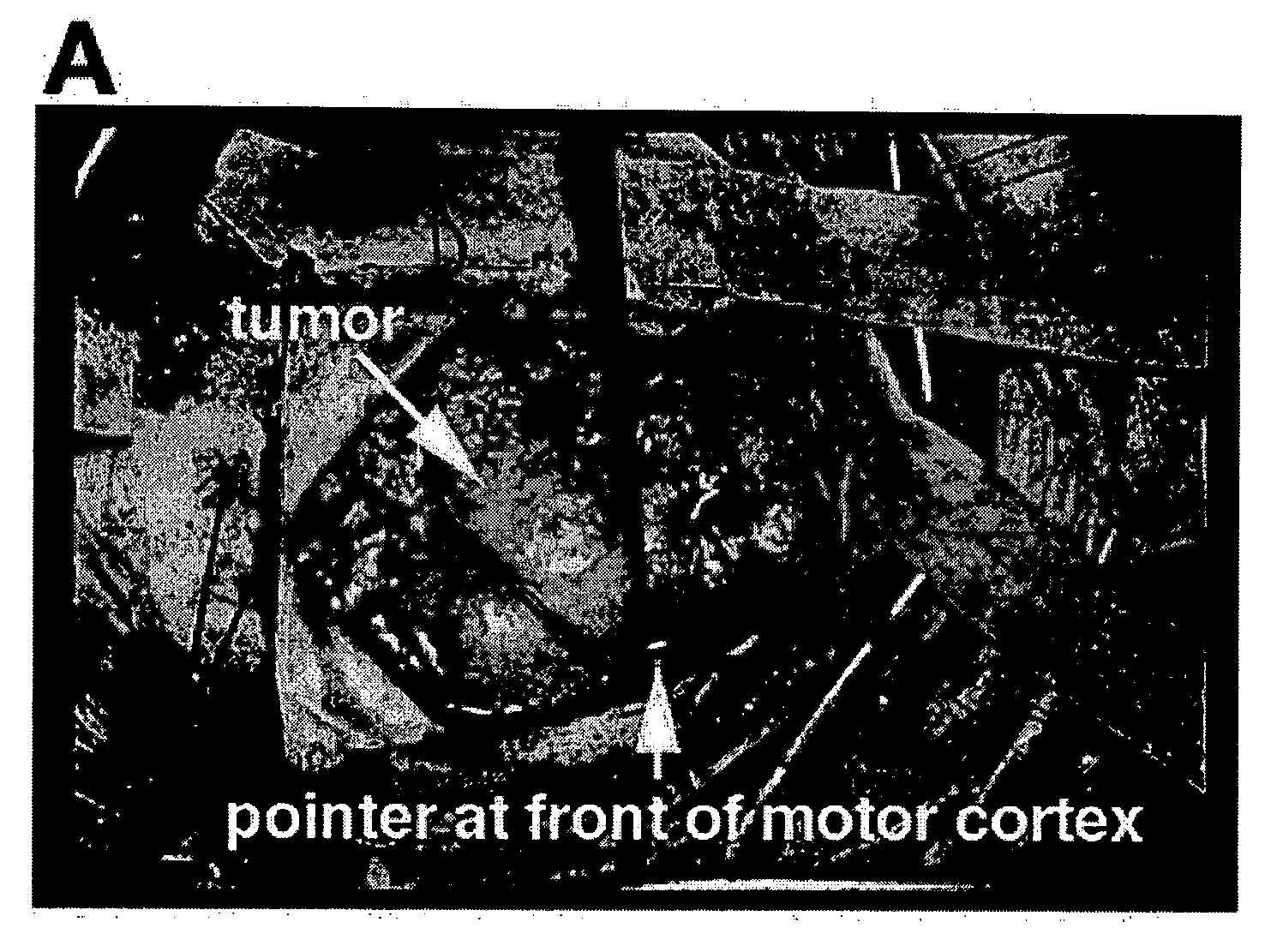
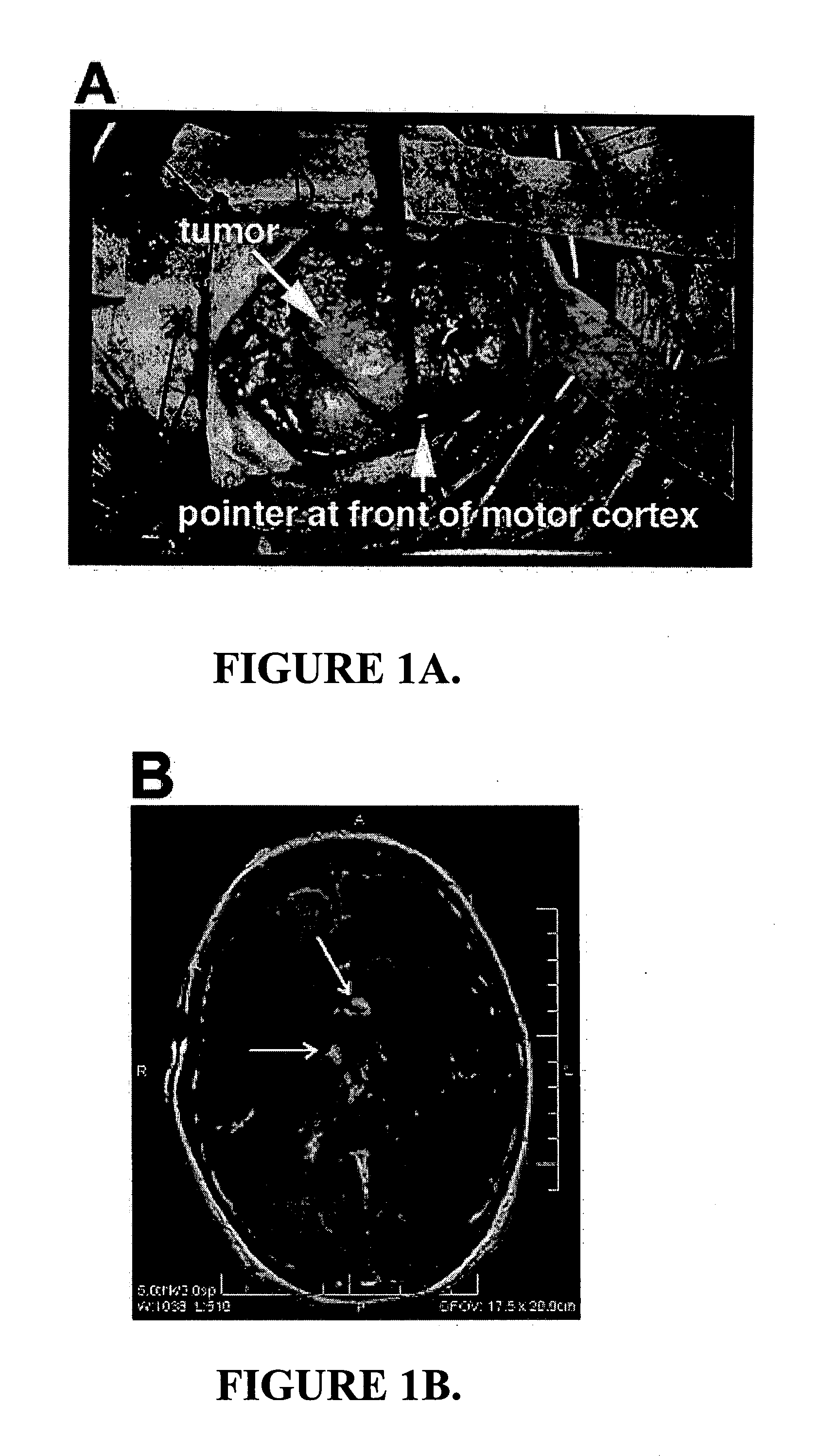
[0107]Animal models. All mouse studies were conducted in accordance with IACUC approved protocols. Subcutaneous xenografts were established in nu / nu (nude) mice using 9L, a rat gliosarcoma cell line (ATCC), and RH30, a rhabdomyosarcoma cell line. The xenografts were established using 1 million 9L or RH30 cells suspended in serum free media and matrigel at a 1:1 ratio. Intracranial xenografts were established by stereotaxic injection of 1 million 9L cells suspended in 10 μl PBS into the brain 3 mm lateral and posterior to the bregma. ND2:SmoA1 medulloblastoma mice, TRAMP prostate cancer mice and Apc1638N intestinal adenoma and adenocarcinoma mice were as previously described. See, Fodde, R., et al., A targeted chain-termination mutation in the mouse Apc gene results in multiple intestinal tumors. Proc. Natl. Acad. Sci. U.S.A., 1994. 91(19): p. 8969-73; Greenberg, N. M., et al., Prostate cancer in a transgenic mouse. ...
PUM
| Property | Measurement | Unit |
|---|---|---|
| diameter | aaaaa | aaaaa |
| wavelength | aaaaa | aaaaa |
| wavelength | aaaaa | aaaaa |
Abstract
Description
Claims
Application Information