
System and Device for in Vivo Procedures

a technology of in vivo procedures and systems, applied in the field of in vivo imaging, can solve problems such as damage to blood vessels, organ functional problems, and cardiac functional problems, and achieve the effects of reducing the risk of cardiac failure, and reducing the safety of patients

Inactive Publication Date: 2008-12-18
GIVEN IMAGING LTD
View PDF99 Cites 36 Cited by
  • Summary
  • Abstract
  • Description
  • Claims
  • Application Information

AI Technical Summary

Benefits of technology

[0013]There is provided, in accordance with some embodiments of the present invention an in-vivo imaging system, device and method which may allow new angles of view in laparoscopy, improving efficacy and safety of the procedure.
[0014]In embodiments of the present invention, data relating to environmental conditions at an in vivo site, or images of an in vivo site, such as a surgical site, may be obtained over a specific period and thus embodiments of the invention enable monitoring of an in vivo site. For example, embodiments of the invention may enable post surgery monitoring of surgical operations, which provides camera or video images of the surgery site during the critical post surgery hours. The system, device and method of the invention may enable an external operator to directly observe changes occurring at an in vivo site, for example to monitor suspected endometriosis, such that pathological occurrences can be detected at their onset and immediately be treated.
[0015]Embodiments of the invention further provide a remote controlled immobilized or moveable in vivo sensing device that can be used for monitoring an in vivo site by utilizing any appropriate sensing device (for example, pH meter, blood detector, imaging device etc.). In the digestive system the device according to an embodiment of the invention may be immobilized or moved for monitoring a site in the digestive system without having to leave an opening in the patient or incise the patient twice for retrieval of the in vivo device at the end of the monitoring period.
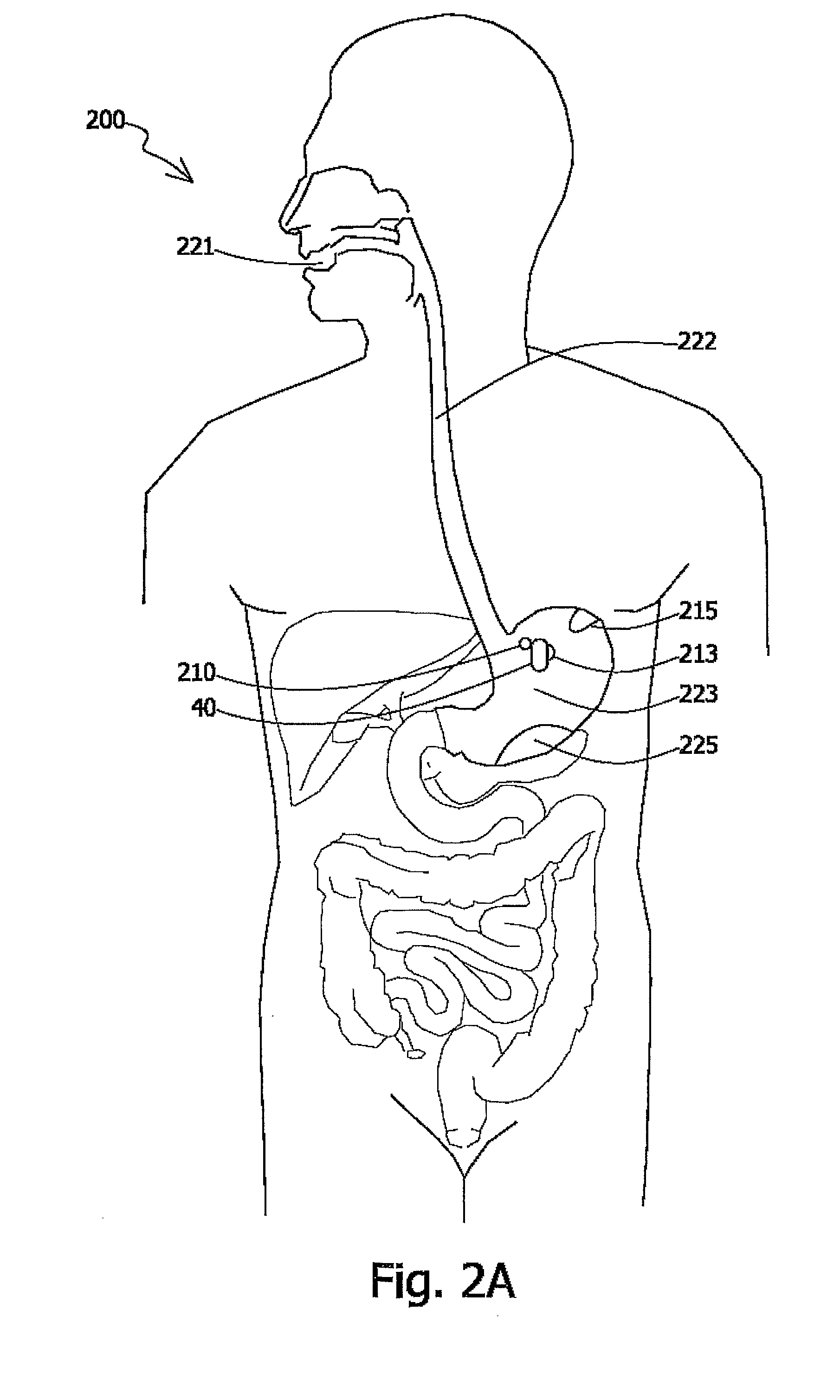
[0018]There is provided, in accordance with some embodiments of the present invention an in-vivo imaging system, device and method for placing an in-vivo sensing device, for example an in-vivo imaging device in the peritoneal cavity at laparoscopy to allow continuous wireless views of in vivo sites and target structures such as the gallbladder, diaphragm and the liver.
[0024]According to one embodiment of the invention an in vivo imaging device may include tissue analysis capabilities. According to one embodiment the device may include an imager, such as a CMOS imager having a plurality of pixel arrays, typically one on top of the other. An imager according to this embodiment may enable the use of different wavelength to detect pathologies.
[0025]According to one embodiment several imaging devices may be used simultaneously in a single procedure and a combined image or separate images transmitted from each of the devices may be displayed simultaneously on the work station display, thus enabling multiple view points and as many viewing angles as necessary, all typically displayed on a single screen or on array of screens in front of the surgeons. Additionally, real images of the lumen may be supplemented with different band images that can give indications on pathologies such as tumors, intraluminal bleeding etc.

Problems solved by technology

During surgery or in the time immediately after surgery patients may experience organ functional problems.
After surgery cardiac functional problems may occur due to build-up of stenotic lesions or other obstructions to the flow of blood through the implanted graft.
However, the introduction of catheters into the vascular system may result in damage to blood vessels.
However, methods of echocardiography do not always result in good quality images after cardiac surgery.
The implants cause internal bleeding, which leads to tissue inflammation and later, scarring and possibly infertility.
Imaging studies, such as ultrasound, can be helpful in studying the pelvis, but still cannot accurately diagnose endometriosis.
Although a bendable scope may enable a wide field of view, it still suffers from limited angles of view and limited camera maneuvering capabilities; it does not enable viewing behind a fold or on both sides of an organ.
Additionally, the use of one imager suffers, inter alia, from the fact that in order to see details the camera must zoom in on a site, in which case the orientation for the surgeon is lost.
Keeping orientation may be at the expense of being able to zoom in on details.
Current endoscopes and instruments are inadequate to perform complex surgeries.
The lack of support for a retroflexed endoscope in the peritoneal cavity makes it hard to reach remote structures and makes the necessary vigorous retraction of tissues and organs such as the liver, stomach, gallbladder or intestines difficult.
When flexible instruments are pushed against such structures the unsupported endoscope is pushed away without improving exposure.

Method used


Image

  • System and Device for in Vivo Procedures
  • System and Device for in Vivo Procedures
  • System and Device for in Vivo Procedures

Examples


Embodiment Construction

[0035]In the following description, various aspects of the present invention will be described. For purposes of explanation, specific configurations and details are set forth in order to provide a thorough understanding of the present invention. However, it will also be apparent to one skilled in the art that the present invention may be practiced without the specific details presented herein. Furthermore, well known features may be omitted or simplified in order not to obscure the present invention.

[0036]Embodiments of the invention can be utilized for monitoring, for example simultaneously, one or more in vivo sites in diverse body systems, as will be exemplified below. In one embodiment there is provided a system for monitoring sites in the GI tract, in which the sensing device is an imaging system.

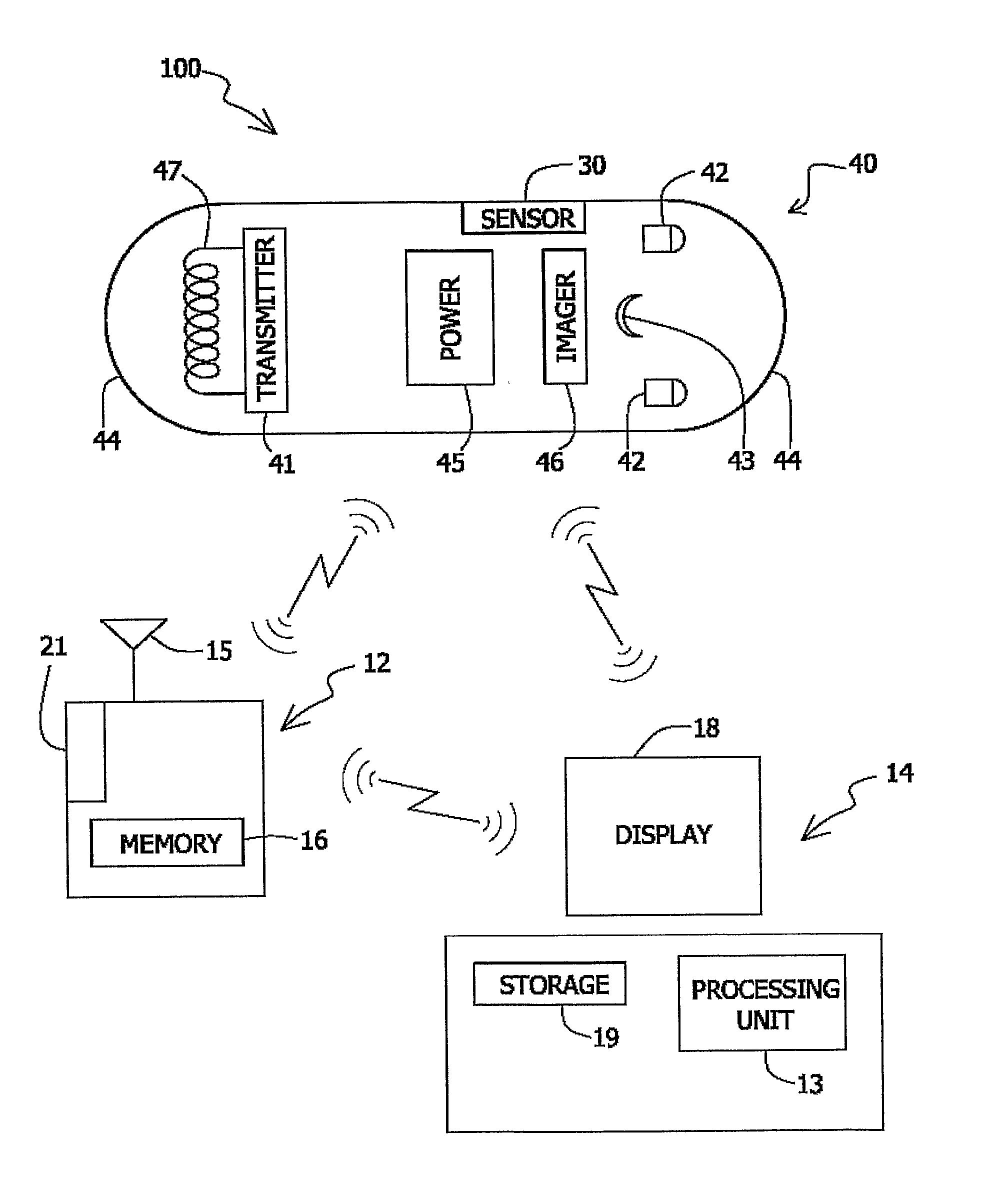
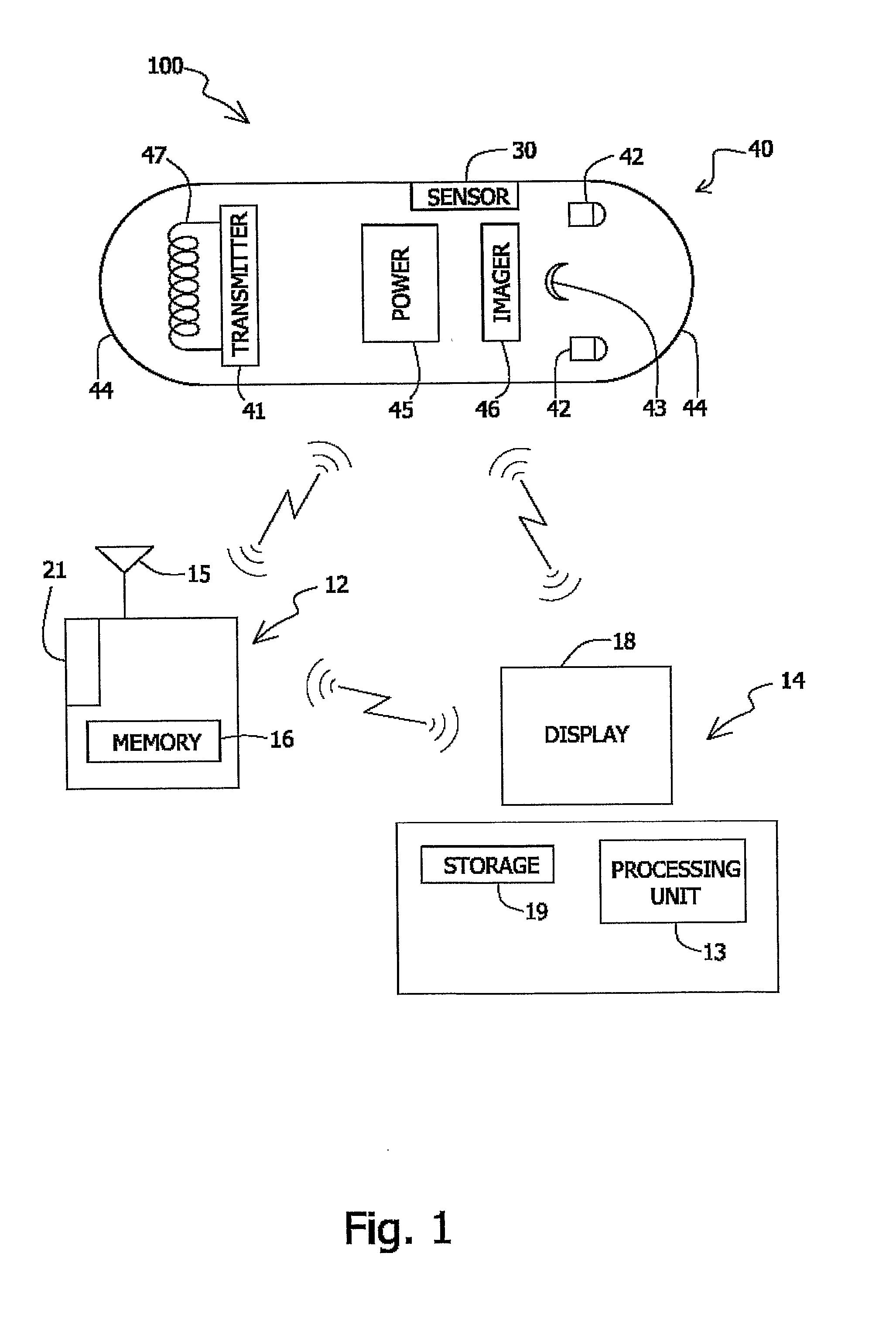
[0037]Reference is made to FIG. 1, which shows a schematic diagram of an in-vivo imaging system 100 according to one embodiment of the present invention. The in-vivo imaging system 100...


PUM


Abstract

The invention relates to a device system and method for providing images of an in vivo site during in vivo procedures, such as laparoscopy wherein the device is capable of illuminating an internal body cavity and has an immobilization unit to fasten the device to a desired location in vivo.

Description

FIELD OF THE INVENTION[0001]The present invention relates to in-vivo imaging. More specifically the invention relates to a system, device and method for monitoring one or more in vivo sites during in vivo procedures.BACKGROUND OF THE INVENTION[0002]In vivo sensing devices, such as thermometers, pH meters, optical scanners, image sensors and so on, can be used for unobtrusively monitoring body systems. During surgery or in the time immediately after surgery patients may experience organ functional problems. For example, during surgery in the gastrointestinal tract the blood pressure at the vicinity of the surgical site is reduced and peristalsis is arrested. After surgery the blood pressure increases and peristalsis is resumed sometimes causing bleeding from the surgical site into the intestine lumen.[0003]Also, for example, in treating coronary artery disease, it is sometimes necessary to bypass coronary arteries with a vascular graft, which is surgically attached to the heart, to c...

Claims


Application Information

Patent Timeline
Patent Type & Authority Applications(United States)
IPC IPC(8): A61B1/06
CPCA61B1/041A61B1/0676A61B1/0684A61B1/2736A61B5/14539A61B5/14546A61B5/6882
Inventor SWAIN, CHRISTOPHER PAULRABINOVITZ, ELISHA
Owner GIVEN IMAGING LTD
Learn More