Titanate nanowire, titanate nanowire scaffold, and processes of making same
a titanate nanowire and nanowire technology, applied in the field of synthetic nanostructures, can solve the problems of lack of macropores to accommodate tissue growth, macropores that are both mechanically tough and macroporous, and natural extracellular matrix is generally too fragile to support weigh
- Summary
- Abstract
- Description
- Claims
- Application Information
AI Technical Summary
Benefits of technology
Problems solved by technology
Method used
Image
Examples
example 1
X-Ray Diffraction (XRD) Patterns
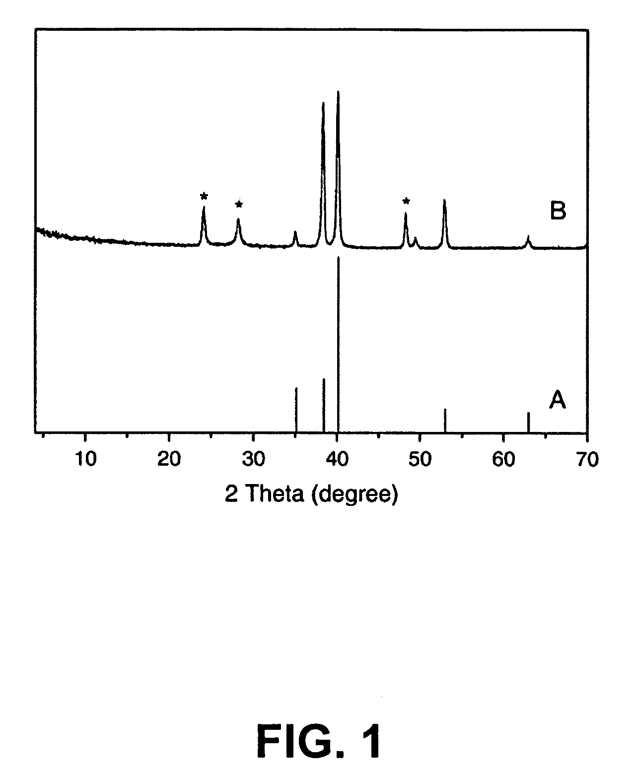
[0122]The plot in FIG. 1A is a typical x-ray diffraction (XRD) pattern of the pure Ti foil. FIG. 1B shows clear diffraction peaks of the titanate on the Ti foil, suggesting that the scaffolds are formed by the titanate nanowires [2,3]. It was observed in our work that after being calcined at 400° C., the scaffolding NWs were transformed to the TiO2—B phase (a=12.1787, b=3.7412, c=6.5249, β=107.0548), and then the TiO2—B can change to the anatase structure at 800° C. (data not shown). Such a series of structural transformations at elevated temperatures agrees well with the results reported by others in the literature [4-6].
example 2
Scanning Electron Microscope Studies of Nanofibers
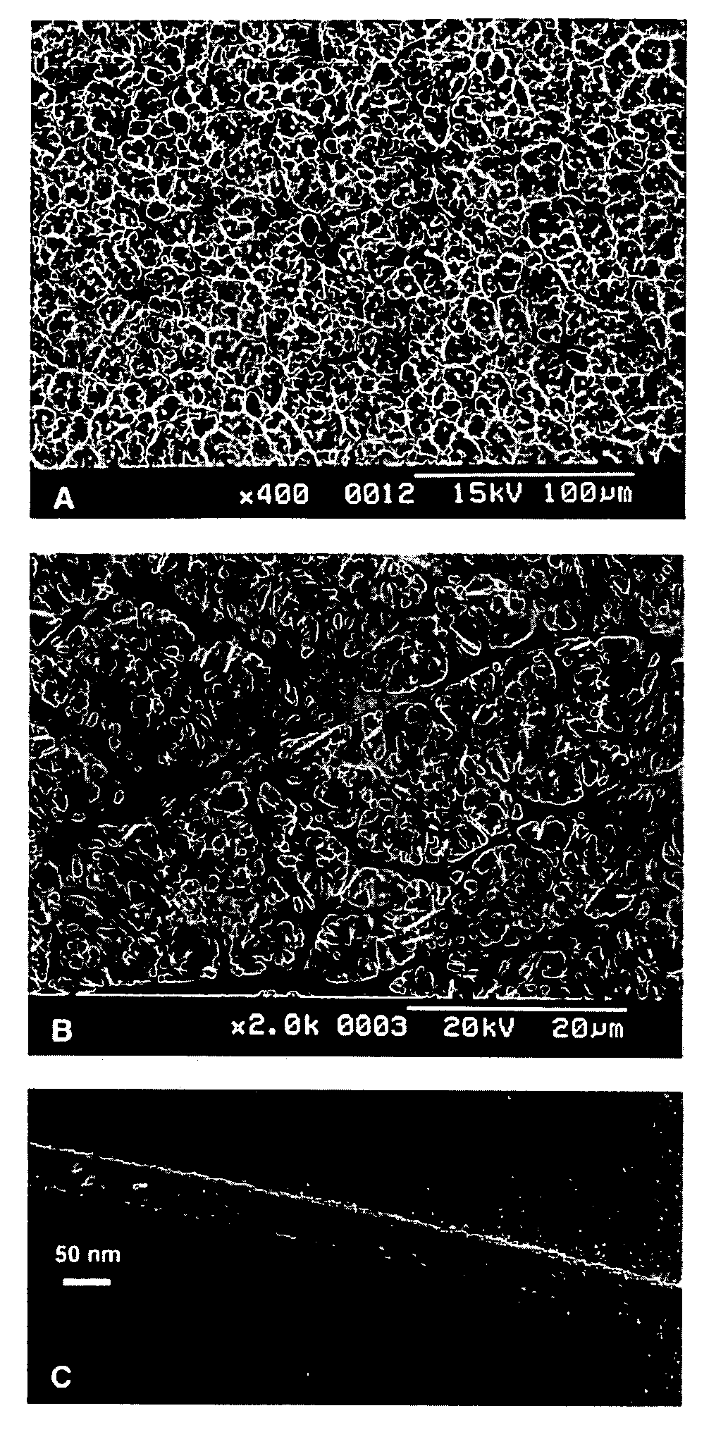
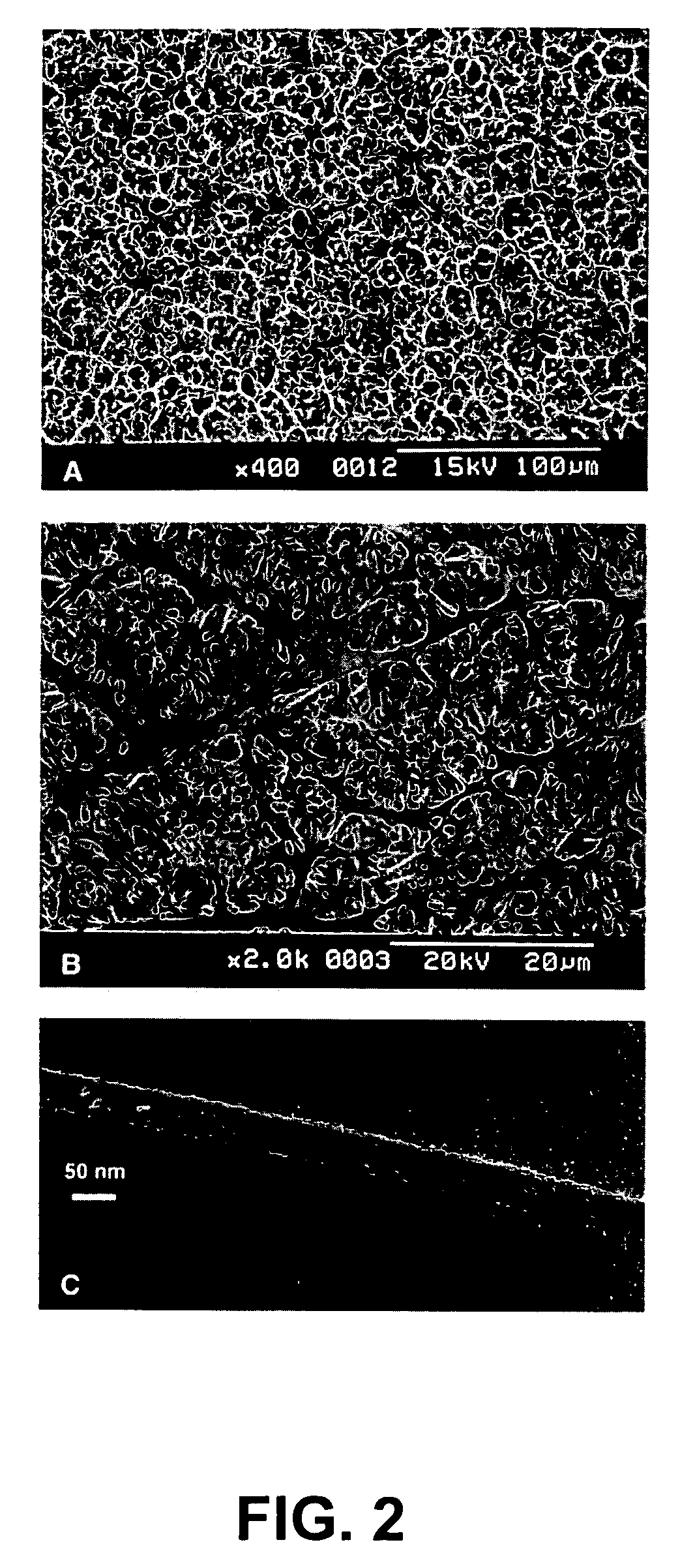
[0123]FIG. 2 demonstrates the SEM photomicrographs of the TiO2 nanofiber scaffolds on the Ti. FIG. 2a provides a low-magnification survey photograph for showing (i) uniformity and (ii) purity of the nanofibers-based scaffolds over the entire surface of the Ti foil.
[0124]At the high magnification, the SEM photographs from a tilted sample reveal details about the nanofibers self-organization into the 3D scaffolds (FIG. 2b). These nanofibers have the length controllable from 5 to 20 μm. FIG. 2c shows a TEM image of a typical nanofiber with the average diameter of about 60 nm.
example 3
Scanning Electron Microscope Studies of Nanofibers on TI Mesh
[0125]For studying whether macroporous scaffolds could be coated on Ti mesh surface [7], a similar synthesis on Ti mesh surface is successfully performed. FIG. 3A depicts an SEM photograph of the scaffold coating on the Ti mesh. A high-resolution SEM photograph (FIGS. 3B and 3C) from the same sample reveals that the nanofibers have self-organized into the 3D scaffolds, very much like that formed on the Ti foil (see FIG. 2b). It is expected that such NW scaffolds on the Ti mesh can be a potentially good biomaterial candidate for regeneration of hard tissues, because the tissue may be able to grow not only into the macroporous bioscaffolds but also the nearly millimeter-level mesh voids.
PUM
| Property | Measurement | Unit |
|---|---|---|
| length | aaaaa | aaaaa |
| length | aaaaa | aaaaa |
| length | aaaaa | aaaaa |
Abstract
Description
Claims
Application Information
 - R&D
- Intellectual Property
- Life Sciences
- Materials
- Tech Scout
- Unparalleled Data Quality
- Higher Quality Content
- 60% Fewer Hallucinations
Browse by: Latest US Patents, China's latest patents, Technical Efficacy Thesaurus, Application Domain, Technology Topic, Popular Technical Reports.
© 2025 PatSnap. All rights reserved.Legal|Privacy policy|Modern Slavery Act Transparency Statement|Sitemap|About US| Contact US: help@patsnap.com


