Antigen Detection

a technology of antigens and fluorescent labels, applied in the field of antigen detection, can solve the problems of human error, human error, and relatively complex apparatus, and achieve the effect of reducing human error

Inactive Publication Date: 2008-12-25
THE UNIV COURT OF THE UNIV OF EDINBURGH +1
View PDF8 Cites 7 Cited by
  • Summary
  • Abstract
  • Description
  • Claims
  • Application Information

AI Technical Summary

Benefits of technology

[0034]Thus, if any red blood cells bind to an antibody bound to the surface of the substrate, this may be detected by a fluorescent signal. By knowing the position of each specific antibody on the substrate, it is possible to identify which antigens are present on the surface of the red blood cells being tested and thus identify the blood group of the sample of blood being tested.
[00...

Problems solved by technology

Additionally, multiplexed typing can be carried out using flow cytometry, but this requires fluorescent labelling and relatively complex apparatus.
Nevertheless, any current systems which rely on detection by eye generally require a relatively large sample of blood and/or a suff...

Method used


Image

  • Antigen Detection
  • Antigen Detection
  • Antigen Detection

Examples


example 1

Preparation of Arrays

[0052]Epoxy silane coated slides were prepared using standard glass microscope slides from Erie Scientific. The slides were cleaned in a caustic ethanol solution comprising:

Sodium Hydroxide 105 g

Water 420 mls

Ethanol 630 mls

[0053]for 2 hours with agitation. The slides were then rinsed twice in deionised water and centrifuged to dryness in an Eppendorf 5810R centrifuge at 1000 rpm for one minute. The slides were then placed in a solution of glycidoxypropyltriethoxy silane (1% v / v in 95:5 ethanol / water) for one hour with constant agitation. After rinsing twice in ethanol the slides were heated in an oven at 383 K for 15 minutes. After cooling, the slides were kept in a desiccated environment. Where used, other slide types were obtained from commercial sources. Gold slides from Erie Scientific or Ssens BV, Hydrogel slides from Schott or Full Moon.

[0054]Antibodies were deposited on these slides using solid pins of either 700 or 200 μm on a Microgrid II spotter from B...

example 2

Microarray Experiments

[0058]Prior to use, the arrays were blocked in Bovine Serum Albumin (BSA); this is generally considered to reduce non-specific binding to the array surface.

[0059]To block, the slides were rinsed briefly in Phosphate Buffered Saline (PBS) pH 7.0 containing 1% Bovine Serum (BSA) and 0.1% Tween 20 by vigorously submerging 10 times. They were then placed into a fresh container of PBS pH 7.0 containing 1% BSA for one hour at room temperature, with constant mixing.

[0060]The slides were rinsed briefly in PBS pH 7.0 (submerged 10 times) and centrifuged to dryness in an Eppendorf 5810R centrifuge at 1000 rpm for one minute.

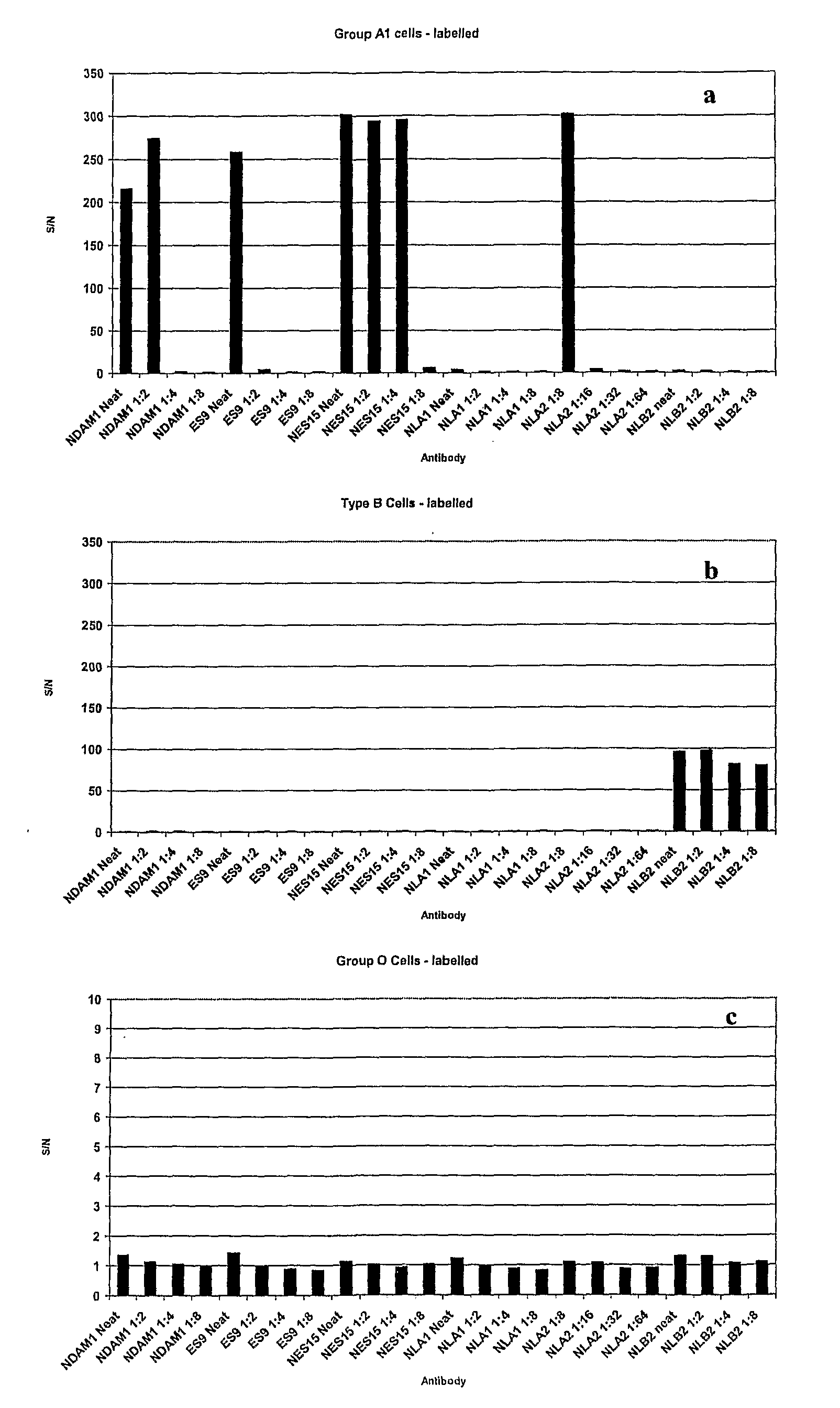
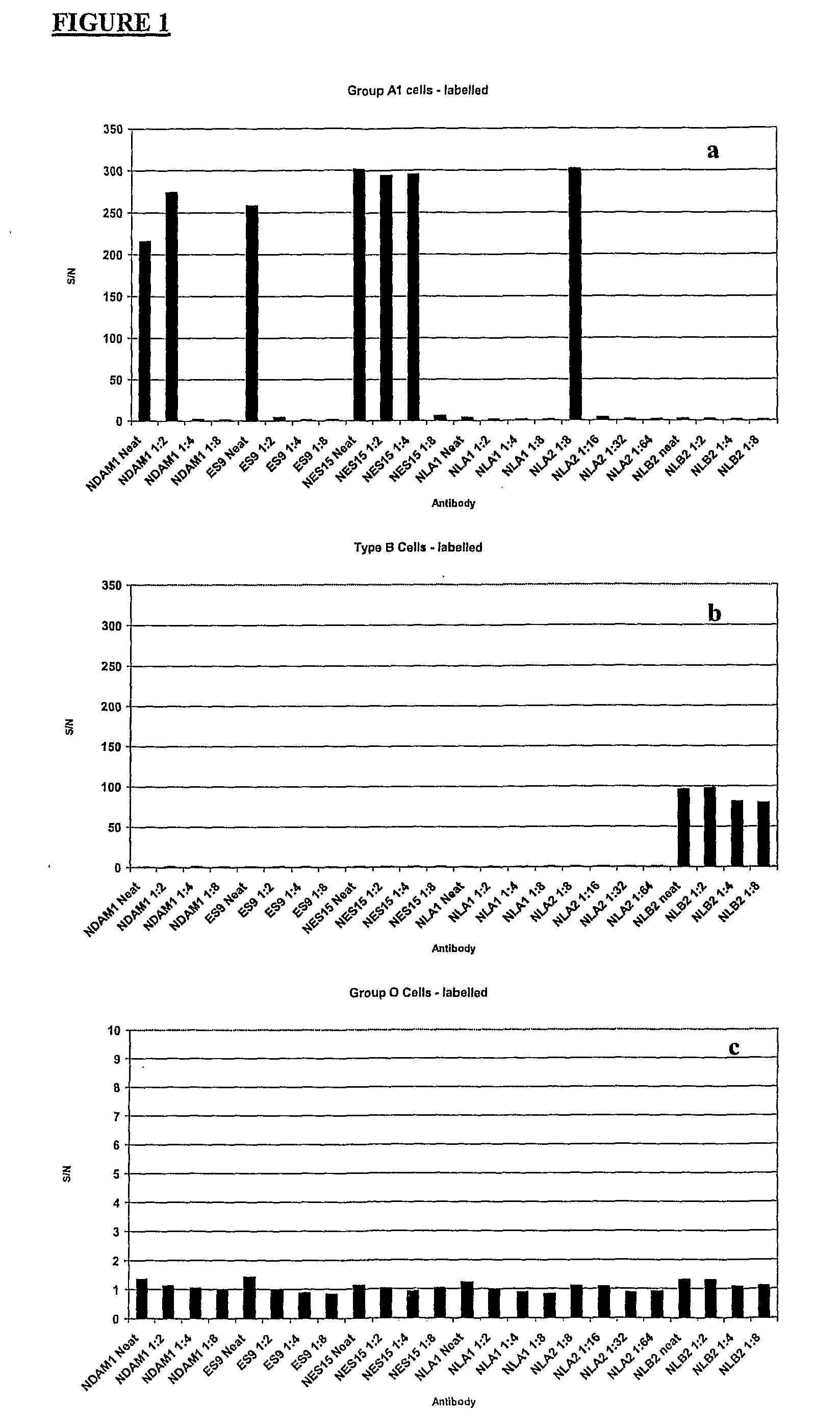
[0061]Blood samples Were incubated on the array using hybridisation chambers from Schleicher and Schuell (approximate capacity: 450 μl). Blood samples were incubated on arrays for 1 hour at room temperature with constant shaking. After incubation, the hybridisation chambers were removed and the slides washed in a mixture of PBS and Tween 20 (1%) by vi...

example 3

Optimisation of Scanning Conditions

[0068]Red Blood Cells have an Absorbance Spectrum as Shown in FIG. 3.

[0069]This absorbance spectrum is typically of oxy-haemoglobin, as would be expected with native red blood cells. Since the present inventors originally thought that haemoglobin species would be responsible for the fluorescence of erythrocytes, they tried to maximise the fluorescent signal by tuning the excitation wavelength to the absorbance spectrum shown in FIG. 3. In their initial experiments, using fluorescein labelled cells, they used scanner settings for fluorescein (excitation 488 nm, emission 530 nm). Since unlabelled red blood cells absorb strongly at 420, 540 and 580 nm, they would expect one of these wavelengths to give the strongest fluorescence when excited. The peak at 420 nm has the strongest absorbance but since commercial microarray scanners do not have lasers which can excite at this wavelength, the lowest excitation available was 488 nm.

[0070]The present invent...


PUM


Abstract

The present invention relates to methods of detecting specific cell surface antigens present in a sample of cells being tested and in particular blood group antigens, which methods do not employ the addition of extrinsic labels to detect said cell surface antigens. Typically detection is carried out using an intrinsic fluorescence capability of the cells being tested.

Description

FIELD OF THE INVENTION[0001]The present invention relates to methods of detecting specific cell surface antigens present in a sample of cells being tested and in particular blood group antigens, which methods do not employ the addition of extrinsic labels to detect said cell surface antigens. Typically detection is carried out using an intrinsic fluorescence capability of the cells being tested.BACKGROUND TO THE INVENTION[0002]Blood typing in the clinical and transfusion fields is typically currently carried out using agglutination assays either in multi well plates, see for example, U.S. Pat. No. 4,770,856, or in card / column format (e.g. U.S. Pat. No. 5,552,064 and U.S. Pat. No. 5,338,689). Additionally, multiplexed typing can be carried out using flow cytometry, but this requires fluorescent labelling and relatively complex apparatus.[0003]U.S. Pat. No. 4,851,210 describes a blood-typing device based on capillary flow through a membrane, which comprises an array of type-specific a...

Claims


Application Information

Patent Timeline
IPC IPC(8): C40B30/04G01N33/00G01N33/569G01N33/53G01N33/554G01N33/567G01N33/80
CPCB82Y15/00G01N33/56966G01N33/80
Inventor CAMPBELL, COLINGHAZAL, PETERPETRIK, JURAJROBB, JANINE SCOTT
Owner THE UNIV COURT OF THE UNIV OF EDINBURGH
Why Eureka
Try Eureka