Dark blood delayed enhancement magnetic resonance viability imaging techniques for assessing subendocardial infarcts

a technology of magnetic resonance imaging and subendocardial infarction, which is applied in the field of black blood viability magnetic resonance imaging (mri) pulse sequences, can solve the problems of limited clinical use impact of such technology, limitations of ecg, and inutility of biomarkers, and achieve good image contrast and great image contrast

Active Publication Date: 2009-01-01
SIEMENS MEDICAL SOLUTIONS USA INC +1
View PDF19 Cites 30 Cited by
  • Summary
  • Abstract
  • Description
  • Claims
  • Application Information

AI Technical Summary

Benefits of technology

[0021]We have now developed a new “dark blood” or “black blood” myocardial viability delayed enhancement imaging technique which can obtain dark blood delayed enhancement images by a timed combination of a selective preparation after the cardial R-wave and a later, non-selective inversion. Such MRI sequences can aid the detection of small subendocardial infarcts.
[0022]One exemplary illustrative non-limiting implementation combines an early slice-selective magnetic preparation of heart tissue, followed by a particular calculated delay to allow blood exchange of the heart, followed by late non-selective inversion (e.g., in the following heartbeat). The timed combination of slice-selective and non-selective preparation decouples the infarct-curve from the blood-curve and enables greater image contrast than is possible for example using two non-selective preparations.
[0023]In one exemplary illustrative non-limiting implementation, to make both blood and non-infarcted myocardium appear black in the image, a preparation is used to cause the relaxation curves of both T1-species to simultaneously cross the zero-line (“be nulled”). In one exemplary illustrative non-limiting implementation, the timed combination of slice-selective and non-selective preparation decouples the infarct from the blood curve and enables good image contrast.

Problems solved by technology

Thus, biomarkers are not useful for the diagnosis of subacute or chronic MI.
The ECG also has limitations: Q waves that form the fundamental basis of the diagnosis of chronic MI may be absent or, if initially present, may disappear at a later time point.
However, practical drawbacks have in the past limited the impact of such technology for general clinical use.
For example, standard CMR is generally more complex as compared to some imaging modalities.
These conditions can limit clinical throughput and the types of patients that can be scanned, and may also increase the complexity and length of CMR training.
Conversely, the presence of predominant myocardial scarring predicts increased operative mortality and the absence of these salutary effects.
Therefore, small subendocardial infarcts are often hard to detect in standard delayed enhancement images.
The infarct may be missed or its size may be underestimated.
However, due to their similar T1 values, the contrast between infarct and blood is still small.
However, a recurring problem has been that the standard classic double-inversion “dark blood” approach does not work in conjunction with delayed enhancement since the contrast agent and since only one T1 species is nulled cannot be used for viability imaging without adding further preparation pulses.
There are therefore challenges associated with using the classic approaches for dark blood viability.
For example, the classic dark blood preparation does not provide T1-weighting of the tissue.
Generally, the simultaneous nulling of blood and normal myocardium would be extremely difficult as there may be insufficient blood exchange between double IR preparation and image readout.
However, to our knowledge, it has not generally been used in a combination of early slice-selective preparation, blood exchange of the heart, and late non-selective preparation in the presence of MR-contrast agent to acquire “black-blood viability” images (note that in this context, “viability” refers to the property of heart tissue, myocardium, of being alive, “viable”, or dead, “non-viable”).

Method used


Image

  • Dark blood delayed enhancement magnetic resonance viability imaging techniques for assessing subendocardial infarcts
  • Dark blood delayed enhancement magnetic resonance viability imaging techniques for assessing subendocardial infarcts
  • Dark blood delayed enhancement magnetic resonance viability imaging techniques for assessing subendocardial infarcts

Examples


example preparation

Selective Saturation Followed By Non-Selective Inversion

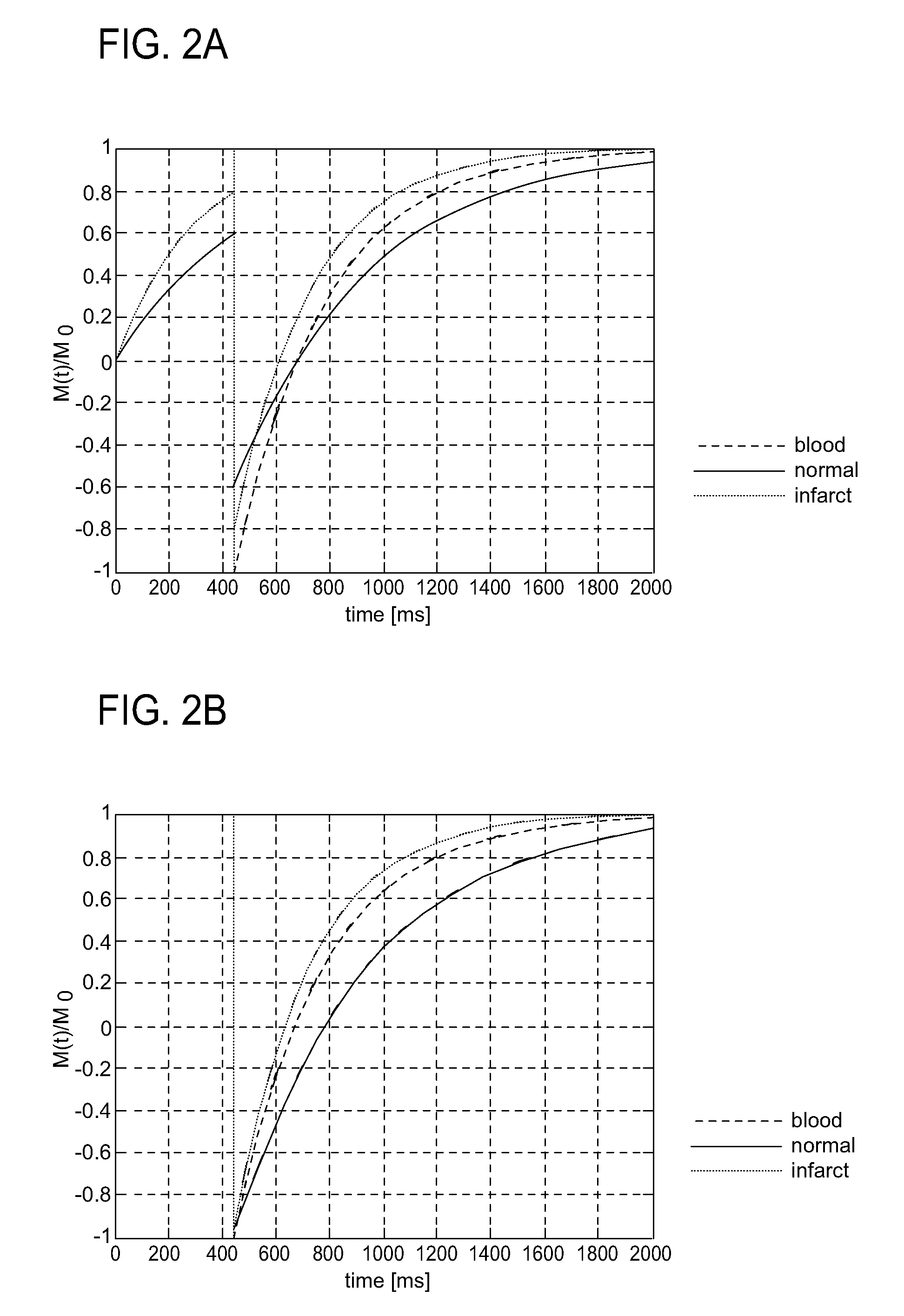
[0067]FIG. 3 shows the magnetization recovery of normal myocardium, infarct, and blood as occurring in an exemplary illustrative non-limiting implementation providing a slice-selective saturation pulse (SS SR) followed by a non-selective inversion. The curves shown are for typical relaxation constants (T1-valued) 10-20 minutes after an administration of a 1.25 mmol / Kg dose of contrast agent. The T1-values used in this and the following simulations are T1 infarct=280 ms, T1 normal=490 ms, and T1 blood=330 ms. These values change as a function of time after contrast agent injection, and modify the timing of the pulses needed to make blood and normal myocardium black.

[0068]In this particular non-limiting illustrative implementation, the slice-selective saturation recovery pulse (SS SR) sets the magnetization of infarct and “normal” to zero. The magnetization of both tissue types recover and is experiencing a non-selective inversio...

example

Non-Selective IR Followed By Timed Classic Double IR

[0083]FIGS. 9 and 10 show additional exemplary illustrative non-limiting implementations providing triple inversion recovery “triple IR” type preparation schemes. The T1-values used in the recovery curves simulations in FIGS. 9 and 10 are T1 infarct=280 ms, T1 normal=490 ms, and T1 blood=330 ms in this particular example.

[0084]In the exemplary illustrative non-implementations shown in FIGS. 9 and 10, a preparation pulse is played at the beginning of the preparation scheme and is later followed by a so-called double-IR preparation. The time between the first preparation and the double-IR preparation is of the essence. The combination of a preparation, an exact time delay, and this classic DIR provides useful results.

[0085]The double-IR preparation is widely used in MRI. We will refer to it as “classic double IR or classic DIR”. This classic DIR preparation consists of a non-selective inversion immediately followed by a slice-selecti...


PUM


Abstract

The technology herein provides a dark blood delayed enhancement technique that improves the visualization of subendocardial infarcts that may otherwise be disguised by the bright blood pool. The timed combination of a slice-selective and a non-selective preparation improves the infarct/blood contrast by decoupling their relaxation curves thereby nulling both the blood and the non-infarcted myocardium. This causes the infarct to be imaged bright and the blood and non-infarct to both be imaged dark. The slice-selective preparation occurs early enough in the cardiac cycle so that fresh blood can enter the imaged slice.

Description

CROSS-REFERENCES TO RELATED APPLICATIONS[0001]This application claims the benefit of priority from provisional application No. 60 / 887,596 filed Jan. 31, 2007, incorporated herein by referenceTECHNOLOGICAL FIELD[0002]The technology herein relates to magnetic resonance imaging (MRI) pulse sequences for use in detecting infarct (dead heart cells or scars) in the human heart, and more particularly to black blood viability magnetic resonance imaging (MRI) techniques that render blood and non-infarcted myocardium dark and infarct bright.BACKGROUND AND SUMMARY[0003]Myocardial infarction (MI) occurs in almost a million people each year in the United States, where coronary heart disease is the leading cause of hospital admissions. According to the joint American College of Cardiology and European Society of Cardiology consensus document concerning the redefinition of MI, the diagnosis of MI is often based on cardiac biomarkers and ECG changes. However, biomarkers are only elevated for 4 to 1...

Claims


Application Information

Patent Timeline
Patent Type & Authority Applications(United States)
IPC IPC(8): A61B5/055
CPCA61B5/055G01R33/54G01R33/5601G01R33/5673G01R33/5607G01R33/5608G01R33/5602
Inventor REHWALD, WOLFGANGSALERNO, MICHAEL
Owner SIEMENS MEDICAL SOLUTIONS USA INC
Why Eureka
Try Eureka