
Multi-modal imaging system and workstation with support for structured hypothesis testing

Inactive Publication Date: 2009-02-12
KONINKLIJKE PHILIPS ELECTRONICS NV
View PDF25 Cites 39 Cited by
  • Summary
  • Abstract
  • Description
  • Claims
  • Application Information

AI Technical Summary

Benefits of technology

[0020]The user can select at least one of a pre-defined post processing analysis and an ad-hoc post processing analysis at a statistical analysis portal. A reporting portal allows a user to customize a data reporting method.
[0023]Another advantage lies in greater flexibility for a user to design and execute studies.
[0026]Another advantage lies in the structured post processing of imaging data to maximize the statistical confidence of the results.

Problems solved by technology

Presently available imaging scanners are not equipped with standardized equipment and techniques that support automated and scientifically rigorous workflow suited to the testing of medical hypotheses.

Method used


Image

  • Multi-modal imaging system and workstation with support for structured hypothesis testing
  • Multi-modal imaging system and workstation with support for structured hypothesis testing
  • Multi-modal imaging system and workstation with support for structured hypothesis testing

Examples


Embodiment Construction

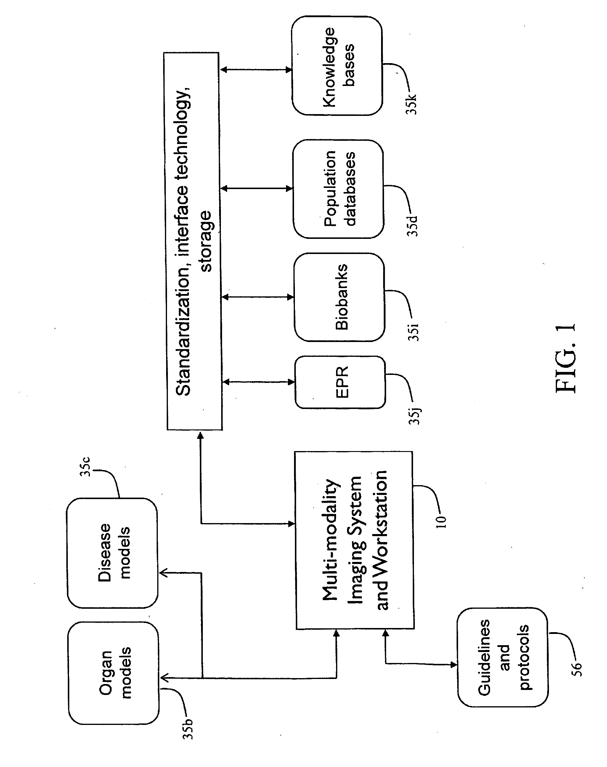
[0037]With reference to FIG. 1, an exemplary context of imaging systems used for diagnostic, therapeutic, and / or research activities is shown.

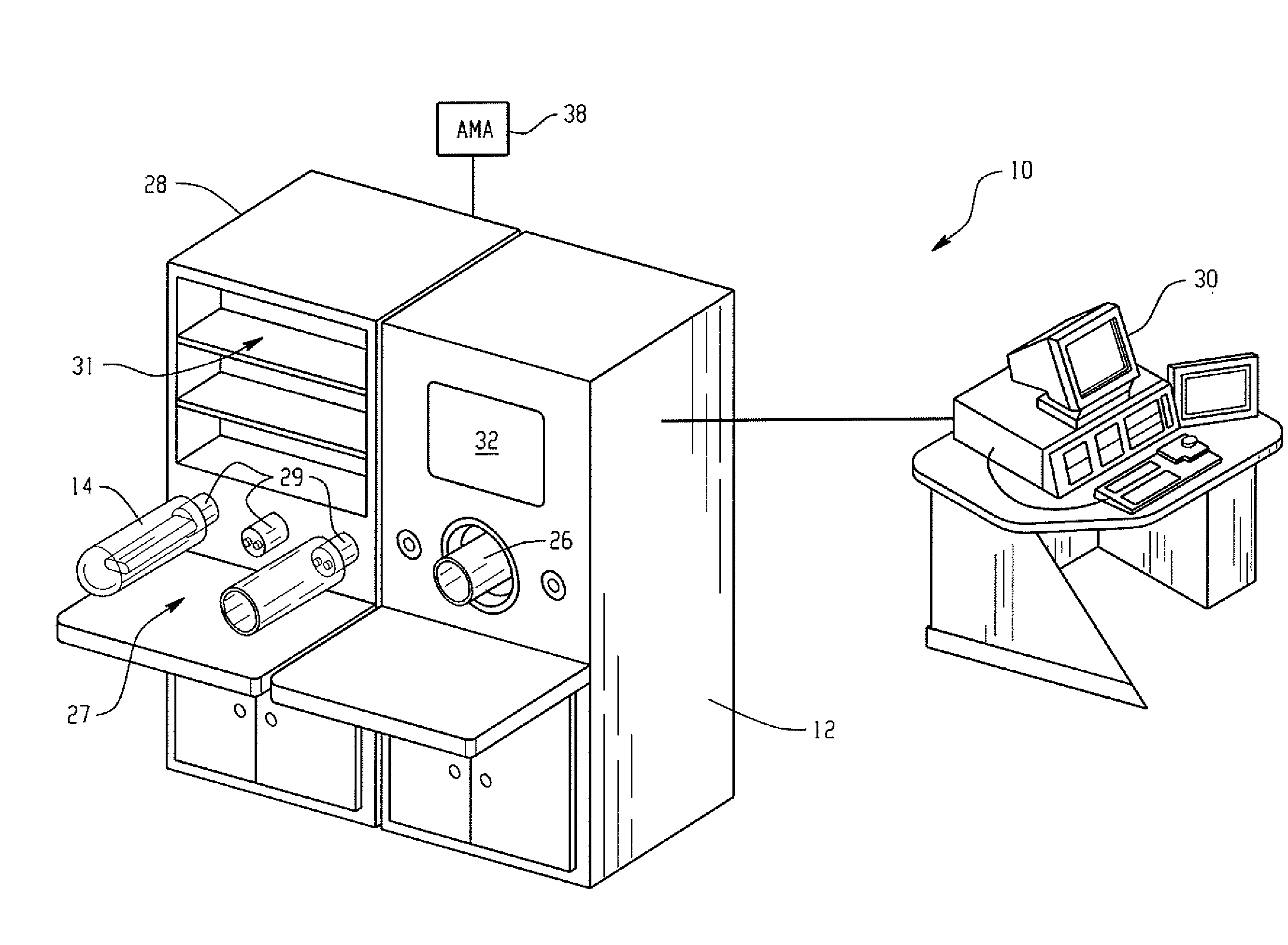
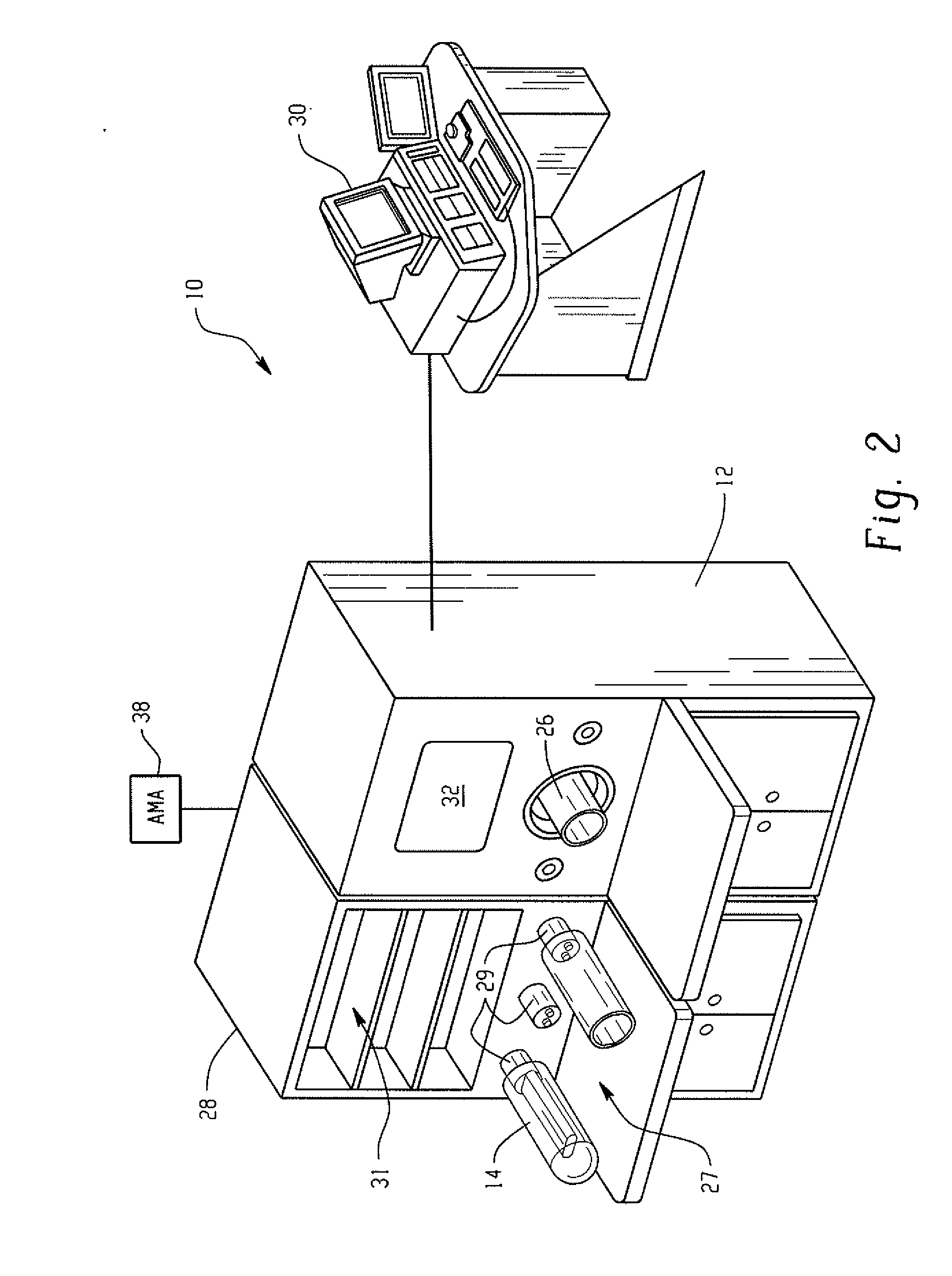
[0038]With reference to FIG. 2 and continuing reference to FIG. 1, an exemplary imaging system 10 is shown. Optional components to facilitate small animal imaging are included on the figure. The present application contemplates a system with modules for positron emission tomography (PET), Computed Tomography (CT), single photon emission computed tomography (SPECT), animal preparation, a research workstation for visualization, image registration, fusion, and analysis capabilities and other imaging and data handling. The various modules are combined within a cover that allows flexible configurations with various combinations of side-by-side, back to back, distributed, and / or in-line configurations, determined by space and throughput issues. A common subject positioner is also contemplated, as well as an animal holder that can be docked and undoc...


PUM


Abstract

Investigation of in vivo models of disease requires imaging studies involving single subjects in single imaging sessions, serial imaging of individuals or groups of subjects, and integration of data across diverse and heterogeneous experimental methodologies. Each type of experiment is preferably supported by various feature sets that can be rigorously applied to produce quantitative, reproducible results. Current imaging scanners are not equipped with standardized capability that supports an automated and scientifically rigorous workflow suited to hypothesis testing. An imaging system includes a research workstation at which a user can design, execute, study, and report imaging plans. Flexibility that comes along with a modular design of the system allows the user to customize workflow parameters for more robust hypothesis testing.

Description

CROSS REFERENCE TO RELATED APPLICATIONS[0001]This application is a continuation of PCT application number PCT / US2007 / 069596 filed May 24, 2007 which claims the benefit of U.S. provisional application No. 60 / 803,755 filed Jun. 2, 2006, which is incorporated herein by reference.BACKGROUND OF THE INVENTION[0002]The present application relates to the in-vivo imaging arts. It finds particular application with workflow and software processing in connection with small animal imaging in a research environment, and will be described with particular reference thereto. It is to be appreciated that the present application also finds use in other clinical and research settings, such as research with human subjects.[0003]Presently available imaging scanners are not equipped with standardized equipment and techniques that support automated and scientifically rigorous workflow suited to the testing of medical hypotheses. Pre-clinical imaging helps to bridge the gap between medical treatment ideas t...

Claims


Application Information

Patent Timeline
IPC IPC(8): A61B1/00A61B5/00A61M16/10A61D99/00
CPCA61B5/4821A61B6/032A61B6/0421A61B6/0442A61B2503/40A61B6/508A61B6/5247G06F19/321A61B6/467A61B5/0035G16H30/20
Inventor ZAGORCHEV, LYUBOMIRBUCKLER, ANDREWJEAN, ERIC
Owner KONINKLIJKE PHILIPS ELECTRONICS NV
Learn More