
Process and apparatus for imaging

a technology of process and apparatus, applied in the field of composition, processes and apparatus for imaging, can solve the problems of inability to process and analyze large numbers of specimens, limited imaging technology, and inability to achieve the effect of large-scale processing and analysis

Active Publication Date: 2009-03-26
BOARD OF RGT THE UNIV OF TEXAS SYST
View PDF10 Cites 37 Cited by
  • Summary
  • Abstract
  • Description
  • Claims
  • Application Information

AI Technical Summary

Benefits of technology

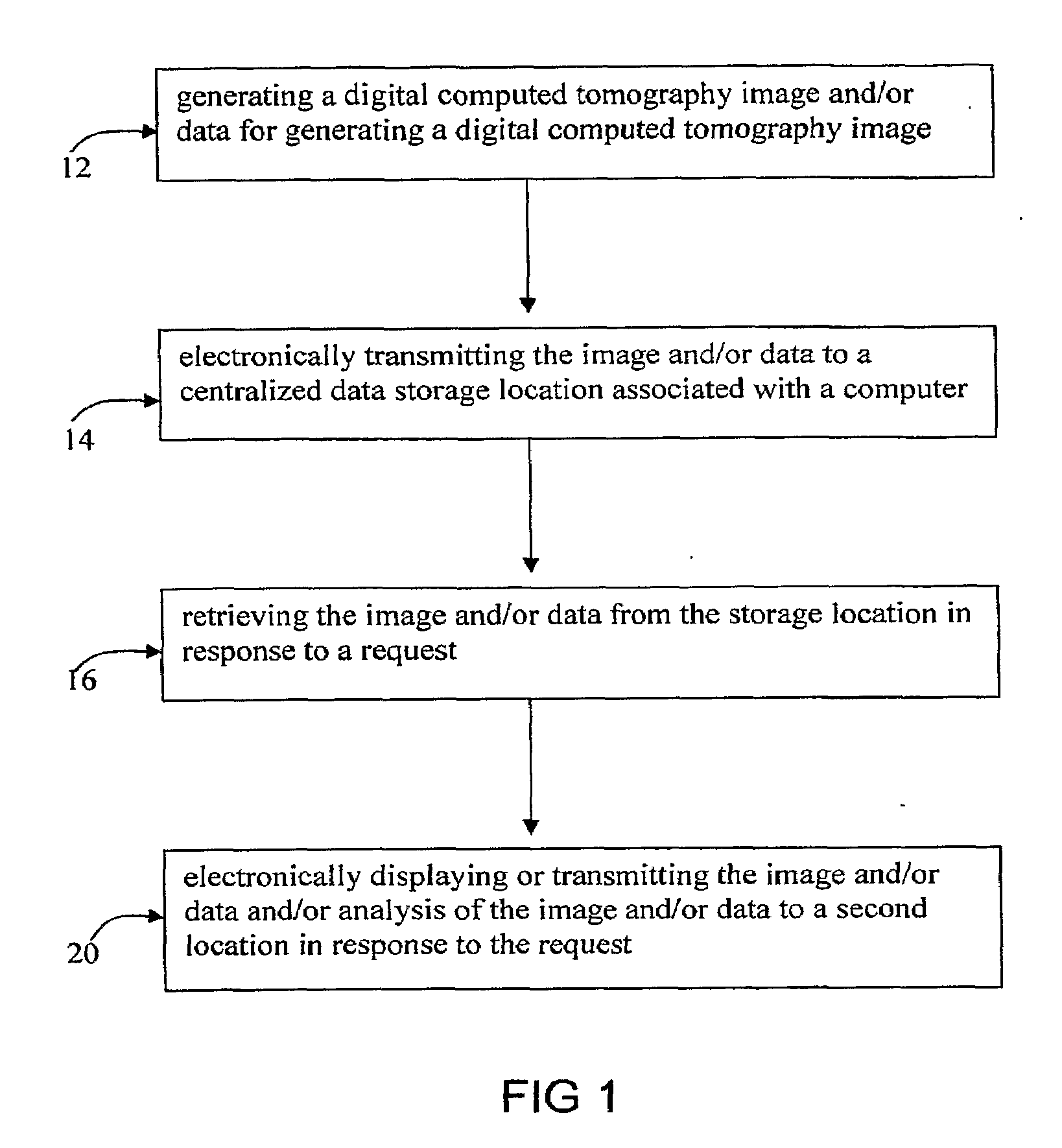
[0018]In a detailed process, the specimen to be scanned is a specimen obtained from an individual animal at a first time. Later, a second specimen is obtained from the same individual animal at a second time. Both the first and second specimens are stained and scanned to produce a microCT image. In general, a procedure is performed subsequent to the first time a specimen is obtained and prior to obtaining the second specimen. In this way, the procedure may be assessed for effectiveness or other effects.

Problems solved by technology

However, in spite of the opportunities presented, there remain limitations in imaging technologies.
Traditional histological methods of screening embryonic animals or other specimens for anatomical and / or molecular variation are time intensive, such that processing and analysis of large numbers of specimens is generally impractical.
This effort is frustrated by multiple mode imaging owing to difficulties in attaining image registration due to positioning inconsistency.
A limitation of existing animal holders is that close contact of the animal with the animal holder is visible in the resulting images, making image post-processing tedious.
An image of animal tissues in proximity with the holder is often obscured or poorly resolved.
Image quality and efficiency suffer since the holder must be identified and subtracted in subsequent image processing.
MicroCT virtual histology throughput has also been hampered by a lack of specimen holders capable of containing multiple specimens.
In addition, microCT virtual histology of whole embryos results in voluminous data.

Method used


Image

  • Process and apparatus for imaging
  • Process and apparatus for imaging
  • Process and apparatus for imaging

Examples


example 1

[0155]Staining methods for use in producing a microCT image according to the present invention are compared with conventional histological sectioning and staining using mouse embryos.

Specimen Preparation

[0156]Pax3:Fkhr transgenic mice (Keller, C., et al., Genes Dev 18, 2608-13, 2004; Keller, C. et al., Genes Dev 18, 2614-26, 2004), as well as wild-type mice are used in an example of an inventive process. Embryos are harvested at E9.5-E12.5 gestational ages, then fixed in 10% buffered formalin overnight at 4° C. Hematoxylin and eosin stained paraffin sections are prepared using established methods (Keller, C., et al., Genes Dev 18, 2608-13, 2004), then visualized at 2× magnification on a Nikon Eclipse 80i microscope. Immunohistochemistry with the Pax7 monoclonal antibody (Developmental Hybridoma Studies Bank, Iowa City, Iowa) is performed as previously described (Keller, C., et al., Ibid). For microCT-based virtual histology embryos are then stained according to an inventive process....

example 2

[0165]Whole embryos at gestational ages with limited epidermal layers are used in particular examples of inventive processes. For instance, midgestation whole embryos (E8-E13.5) that lack significant epidermal development may be used, and skinned embryos up to E19 can be satisfactorily stained as well. Embryos as stained as described in Example 1. Isosurfaces and sagittal crossections of a time series of E9.5, E10.5, E11.5, and E12.5 embryos are performed, scanned at 8 micron isometric resolution. At these resolutions, features such as the developing brain vesicles, neural tube, heart chambers, and liver can be clearly delineated. Due to the increased lipid content of the liver, attenuation of osmium-stained hepatic tissue results in the highest opacity and brightness.

example 3

Rapid 27 Micron Resolution Scans

[0166]A GE eXplore RS small animal scanner is used to perform scans of wild-type E12.5 embryos at 27 micron isometric resolution in approximately two hours. The embryos are stained as described in Example 1. A comparison of 8 micron and 27 micron scans of the same embryo shows that while 8 micron sections display considerably higher spatial resolution, the 27 micron sections are nonetheless adequate to distinguish features such as the semicircular canal, the neural tube central canal, and the cardiac chambers. From the perspective of high throughput phenotyping, the resolution of these 27 micron microCT scans are in the range of magnetic resonance microscopy, but at a nearly six-fold time savings. Furthermore, these 2 hour, 27 micron resolution scans are adequate to perform high quality segmentation analysis of major organ compartments, an advantage for computer-based, automated phenotyping. A caveat is that the small lumens within some organs, such a...


PUM


Abstract

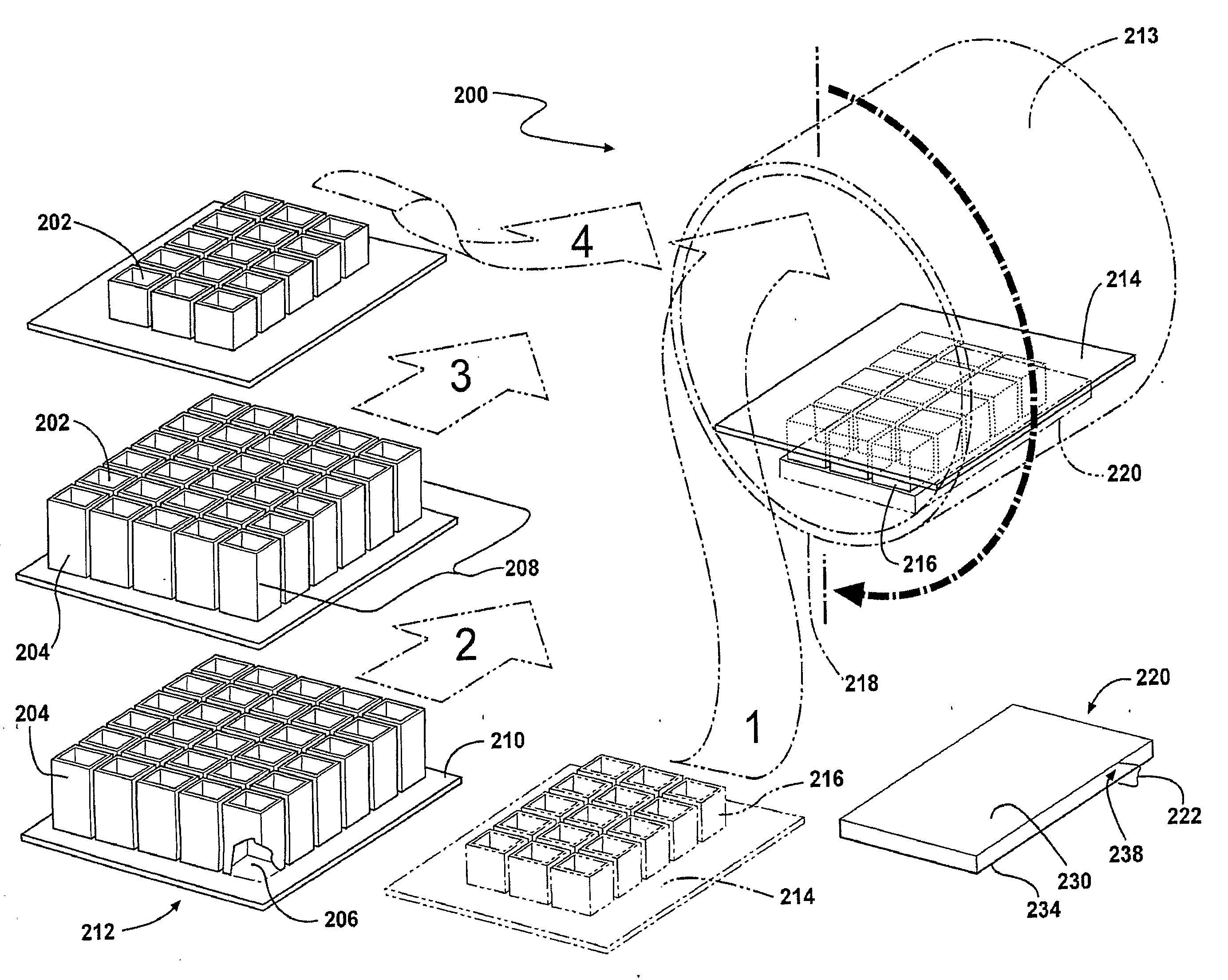
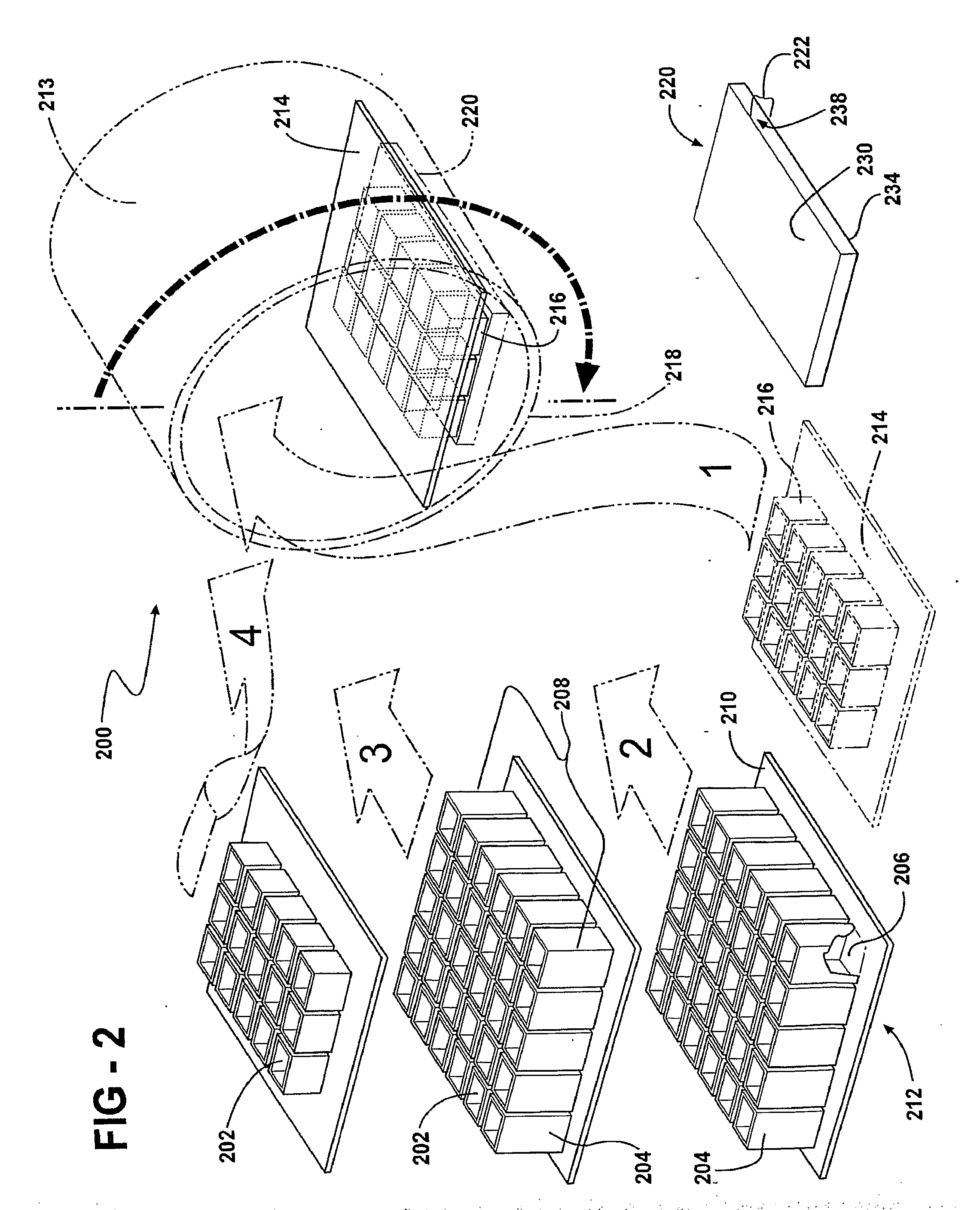
Processes for producing a microCT image for virtual histology using x-ray microscopic computed tomography are described along with processes for rapid and inexpensive high-throughput methods of high resolution imaging for screening an ex vivo embryo for phenotype using computed tomography imaging. Staining of particular components of specimens with one or more staining agents is described which contributes to high quality image generation and identification of anatomical structures as well as localization of molecular targets. Inventive animal and specimen holders are detailed which allow for reduced post-imaging processing of generated images. In particular, animal and specimen holders are provided which include a highly transparent bed or liner which separates the animal or specimen from a less transparent structure. A further animal holder is provided for placing and / or maintaining the animal in a desired position during an imaging procedure or multiple imaging procedures including a bed conforming to the animal's body.

Description

RELATED APPLICATIONS[0001]This application is a non-provisional application that claims priority benefit of U.S. Provisional application Ser. No. 60 / 762,327 filed Jan. 26, 2006; the contents of which are incorporated herein by reference.FIELD OF THE INVENTION[0002]The present invention relates in general to compositions, processes and apparatus for imaging, and in particular to improved preparation, collection and processing of specimen images, especially those obtained from X-ray microscopic computed tomography.BACKGROUND OF THE INVENTION[0003]Developments in imaging technologies have traditionally contributed to breakthroughs in scientific understanding. While the earliest imaging tools were primarily directed to resolving gross anatomical structures, modern imaging techniques allow visualization of very fine structural details as well as molecular information in some cases.[0004]The development of a number of new imaging modalities, along with evolution of molecular and cellular ...

Claims


Application Information

Patent Timeline
Patent Type & Authority Applications(United States)
IPC IPC(8): A61B6/00H05G1/00
CPCG01N1/30G01N23/046G01N2223/612G01N2223/419A61B6/508A01K1/031
Inventor KELLER, CHARLESBAHADUR, ALI N.
Owner BOARD OF RGT THE UNIV OF TEXAS SYST
Learn More