Phage Display By Novel Filamentous Bacteriophage

a filamentous bacteriophage and phage technology, applied in the field of new phage vectors and phage display methods, can solve the problems of extremely difficult to produce mutants while retaining this function, and achieve the effect of efficient production of random peptides

Inactive Publication Date: 2009-04-23
UCHIYAMA FUMIAKI
View PDF0 Cites 20 Cited by
  • Summary
  • Abstract
  • Description
  • Claims
  • Application Information

AI Technical Summary

Benefits of technology

[0015]The inventor has discovered filamentous bacteriophage vectors which allow a peptide to be inserted at the interior of the N-terminal domain of a pIII protein, and have created novel displays of peptides. Insertion mutagenesis using single-stranded DNA from the M13K07 phage was employed to search for sites at the interior of the N-terminal domain of pIII where peptide insertion is possible. As a result, mutant bacteriophages having a peptide inserted at one such site, located between the proline residue at position 11 and the histidine residue at position 12 on mature pIII, were obtained. It was possible to insert a peptide having up to 30 amino acid residues at this insertion site in the mutant phages without a loss in the phage infection and growth functions.
[0018]Also, concerning the peptide libraries obtained, the frequencies of amino acid residues within random sequences in 171 appropriately selected clones were analyzed in order to investigate the diversity of the amino acid sequences. The results showed that there was a high positive correlation between the amino acid frequencies predicted from codons which encode the random sequences and the measured amino acid frequencies in random sequences from clones. This indicates that the production of libraries using the M13yt42 vector is not readily subject to a biological bias, which is a major advantage in the expression of random peptides. N-terminal fusion-type phage libraries up until now were characterized by a high frequency of proline residues in cyclic random sequences due to disulfide linkages, whereas the amino acid frequencies within the present libraries are characterized by the frequent appearance of a single glycine residue. Because the M13yt42 vector is able in this way to display a peptide at the interior of the N-terminal domain of pIII, along with the above-indicated advantage in terms of peptide expression, it is possible to display random peptide sequences which, unlike N-terminal fusion-type M13 libraries up until now, are rich in glycine and to some degree conformationally constrained.
[0062]The phage display method of the invention is able not only to efficiently produce random peptides, but also to provide novel sources in peptide conformation.

Problems solved by technology

Because pIII carries out in this way the essential function of phage infection, producing mutants while retaining this function is thought to be extremely difficult.

Method used


Image

  • Phage Display By Novel Filamentous Bacteriophage
  • Phage Display By Novel Filamentous Bacteriophage
  • Phage Display By Novel Filamentous Bacteriophage

Examples


example 1

DNA Protocols Used in the Present Specification

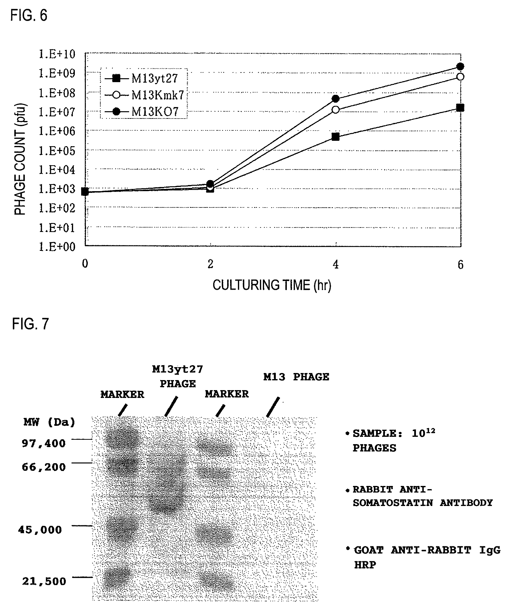
[0094]Unless noted otherwise in the specification, the following protocols were used in carrying out the experiments. The culturing of M13 phages, the preparation of M13 phages, the extraction of DNA from E. coli and the M13 phages, enzyme reactions using DNA and oligonucleotides, and the transformation of E. coli were all carried out in accordance with the manufacturer's protocols for the materials used. In the absence of manufacturer's protocols, the protocols described in Molecular Cloning: A Laboratory Manual (Cold Spring Harbor Laboratory Press) were followed.

[0095]DNA base sequence determinations were carried out with special-purpose kit reagents using an ALF DNA sequencer (Amersham Bioscience) or an Open Gene DNA sequencer.

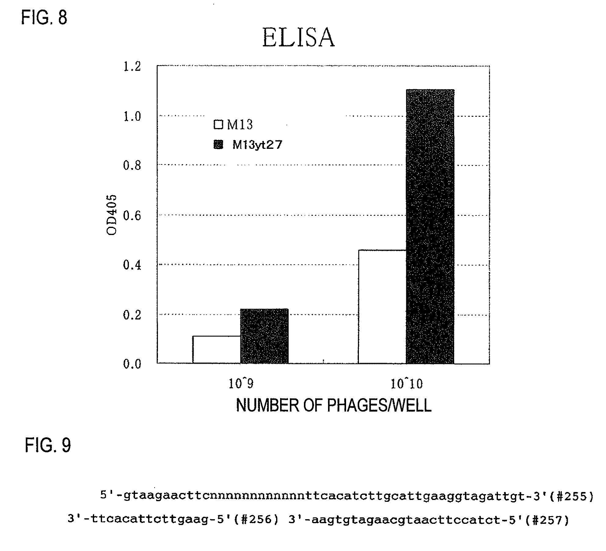
[0096]The oligonucleotides were custom synthesized by Greiner Japan KK and Genenet Co., Ltd. Base sequences for each oligonucleotide are shown in FIG. 14. The symbol # indicates the oligonucleotide number.

example 2

Construction of the Vector M13yt6 for Expressing Random

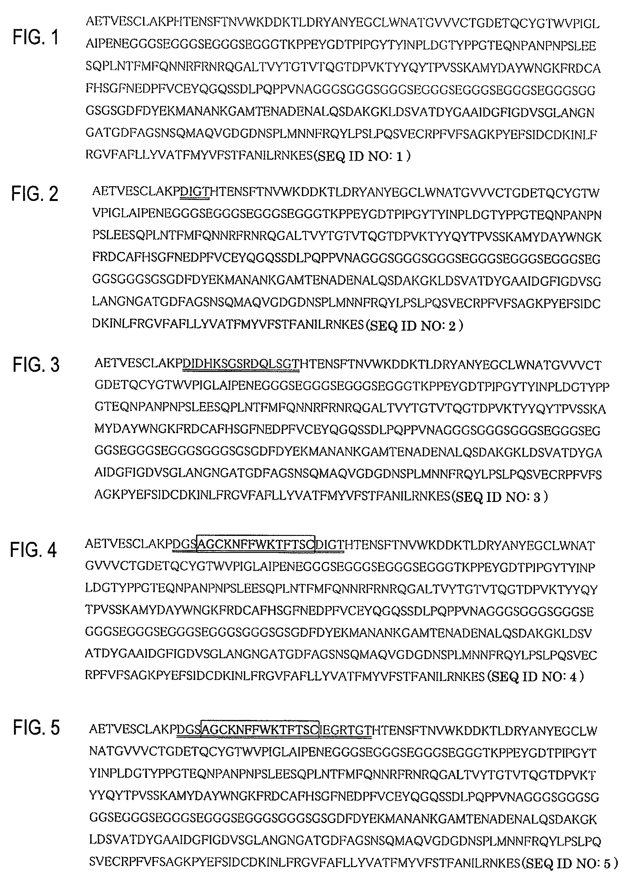
[0097]Peptides in a Filamentous Bacteriophage Using the single-stranded DNA (abbreviated below as “ssDNA”) of the M13KO7 phage, the oligonucleotide #186 shown in FIG. 14, and a Mutant K kit (Takara 6060; Takara Bio), mutation was carried out in vitro in accordance with the kit protocol. A phage solution, RF DNA and ssDNA were prepared from single plaques obtained by transformation. Restriction enzyme cleavage of the RF DNA at KpnI or EcoRV resulted in the identification of 8.7 Kb DNA fragments in each case. Restriction enzyme cleavage at two places, i.e., KpnI and HindIII or EcoRV and HindIII, resulted in the identification of DNA fragments of 5.7 Kb and 3.0 Kb in each case. In addition, the DNA base sequence at the mutation site in ssDNA was determined, and the M13yt6 phage thereby identified.

example 3

Preparation of M13yt6 Vector DNA Fragments Double-Cleaved by Restriction Enzymes EcoRv and KpnI

[0098]The M13yt6 vector was cleaved by the restriction enzyme EcoRV (Takara Bio), then cleaved by the restriction enzyme KpnI (Takara Bio). The resulting 8.7 Kb EcoRV-KpnI double-cleaved DNA fragments were isolated by electrophoresis using 0.8% agarose gel, following which the DNA fragments were collected from the agarose gel. The concentration of the collected DNA was determined by comparing the degree of color development induced by 0.5 μg / ml of ethidium bromide with that of control DNA (λ HindIII marker: TOYOBO).


PUM

PropertyMeasurementUnit
concentrationaaaaaaaaaa
temperatureaaaaaaaaaa

Abstract

[Problems] To provide a phage display vector which can be fused in the inside of pIII and a phage display method using the vector.[Means for Solving Problems] Disclosed is a phage display method which is characterized by using a mutant pIII protein having an amino acid residue inserted between a proline residue at position 11 and a histidine residue at position 12 in an M13 phage pIII protein as depicted in SEQ ID NO:1. The method enables to produce a random peptide with high efficiency and to provide a novel source in peptide conformation.

Description

TECHNICAL FIELD[0001]The present invention relates to a novel phage vector and a phage display method using the vector.BACKGROUND[0002]When peptides are fused to the N-terminus of the pIII protein of a M13 phage, the peptides are displayed on M13 phage particles. This is known as the “phage display technique.” By having the peptides that are fused be random peptides, a peptide library can be constructed, which provides a source for screening ligands (Scott, J. K. and Smith, G. P. (1990): “Searching for peptide ligands with an epitope library,” Science 249, 386-90).[0003]The phage display technique can in theory provide an enormous number of small peptides, and the peptide libraries thereby obtained generate diversity in the primary structure of the peptides. Many developments are underway in M13 phage display technology. Indeed, a variety of vectors for peptide display have already been developed (Smith, G. P. and Petrenko, V. A. (1997): “Phage Display,” Chem. Rev. 97, 391-410).[000...

Claims


Application Information

Patent Timeline
Patent Type & Authority Applications(United States)
IPC IPC(8): C40B40/02C07K14/005C07H21/04C12N15/63C12N1/21C40B40/10C12N7/01
CPCC07K14/005C12N2795/14122C12N15/1037
Inventor UCHIYAMA, FUMIAKI
Owner UCHIYAMA FUMIAKI
Why Eureka
Try Eureka