
Methods and compositions for protein labeling using lipoic acid ligases

a technology of lipoic acid ligase and protein, applied in the field of methods and compositions for protein labeling using lipoic acid ligase, can solve the problems of perturbing the function of the protein of interest, using such probes, and difficulty in targeting the probes with very high specificity to particular proteins of interes

Active Publication Date: 2009-06-11
MASSACHUSETTS INST OF TECH
View PDF7 Cites 7 Cited by
  • Summary
  • Abstract
  • Description
  • Claims
  • Application Information

AI Technical Summary

Benefits of technology

[0004]According to one aspect of the invention, methods for labeling a target protein are provided. the methods include contacting a fusion protein with a lipoic acid analog, and allowing sufficient time for the lipoic acid analog to be conjugated to the fusion protein via an acceptor polypeptide, in the presence of a lipoic acid ligase or mutant thereof, wherein the fusion protein is a fusion of the target protein and the acceptor polypeptide. In some embodiments, the lipoic acid analog comprises an alkyl azide, or an alkyne carboxylic acid, an aryl azide photoaffinity probe, or a fluorophore substrate. In certain embodiments, the lipoic acid analog is detectably labeled. In some embodiments, the lipoic acid analog is directly detectable. In some embodiments, the directly detectable label is coumarin, fluorescein, an aryl azide, a diazirine, a benzophenone, a resorufin, a xanthene-type fluorophore, a chloroalkane, a metal-binding ligand, or a derivative thereof. In some embodiments, the detectable label is coumarin. In certain embodiments, the lipoic acid analog is labeled with an indirectly detectable label. In some embodiments, the indirectly detectable label is an enzyme. In some embodiments, the lipoic acid analog is labeled with a membrane impermeant label. In certain embodiments, the lipoic acid analog is labeled after conjugation to the fusion protein. In some embodiments, the lipoic acid analog is labeled with a cyclooctyne conjugate. In some embodiments, the cyclooctyne conjugate is detectably labeled. In certain embodiments, the detectable label is coumarin, fluorescein, a aryl azide, a diazirine, a benzophenone, a resorufin, a xanthene-type fluorophore, a chloroalkane, a metal-binding ligand, or a derivative thereof. In some embodiments, the target protein is a cell surface protein. In some embodiments, the fusion protein is in a cell. In some embodiments, the cell expresses the lipoic acid ligase or mutant thereof. In certain embodiments, the cell is a eukaryotic cell. In some embodiments, the cell is a bacterial cell. In certain embodiments, the acceptor polypeptide comprises an amino acid sequence of any one of SEQ ID NO: 1, 2, 3, 4, 5, 6, 7, 8, 9, or 10, or a functional variant thereof. In some embodiments, the functional variant of any one of SEQ ID NO: 1, 2, 3

Problems solved by technology

The major bottleneck to using such probes inside cells, however, is the difficulty of targeting the probes with very high specificity to particular proteins of interest, given the chemical heterogeneity of the cell interior.
Because GFPs are genetically encoded, their labeling is absolutely specific and GFP variants have proven extremely useful for in vivo studies of protein localization, however, they still have severe limitations such as their large size (˜235 amino acids), which can perturb the function of the protein of interest, and the fact that they are not very bright and only amenable to optical microscopy.
This method has yielded exciting new biological information, but suffers from poor specificity, and cell toxicity.

Method used


Image

  • Methods and compositions for protein labeling using lipoic acid ligases
  • Methods and compositions for protein labeling using lipoic acid ligases
  • Methods and compositions for protein labeling using lipoic acid ligases

Examples


example 1

Introduction

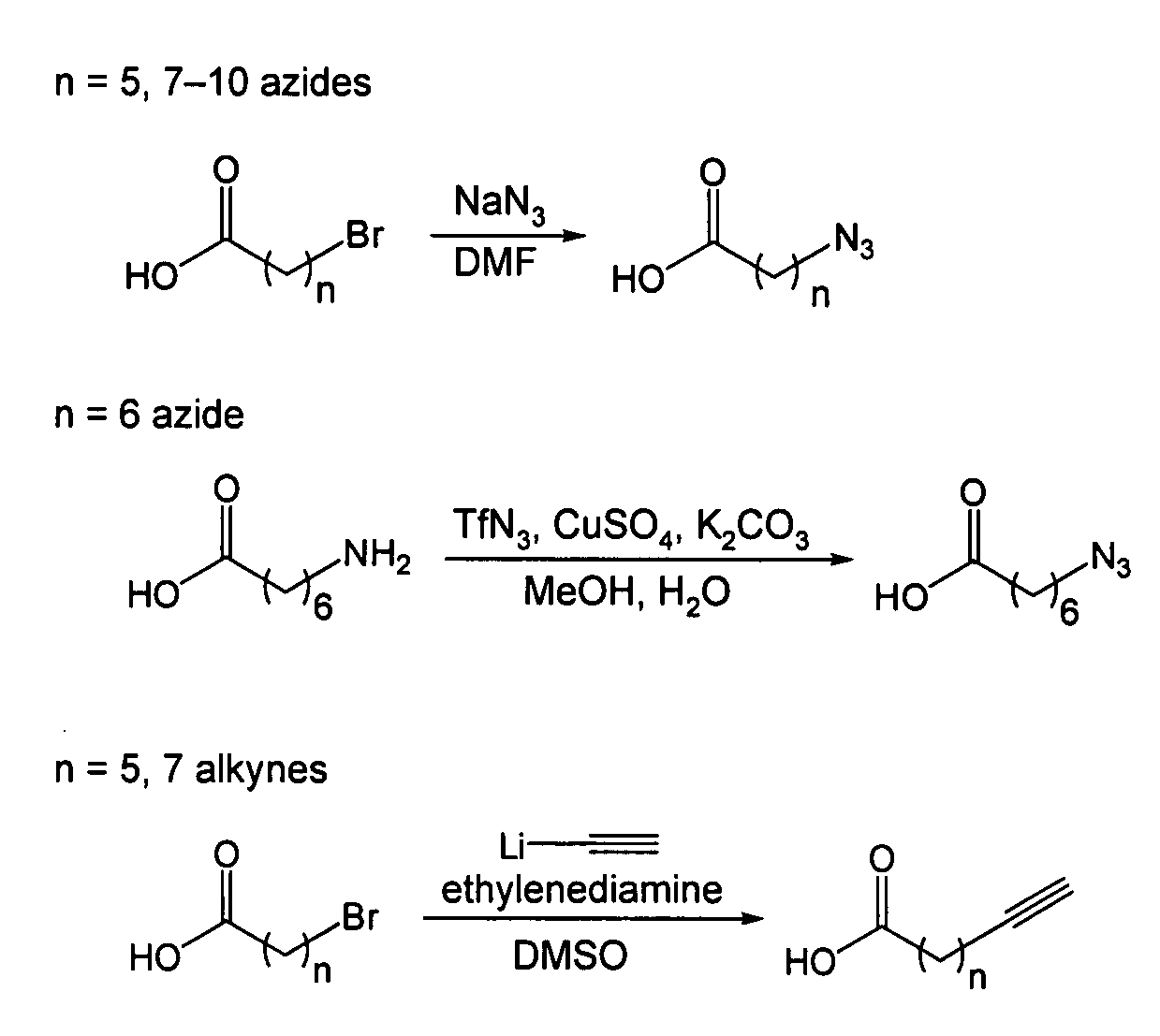
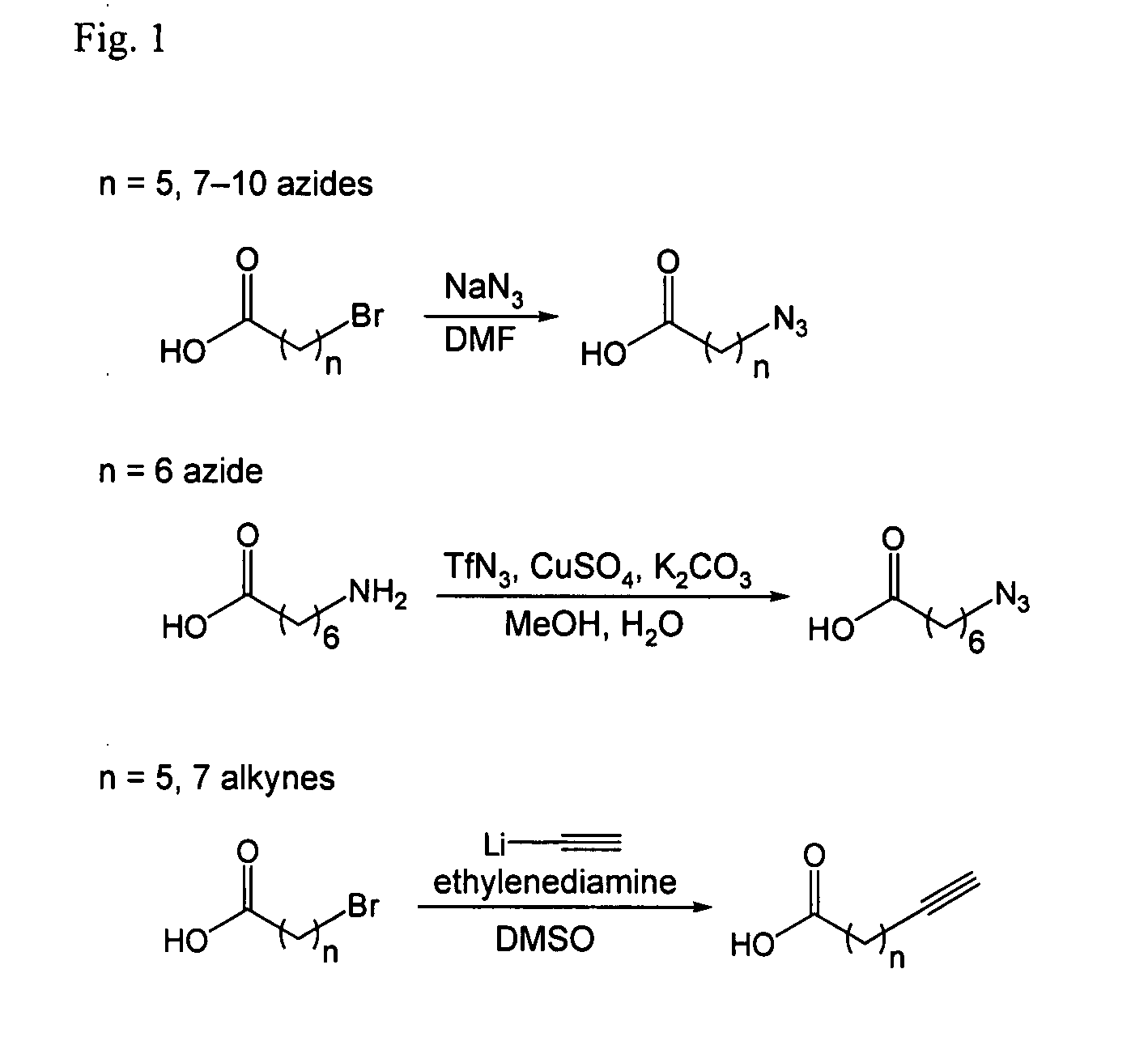
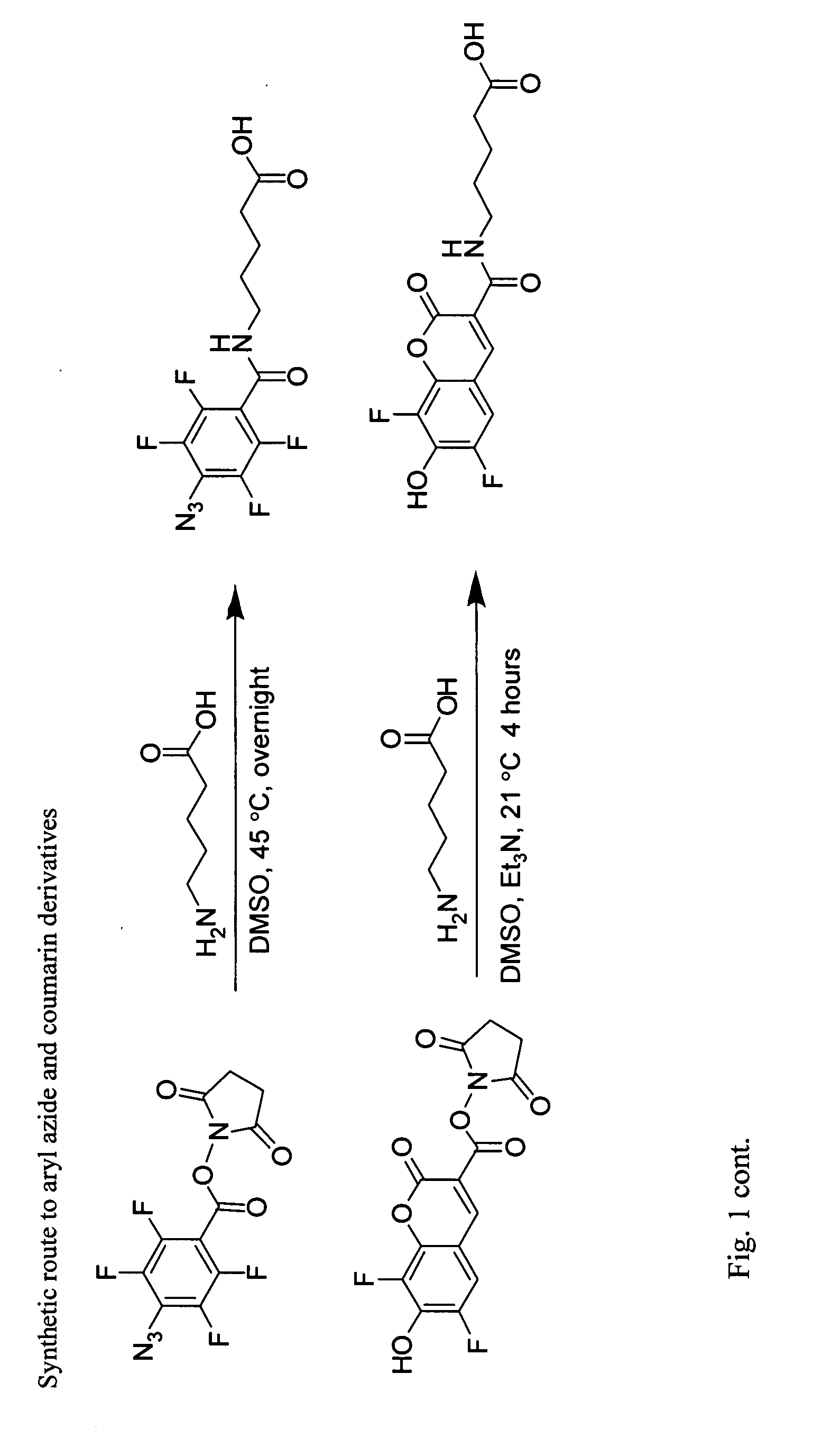
[0125]The following are general synthetic methods used in the experiments described herein, including those of Example 2.

General Synthetic Methods

[0126]Reagents were purchased from Sigma-Aldrich (St, Louis, Mo.), Alfa Aesar (Ward Hill, Mass.), TCI America (Portland, Oreg.), Invitrogen (Carlsbad, Calif.), or GE Healthcare and used without further purification. Analytical thin layer chromatography was performed using 0.25 mm silica gel 60 F254 plates and visualized with ninhydrin or bromocresol. Flash column chromatography was carried out using silica gel (ICN SiliTech 32-63D). Mass spectra were recorded on an Applied Biosystems 200 QTRAP Mass Spectrometer (foster City, Calif.) using electrospray ionization. HPLC was performed on a Varian Prostar Instrument (Palo Alto, Calif.) equipped with an autosampler and photo-diode-array detector. For analytical HPLC, a reverse-phase 250×4.6 mm Microsorb-MV 300 C18 column was used. For preparative HPLC, a reverse-phase 250×10 mm Micr...

example 2

Introduction

[0174]Live cell imaging is a powerful method for studying protein dynamics at the cell surface, but conventional probes, such as antibodies and fluorescent ligands, are bulky, interfere with protein function (1,2), or dissociate after internalization (3,4). To overcome these limitations, we developed a method to covalently tag any cell surface protein with any chemical probe with remarkable specificity. Through rational design, we re-directed a microbial lipoic acid ligase (LplA) (5) to specifically ligate an alkyl azide to an engineered LplA acceptor peptide (LAP) tag. The alkyl azide is then selectively derivatized with a cyclooctyne (6) conjugated to any probe of interest. We demonstrate the utility of this method by first labeling LAP fusion proteins expressed on the surface of living mammalian cells with Cy3, AlexaFluor568, and biotin. Next, we combined LAP-tagging with our previously reported tagging method (7,8) to simultaneously monitor the dynamics of two recept...


PUM

PropertyMeasurementUnit
Fractionaaaaaaaaaa
Fractionaaaaaaaaaa
Fractionaaaaaaaaaa

Abstract

The invention provides compositions and methods of use thereof for labeling peptide and proteins in vitro or in vivo. The methods described herein employ lipoic acid ligase or mutants thereof, and lipoic acid analogs recognized by lipoic acid ligase and lipoic acid ligase mutants.

Description

GOVERNMENT SUPPORT[0001]This invention was made with government support awarded by the National Institutes of Health under Grant Number R01 GM072670-01. The Government has certain rights in the invention.BACKGROUND OF THE INVENTION[0002]Biophysical probes such as fluorophores, spin labels, and photoaffinity tags have greatly improved the understanding of protein structure and function in vitro, and there is great interest in using them inside cells to study proteins within their native context. The major bottleneck to using such probes inside cells, however, is the difficulty of targeting the probes with very high specificity to particular proteins of interest, given the chemical heterogeneity of the cell interior. The most prominent method for labeling cellular proteins is to genetically encode green fluorescent protein (GFP) or one of its variants as a fusion to the protein of interest. Because GFPs are genetically encoded, their labeling is absolutely specific and GFP variants ha...

Claims


Application Information

Patent Timeline
IPC IPC(8): C07K14/245C07D339/04
CPCC12N9/93G01N33/532G01N33/533C07K4/00C12N15/62C12N9/1241G01N2333/9015
Inventor TING, ALICE Y.SUAREZ, MARTA FERNANDEZBARUAH, HEMANTACHOI, YOON-AA
Owner MASSACHUSETTS INST OF TECH
Learn More