Regenerative Dot cells

a dot cell and regenerative technology, applied in the field of regenerative dot cells, can solve the problems of heart tissue necrosis, scarring can happen in different tissues and organs, and patients with large scars have life-long psychological and physical burdens, and achieve the effect of high regenerative

Inactive Publication Date: 2009-06-18
THE BOARD OF TRUSTEES OF THE LELAND STANFORD JUNIOR UNIV
View PDF2 Cites 11 Cited by
  • Summary
  • Abstract
  • Description
  • Claims
  • Application Information

AI Technical Summary

Benefits of technology

[0010]Methods and compositions are provided for the isolation, culture and use of mammalian Dot cells. Dot cells are highly regenerative, and find use in a variety of situations for repair of skin wounds, cardiac damage and blood vessel damage. Dot cells are very small cells, having an average size of about 0.5 μm, and having an undefined nuclear structure and generally lacking intracellular organelles. Dot cells may be isolated from fetal or adult tissues, and are found in tissue including, without limitation, fetal dermal tissue, blood, and bone marrow. The cells are characterized as expressing one or more markers selected from E-cadherin, integrin β1, CXCR4, CD90 and CD34, and may be selected on the basis of such expression patterns. Dot cells also express transcription factors characteristic of stem cells, including Oct / 4, Nanog and Sox-2

Problems solved by technology

As a result of such damage, scarring can happen in different tissue and organs, such as skin, liver and heart.
In skin, patients with large scars have life long psychological and physical burdens, since no proven therapy for scarring exists.
In cardiovascular diseases, an acute heart attack may be induced by ischemia of cardiomyocytes, which can result in the necrosis of heart tissue and leads to scar formation in heart.
While stem cells are believed to have great potential for regeneration of damaged tissues, some reports have been contradictive, for example in the use of adult bone marrow stem cells to regenerate cardiomyocytes.
Further, conventional bone marrow stem cells have been reported to lose the ability to regenerate tissue after in vitro culture.
However, although attempts have been made to prospectively isolate stromal stem cells with multi-lineage potential, the results are still unclear or controversial.
Thus, the biological significance of fusion-derived tissue regeneration may be low under normal physiological conditions.

Method used


Image

  • Regenerative Dot cells
  • Regenerative Dot cells
  • Regenerative Dot cells

Examples


example 1

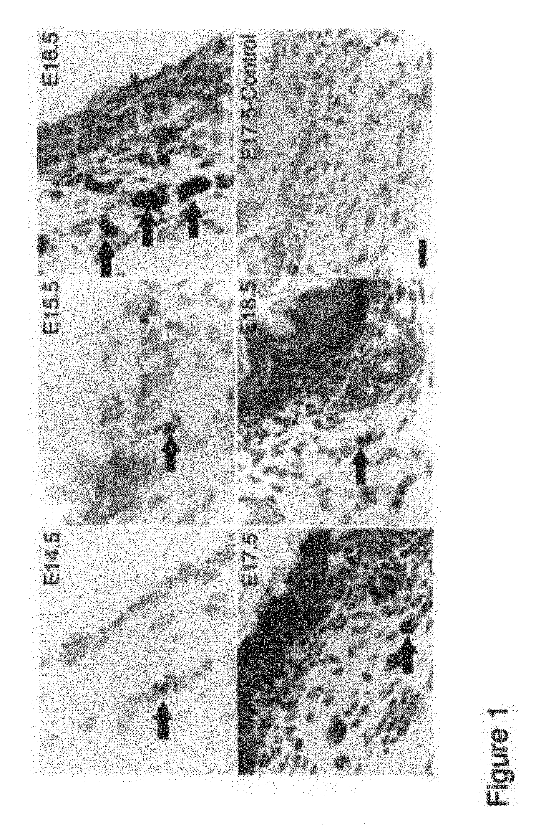
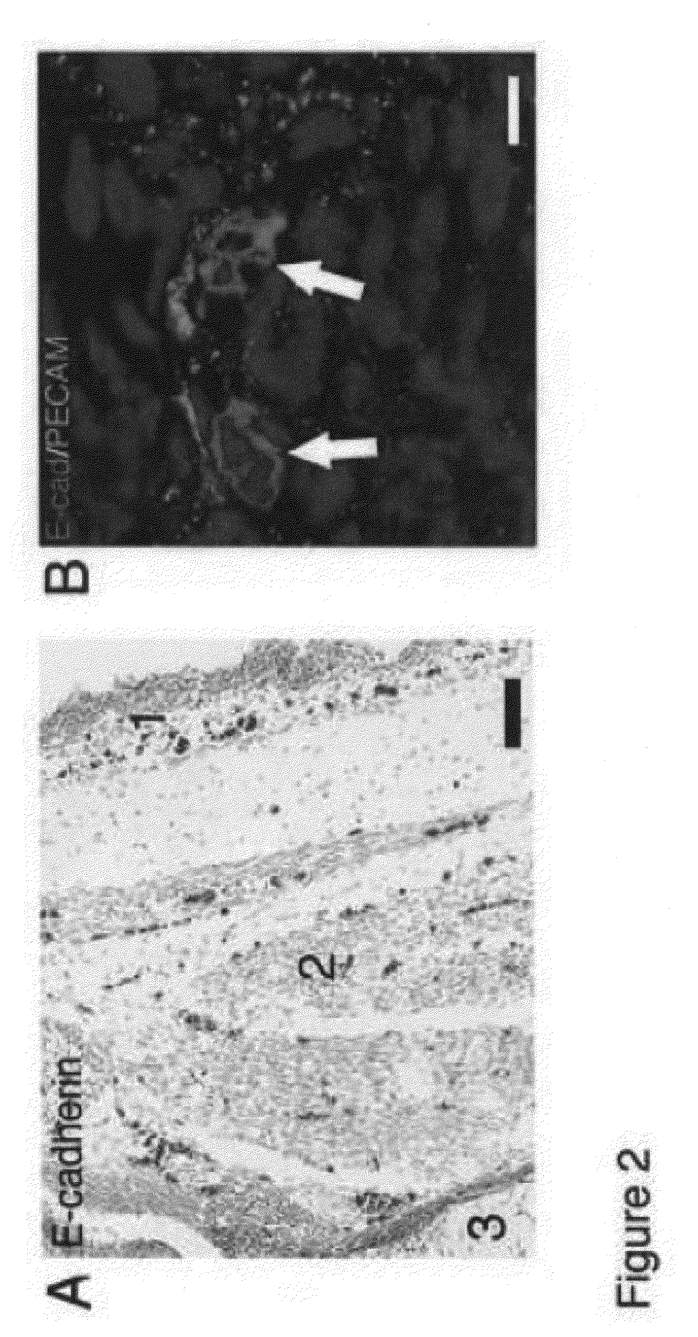
[0123]Wounds in fetal skin heal without scar, however the mechanism is unknown. We have identified a novel group of E-cadherin positive cells in the blood of fetal and adult mice and named them “Dot cells”. The percentage of Dot cells in E16.5 fetal mice blood is more than twenty times higher compared to adult blood. Dot cells also express integrin β1, CD184, CD34, CD13low and Sca1 low, but not CD45, CD44, and CD117. Dot cells have a tiny dot shape between one to seven μm diameters with fast proliferation in vitro. Most of Dot cells remain positive for E-cadherin and integrin β1 after one month in culture. Transplantation of Dot cells to adult mice heals skin wounds with less scar due to reduced smooth muscle actin and collagen expression in the repair tissue. Tracking GFP-positive Dot cells demonstrates that Dot cells migrate to wounds and differentiate into dermal cells, which also express strongly to FGF-2, and later lose their GFP expression. Our results indicate that Dot cells ...

example 2

Dot Cells: Primitive Marrow and Circulating Regenerative Fusogenic Cells

[0153]We have herein identified a novel group of circulating and bone-marrow-derived DNA and protein positive particles and termed them “Dot cells”. The morphology of blood-derived Dot cells does not match the definition of conventional eukaryotic cells. Their size ranges from 0.1 to 2 μm, and they do not have nuclear membrane or intracellular organelles. Blood-derived Dot cells have a unique self-renewal mechanism by release of grouped new Dot cells from colonies. When present with differentiated cells, Dot cells aggregate and fuse together. The fused aggregates then undergo cellular transdifferentiation. Both the nuclear and cytoplasmic components of the newly differentiated cells are derived from Dot cell fusion. In vitro expanded Dot cells form spheroids, which release fused or grouped Dot cells that contribute to tissue regeneration during in vivo wound healing. Dot cells are a newly identified cellular pop...

example 3

Dot Cell Differentiation

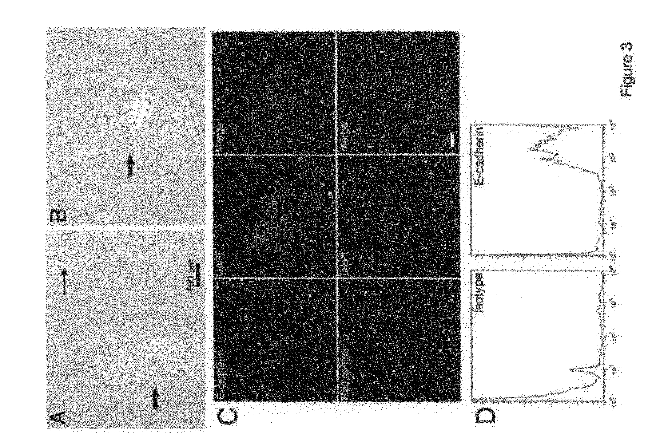
[0182]FIG. 16 shows FACS analysis of surface markers on either freshly sorted blood-derived Dot cells or the in vitro expanded Dot cells. Freshly sorted Dot cells express E-cadherin, integrin β1 / CD29, CXCR4 / CD184, CD90 and CD34. Only a small amount of freshly sorted Dot cells express Sca1. However, after one month in culture, the number of Dot cells that express E-cadherin, integrin β1 and CD184 is significantly reduced, and the number that expresses CD90 and CD34 remains the same. Interestingly, a significant amount of Dot cells start to express c-kit and CD45, but none express Sca1. The change of surface markers may correlate with the increased variety of Dot cell shapes in culture.

[0183]As shown in FIG. 17, Dot cells differentiate into neuron-like cells in vitro. Dot cells were cultured and they have spontaneous differentiation. FIG. 17A shows the morphology of glial cells. 17B shows that Dot cells differentiate into a neuron-like morphology.

[0184]Shown in...


PUM

PropertyMeasurementUnit
sizeaaaaaaaaaa
diameteraaaaaaaaaa
diameteraaaaaaaaaa

Abstract

Methods and compositions are provided for the isolation, culture and use of highly regenerative somatic mammalian cells. The cells are very small, and have an undefined nuclear structure. The cells may be isolated from fetal or adult tissues, and are found in tissue including, without limitation, fetal dermal tissue, blood, and bone marrow. The cells are characterized as expressing one or more markers selected from E-cadherin, integrin β1, CXCR4, CD90 and CD34, and may be selected on the basis of such expression patterns.

Description

GOVERNMENT RIGHTS[0001]This invention was made with Government support under federal grant 1012070, 119, HAERX awarded by the Packard Foundation. The Government has certain rights in this invention.[0002]Tissue damage can be caused by traumatic injury, ischemic and fibrotic disease. As a result of such damage, scarring can happen in different tissue and organs, such as skin, liver and heart. In skin, patients with large scars have life long psychological and physical burdens, since no proven therapy for scarring exists. In cardiovascular diseases, an acute heart attack may be induced by ischemia of cardiomyocytes, which can result in the necrosis of heart tissue and leads to scar formation in heart. Therefore, finding therapy to regenerate damaged tissue or organs is of great interest in clinical medicine. While stem cells are believed to have great potential for regeneration of damaged tissues, some reports have been contradictive, for example in the use of adult bone marrow stem c...

Claims


Application Information

Patent Timeline
Patent Type & Authority Applications(United States)
IPC IPC(8): A61K35/28C12N5/00C12N5/08C12N5/06C12N5/02A61K35/14A61K35/12C12N5/074
CPCA61K35/12C12N5/0607A61K2035/124
Inventor KONG, WUYILI, SHAOWEILORENZ, PETER
Owner THE BOARD OF TRUSTEES OF THE LELAND STANFORD JUNIOR UNIV
Why Eureka
Try Eureka