Method of assessing metastatic carcinomas from circulating endothelial cells and disseminated tumor cells

a metastatic carcinoma and tumor cell technology, applied in the field of cancer prognosis and survival in metastatic cancer patients, can solve the problems of difficult detection and elimination, inability to treat all patients successfully, and inability to improve the survival rate of cancer patients over the past two decades

Inactive Publication Date: 2009-07-30
JANSSEN DIAGNOSTICS LLC
View PDF19 Cites 68 Cited by
  • Summary
  • Abstract
  • Description
  • Claims
  • Application Information

AI Technical Summary

Benefits of technology

[0023]More specifically, the present invention provides the apparatus, methods, and kits for assessing patient survival, the time to disease progression, and response to therapy in MBC. The accurate cell enumeration provides a basisi for prediction of survival, based upon a threshold comparison of the number of cells in blood.

Problems solved by technology

Despite efforts to improve treatment and management of cancer patients, survival in cancer patients has not improved over the past two decades for many cancer types.
Unfortunately, the metastatic colonies are difficult to detect and eliminate and it is often impossible to treat all of them successfully.
These and other debris antigens are thought to be derived from destruction of both circulating and non-circulating tumor cells, and thus their presence may not always reflect metastatic potential, especially if the cells rupture while in an apoptotic state.
Assessing enzyme activity in this type of analysis can involve time-consuming laboratory procedures such as gel electrophoresis and Western blot analysis.
However in both cases, the base levels during remission, or even in healthy normals, are relatively high and may overlap with concentrations found in patients, thus requiring multiple testing and monitoring to establish patient-dependent baseline and cut-off levels.
However, PSA or the related PSMA testing leaves much to be desired.
For example, elevated levels of PSA weakly correlate with disease stage and appear not to be a reliable indicator of the metastatic potential of the tumor.
The aforementioned studies do not provide for consistent data with a long follow-up period or at a satisfactory specificity.
Accordingly, these efforts have proven to be somewhat futile as the appearance of mRNA for antigens in blood have not been generally predictive for most cancers and are often detected when there is little hope for the patient.
These results suggest that tumor cells were shed into the bloodstream (possibly during surgical procedures or from micro metastases already existing at the time of the operation), and resulted in poor patient outcomes in patients with colorectal cancer.
As mentioned, these detection ranges are based on unreliable conversions of amplified product to the number of tumor cells.
Further, PCR-based assays are limited by possible sample contamination, along with an inability to quantify tumor cells.
Most importantly, methods based on PCR, flowcytometry, cytoplasmic enzymes and circulating tumor antigens cannot provide essential morphological information confirming the structural integrity underlying metastatic potential of the presumed CTC and thus constitute functionally less reliable surrogate assays than the highly sensitive imaging methods embodied, in part, in this invention.
Unfortunately, the same spreading of malignant cells continues to be missed by conventional tumor staging procedures.
But these invasive techniques are deemed undesirable or unacceptable for routine or multiple clinical assays compared to detection of disseminated epithelial tumor cells in blood.
The great variation in the reported ranges of CECs from 1 to 1000 per mL makes the interpretation of these reported results quite difficult if not impossible.
In addition little attention is paid to the characterization of the assays used to enumerate CECs.
Currently available prognostic protocols have not demonstrated a reliable means for correlating circulating endothelial cells (CEC) and / or disseminated tumor cells (DTC) to predict progression free- or overall survival in patients with cancers such as metastatic breast cancer (MBC).

Method used


Image

  • Method of assessing metastatic carcinomas from circulating endothelial cells and disseminated tumor cells
  • Method of assessing metastatic carcinomas from circulating endothelial cells and disseminated tumor cells
  • Method of assessing metastatic carcinomas from circulating endothelial cells and disseminated tumor cells

Examples


example 1

Circulating Endothelial Cells (CEC) and Circulating Tumor Cells (CTC) in Patients with Metastatic Colorectal Cancer

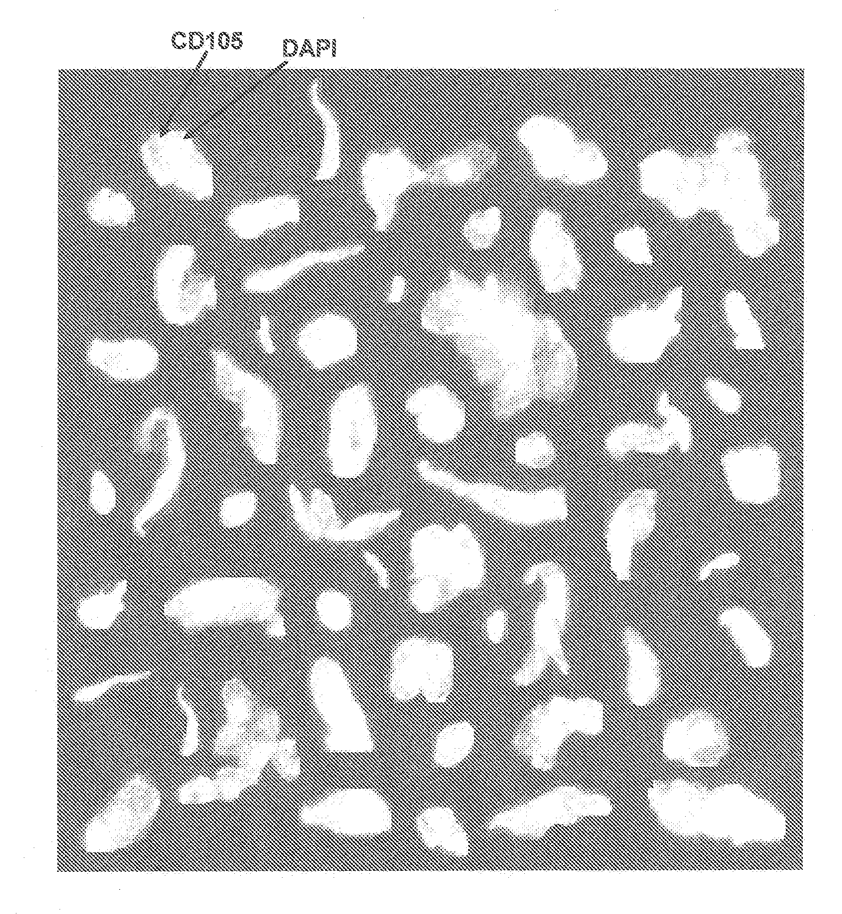
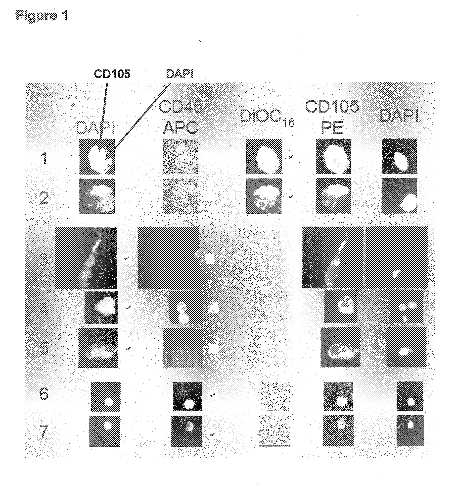
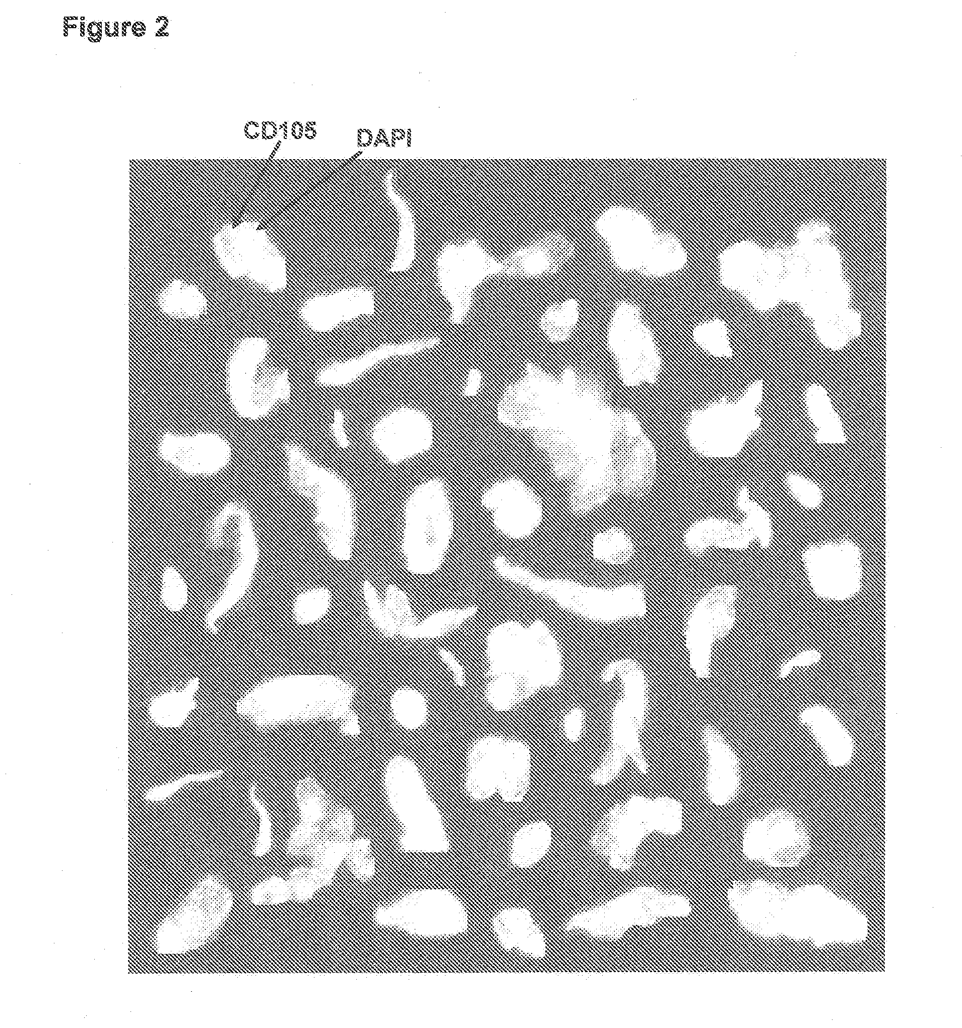
[0039]Lack of validated surrogate endpoints is an impediment to developing new cancer therapy. We hypothesized that CTC and CEC could predict outcome in pts undergoing treatment for metastatic colorectal cancer. Eligible patients for this multicenter study had metastatic colorectal cancer, and were initiating 1st, 2nd, or 3rd-line systemic therapy. Blood was obtained at baseline and 3-4 weeks after treatment initiation for enumeration of CTC / CEC. CTC / 7.5 ml and CEC / 4 ml of blood were measured with the CellTracks® System. CTC were immunomagnetically enriched targeting CD326 (EpCAM), stained with DAPI, cytokeratin 8,18,19, and CD45. CEC expressing CD146 were immunomagnetically enriched and stained with DAPI, CD105, and counterstained with CD45. Cell morphology was confirmed in all cases.

[0040]In 139 controls CTC were virtually absent (0 CTC in 135 and 1 CTC in 4). For 131...

example 2

Circulating Endothelial Cells in Peripheral Blood of Healthy Subjects and Patients with Metastatic Carcinomas

[0042]In order to determine accuracy, precision, and linearity of endothelial cell enumeration in blood and compare CECs in healthy subjects and patients with metastatic carcinomas.

[0043]Blood was drawn in preservative tubes from controls and patients with metastatic carcinomas at multiple geographic locations. Samples were maintained at room temperature, shipped to a central laboratory, and processed within 72 hours of blood collection. All patients and healthy individuals were enrolled using approved protocols and provided informed consent. The healthy individuals used for comparison with the patients had no known illness or fever at the time of draw, no history of malignant disease, and were 35 years of age or older to provide a cohort age-matched with the cancer population.

[0044]The CellTracks® System (Immunicon, Huntingdon Valley, Pa.) used for endothelial cell enumerati...

example 3

Peri-Operative Assessment of Circulating Tumor Cells in Blood, Disseminated Tumor Cells in Bone Marrow, and Tissue Gene Signatures in Patients with Primary Breast Cancer

[0059]Approximately 30% of the 200,000 women diagnosed annually with breast cancer will recur. Without a validated assay to identify these patients, all become candidates for adjuvant therapy. Both Real-time RT-PCR analysis of primary tissue and detection of disseminated tumor cells (DTC) in bone marrow by immunohistochemistry (IHC) purportedly aid in identifying these patients. This study demonstrates that the automated immunomagnetic fluorescent detection systems used to detect ‘circulating’ tumor cells (CTC) in blood could also be used to quantify DTCs in marrow. Incidence of CTCs, DTCs and gene signatures in matched specimens were also compared.

[0060]30 ml blood and 3 ml bone marrow specimens were collected in a preservative peri-operatively from 33 consented primary breast cancer patients stage 0-III. 31 healthy...


PUM

PropertyMeasurementUnit
magnetic fieldaaaaaaaaaa
enzyme activityaaaaaaaaaa
gel electrophoresisaaaaaaaaaa

Abstract

A method for assessing cancer in test subjects is described based upon enumeration of circulating endothelial cells and/or disseminated tumor cells in a test subject. This method is used to quantify disseminated tumor cells. Correlations with circulating tumor cells provides prognostic information with high accuracy in assessing the risk of recurrence in patients with primary breast cancer.

Description

CROSS-REFERENCE TO RELATED APPLICATIONS[0001]This is a non-provisional application which claims priority to U.S. Provisional Applications 60 / 686,701, filed Jun. 2, 2005, and 60 / 686,705, filed Jun. 2, 2005. This application also claims priority to U.S. application Ser. No. 11 / 202,875, filed Aug. 12, 2005. Each of the aforementioned applications is incorporated in full by reference herein.BACKGROUND[0002]1. Field of the Invention[0003]The invention relates generally to cancer prognosis and survival in metastatic cancer patients, based on the presence of morphologically intact circulating cancer cells (CTC) in blood. More specifically, diagnostic methods, reagents and apparatus are described that correlate the presence of circulating cancer cells in 7.5 ml of blood of metastatic breast cancer patients with time to disease progression and survivability. Circulating tumor cells are determined by highly sensitive methodologies capable of isolating and imaging 1 or 2 cancer cells in approx...

Claims


Application Information

Patent Timeline
Patent Type & Authority Applications(United States)
IPC IPC(8): A01N1/02C12Q1/02
CPCG01N33/574G01N33/57492G01N33/57415
Inventor CONNELLY, MARK CARLEDOYLE, GERALD V.RAO, GALLA CHANDRATERSTAPPEN, LEON W.M.M.
Owner JANSSEN DIAGNOSTICS LLC
Why Eureka
Try Eureka