Method to measure and characterize microvesicles in the human body fluids

a human body fluid and microvesicle technology, applied in the field of cancer diagnosis, can solve the problems of no prior art on a method and low efficiency of currently available methods for obtaining exosomes, and achieve the effects of low efficiency, diagnosis and follow-up, and simple and reliabl

Inactive Publication Date: 2009-09-03
EXOSOMICS SPA
View PDF3 Cites 47 Cited by
  • Summary
  • Abstract
  • Description
  • Claims
  • Application Information

AI Technical Summary

Benefits of technology

[0007]A central problem in obtaining useful in vivo data on exosomes is the low level of efficiency of currently available methods to obtain exosomes in order to quantify and characterize them from human body fluids, particularly from plasma. The body fluids may also be ascite, cerebral fluids, bone marrow, urine, faeces or bronco-alveolar washing. To provide a solution to these currently faced problems, this disclosure provides a simple a reliable method to detect and quantify exosomes from body fluids, especially from human plasma. According to this disclosure an ELISA based test (called ExoTest) allows quantific

Problems solved by technology

A central problem in obtaining useful in vivo data on exosomes is the low level of efficiency of currently available methods to obtain exosomes in order to quantify and characterize them from human body f

Method used


Image

  • Method to measure and characterize microvesicles in the human body fluids
  • Method to measure and characterize microvesicles in the human body fluids
  • Method to measure and characterize microvesicles in the human body fluids

Examples


example 1

1. Sources of Exosomes

A. Cell Culture Supernatants

[0043]We used two types of human tumor cell lines, i.e. melanoma and colon carcinoma. MeI 501 and MeI BS are two metastatic melanoma cell lines obtained from melanoma lesions of patients, surgically resected (Istituto Nazionale dei Tumori, Milan, Italy). We also used two colorectal carcinoma cell lines Colo206 (generated from liver metastasis of colorectal cancer patient) and 1869 col (provided by Dr. Maccalli, Istituto Superiore di Sanita′). All cell lines were cultured in RPMI 1640 medium supplemented with 100 IU / ml penicillin, 100 μg / ml streptomycin (Gibco), 2 mM glutamine I(Gibco) and 10% fetal calf serum (FCS) (Invitrogen, Milan, Italy). Human monocytes-derived macrophages (MDM) were obtained from buffy coats of healthy blood donors using CD14 magnetic beads (Milteny Biotec, Germany) and GM-CSF (500 U / ml) for 5 days in culture.

B. Human Tumor SCID-Mouse Plasma

[0044]CB.17 SCID / SCID female mice (Harlan, S. Pietro al Natisone, Italy...

example 2

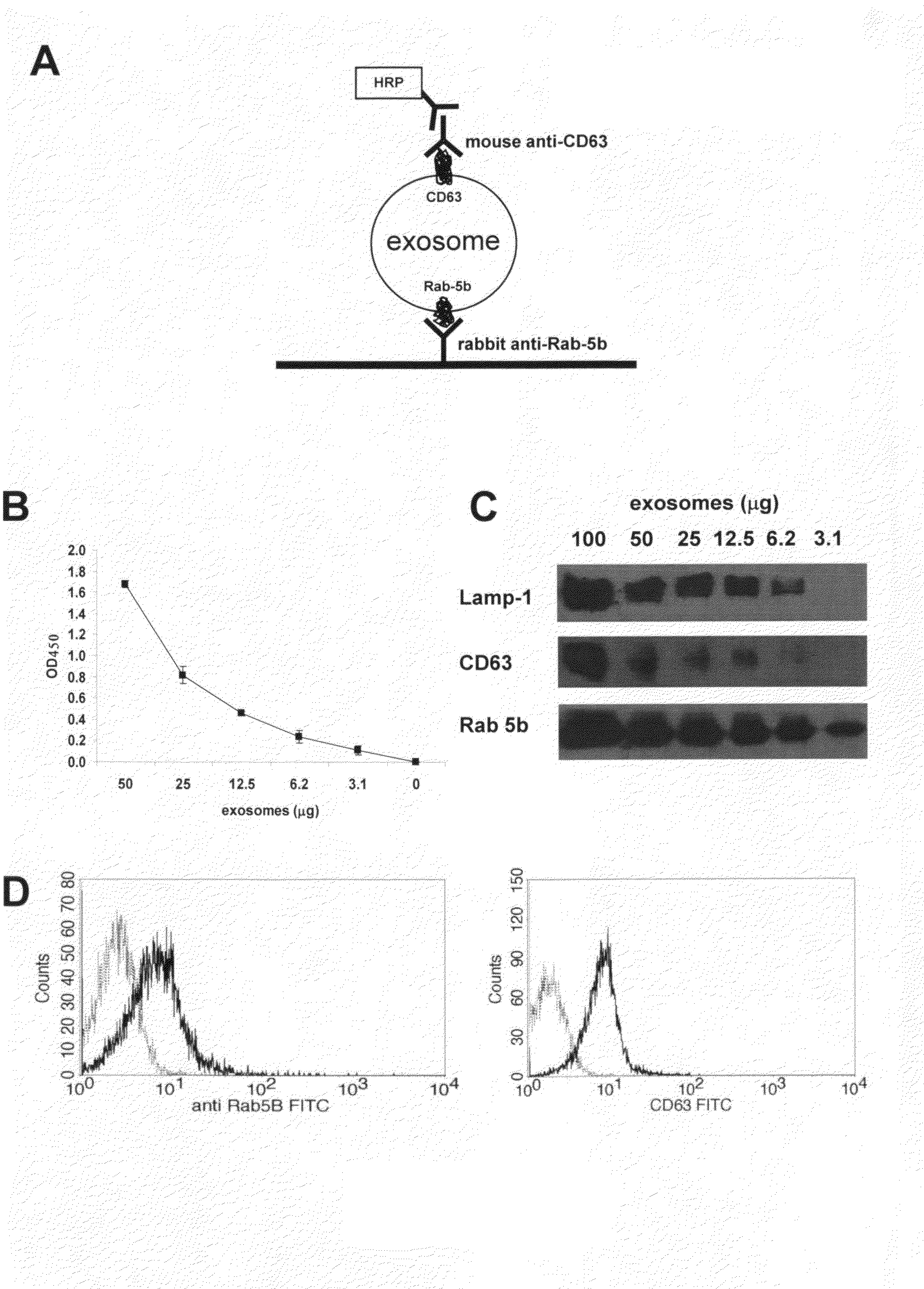
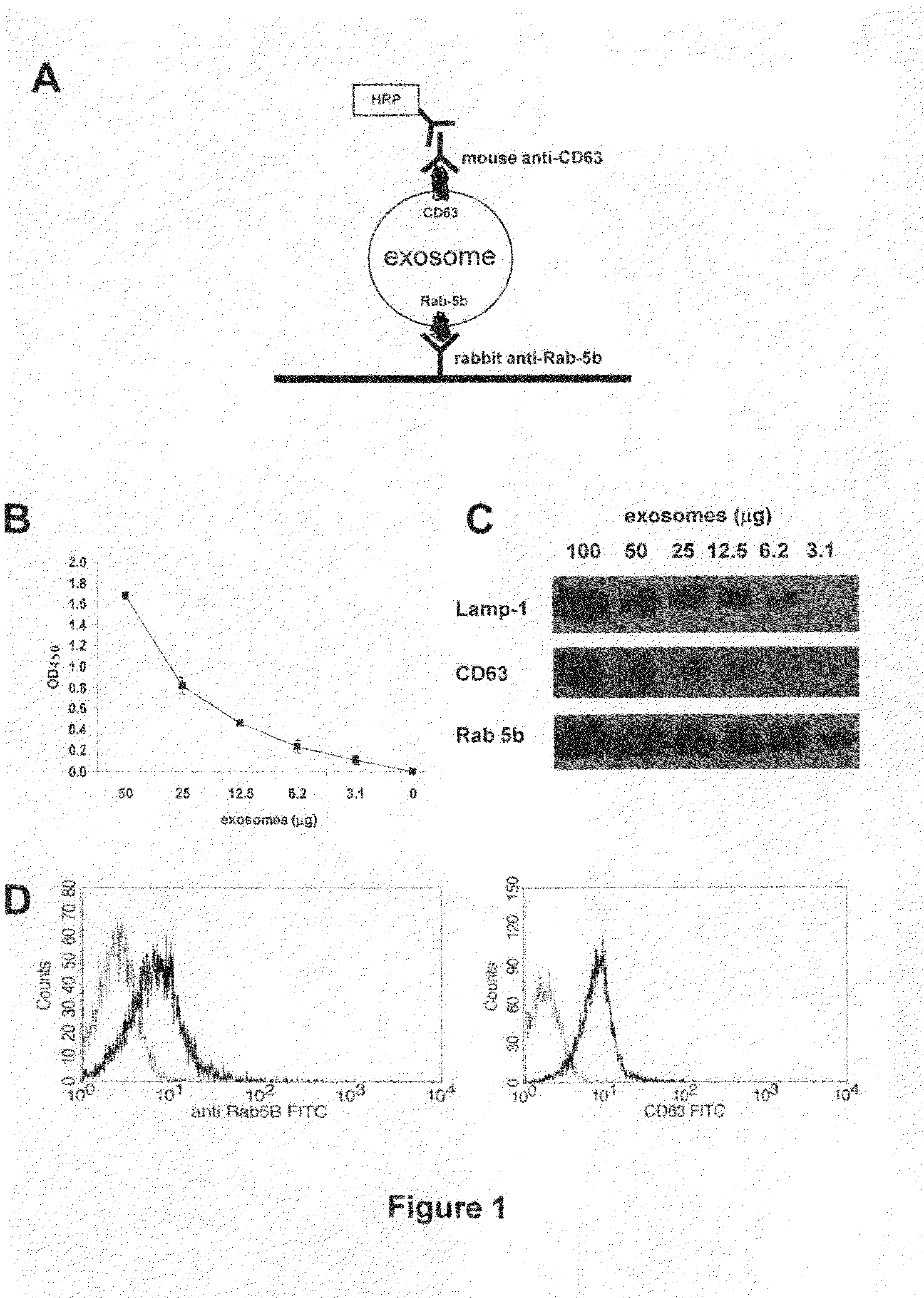
ExoTest Provides Quantification of Exosomes Present in Cell Culture Preparations and has Higher Sensitivity for Exosome Protein Detection than Western Blot Analysis

[0052]Culture supernatants of melanoma and colon carcinoma cell lines were processed following the standard procedure to obtain purified exosomes as described above. The sandwich ELISA set up to detect exosomes (ExoTest, see FIG. 1A) was performed on exosome preparations obtained from cell culture supernatants by differential centrifugation. ExoTest was able to provide a quantification of the exosomes present in the cell culture supernatants, being CD63+ exosomes detectable in dose-dependent manner (FIG. 1B). Negative controls, represented by fractions derived from pellet obtained after the 10 000 g centrifugation, exosomes purified from cell culture medium alone and by only secondary antibody resulted in a barely measurable optical density (OD=0.07+ / −0.01). Intra- and inter-test variability were calculated on six replica...

example 3

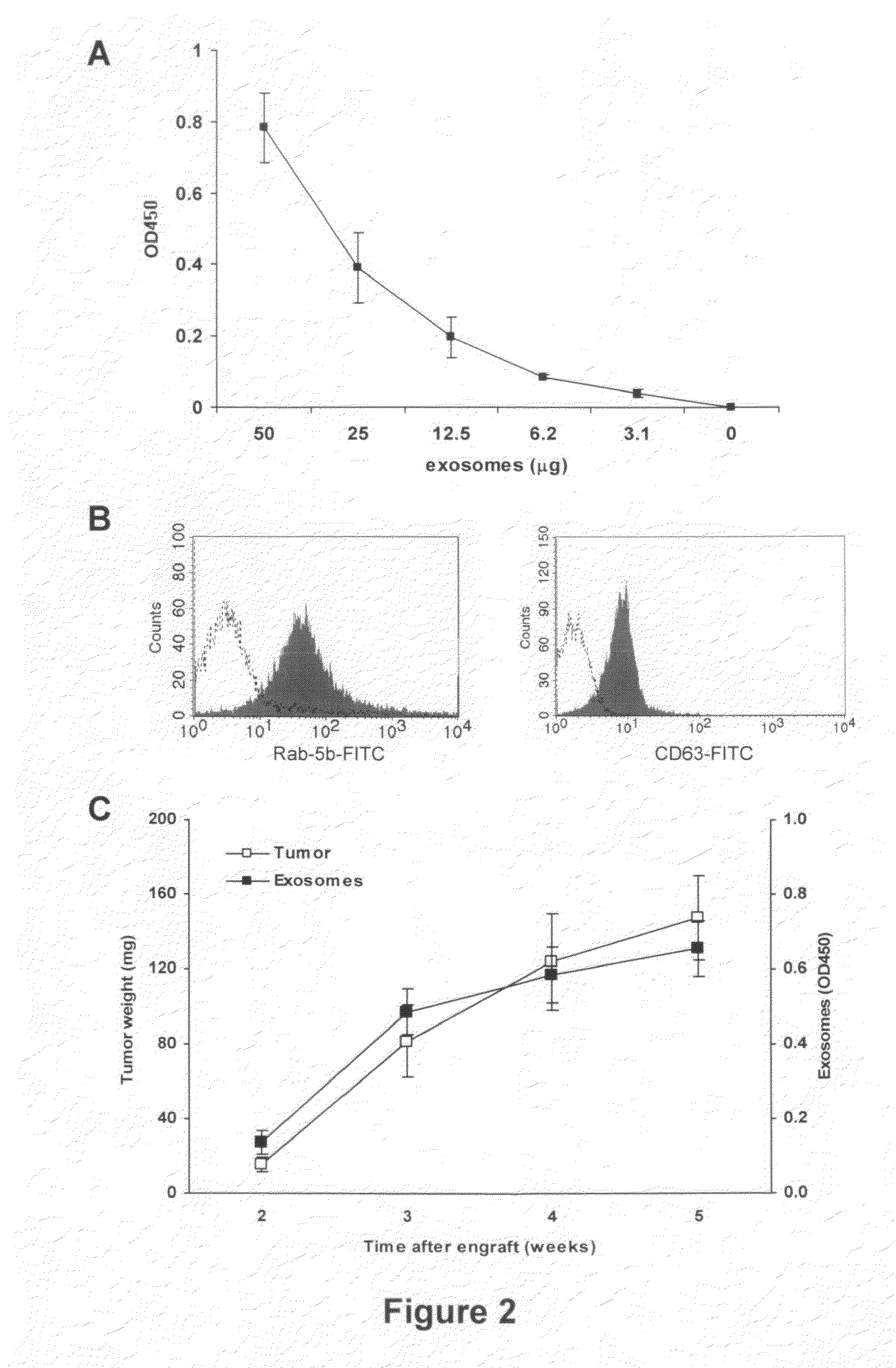
Comparison of the Results of Qualification and Quantification of In Vivo Exosome Preparation of SCID Mice Engrafted with Human Tumors by FACs, WB and ExoTest

[0054]In order to verify the possibility of detecting human tumor-derived exosomes in vivo by using ExoTest, we first performed experiments in which human melanoma (Me501) and colon carcinoma cell lines were injected subcutaneously in SCID mice. In the first experiments, plasma samples obtained at sacrifice from SCID mice carrying engrafted tumors (100-500 mg) were subjected to the exosome purification procedure. As for the exosomes purified from cell culture supernatants, also exosomes purified from mice plasma were clearly and specifically detectable by ExoTest (FIG. 2A) and FACS (FIG. 2B). Exosome preparations obtained from plasma of control SCID mice (not engrafted with human tumors) resulted in background optical densities comparable to the blank samples (OD=0.08±0.03), thus suggesting absence of exosomes in the immunocompr...


PUM


Abstract

This disclosure provides a method to capture, detect, characterize and quantify human exosomes in small volumes of human body fluids by using a sandwich ELISA test. This method allows a full characterization of an exosome preparation, thus providing a tool to distinguish a disease-related condition from a healthy state, by the use of a non-invasive assay. In fact, this method may be useful in screening, diagnosis and prognosis of tumors, with a simple plasma sample. At the same time measurement of circulating exosomes may provide information on the level of tumor mass present in a patient. The method provided here is suitable to evaluate presence of some infectious and/or transmissible agents, such as viral proteins or prion proteins, within circulating exosomes.

Description

PRIORITY[0001]This application claims priority of the U.S. provisional application No. 61 / 062,528 filed on Jan. 25, 2008FIELD OF THE INVENTION[0002]The present invention relates generally to the field of cancer diagnosis. More specifically, the invention relates to a method to quantify and qualify exosomes in the human body fluids.BACKGROUND[0003]Exosomes are microvesicles of a size ranging between 30-120 nm, actively secreted through an exocytosis pathway normally used for receptor discharge and intercellular cross-talk. Exosomes may be detected in cell culture supernatants and some body fluids, following multistep ultracentrifugation. In addition to major hisotcompatibility complex proteins (MHCI, MHC II) and proteins involved in antigen presentation, exosomes may carry membrane and cytocolic proteins involved in many cellular functions. Exosomes are secreted under specific physiological conditions from different cell types such as dendritic cells (DC), lymphocytes, mast cells and...

Claims


Application Information

Patent Timeline
IPC IPC(8): G01N33/53C12Q1/70
CPCG01N33/567G01N33/56988G01N2800/2828G01N2333/16G01N2333/18G01N33/5743
Inventor FAIS, STEFANOLOGOZZI, MARIANTONIA
Owner EXOSOMICS SPA
Why Eureka
Try Eureka