Wave field microscope with sub-wavelength resolution and methods for processing microscopic images to detect objects with sub-wavelength dimensions

Active Publication Date: 2009-10-22
UNIVERSITY OF HEIDELBERG
View PDF8 Cites 59 Cited by
  • Summary
  • Abstract
  • Description
  • Claims
  • Application Information

AI Technical Summary

Benefits of technology

[0063]The microscopical system may further comprise an incubation camber enclosing the sample holder, the illumination and the detection system with the exception of the light source and the light detector. The incubation chamber may be capable of maintaining a stable temperature.
[0

Problems solved by technology

Object structures which are smaller than half the wavelength (250 nanometers for green light) can no longer be resolved, because of the blurred image.

Method used


Image

  • Wave field microscope with sub-wavelength resolution and methods for processing microscopic images to detect objects with sub-wavelength dimensions
  • Wave field microscope with sub-wavelength resolution and methods for processing microscopic images to detect objects with sub-wavelength dimensions
  • Wave field microscope with sub-wavelength resolution and methods for processing microscopic images to detect objects with sub-wavelength dimensions

Examples


Embodiment Construction

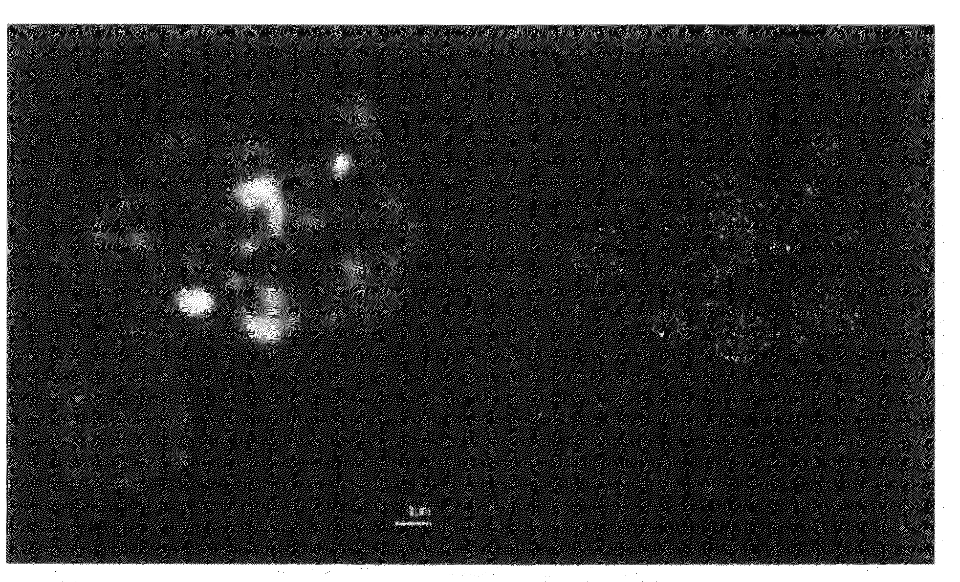
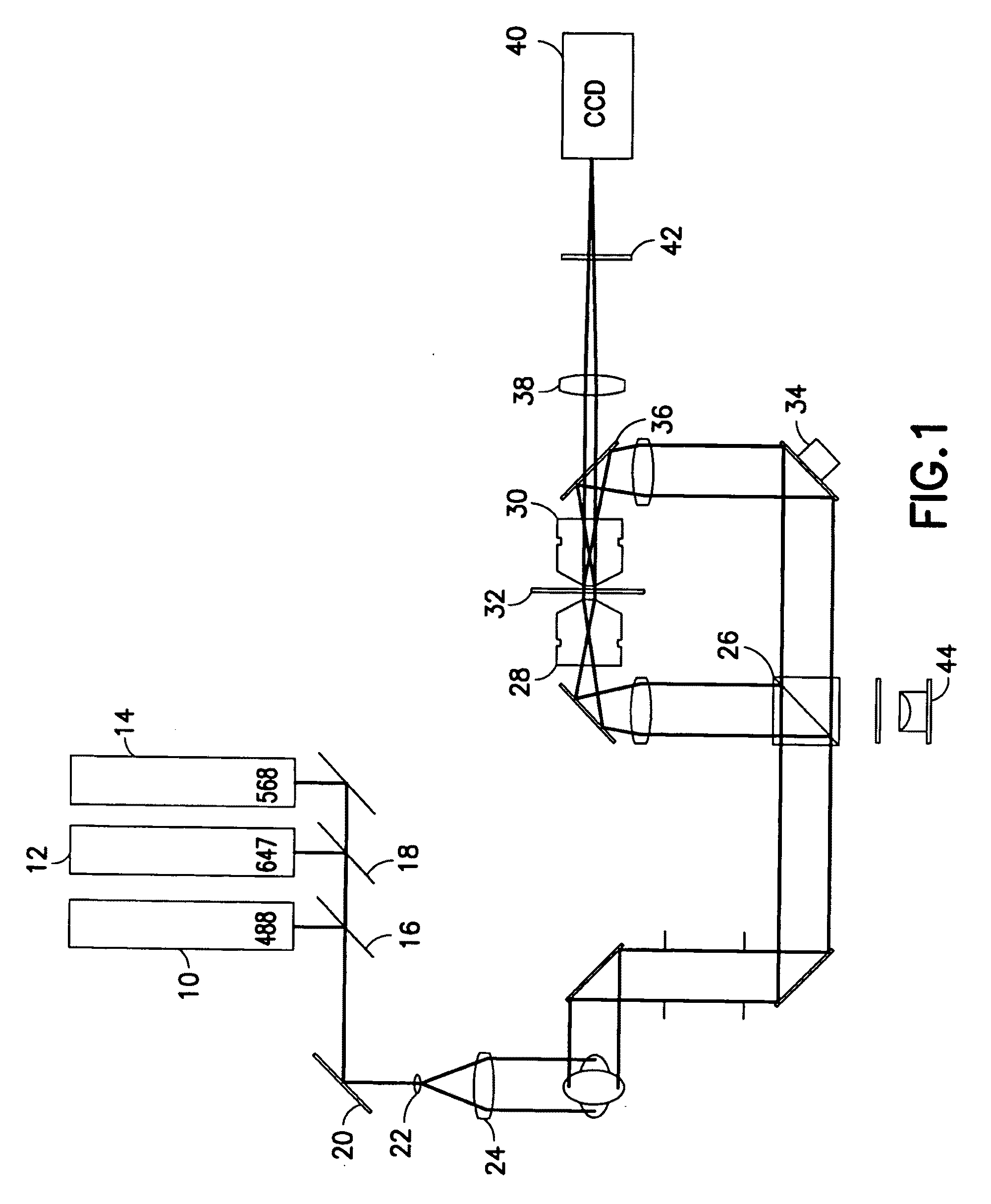
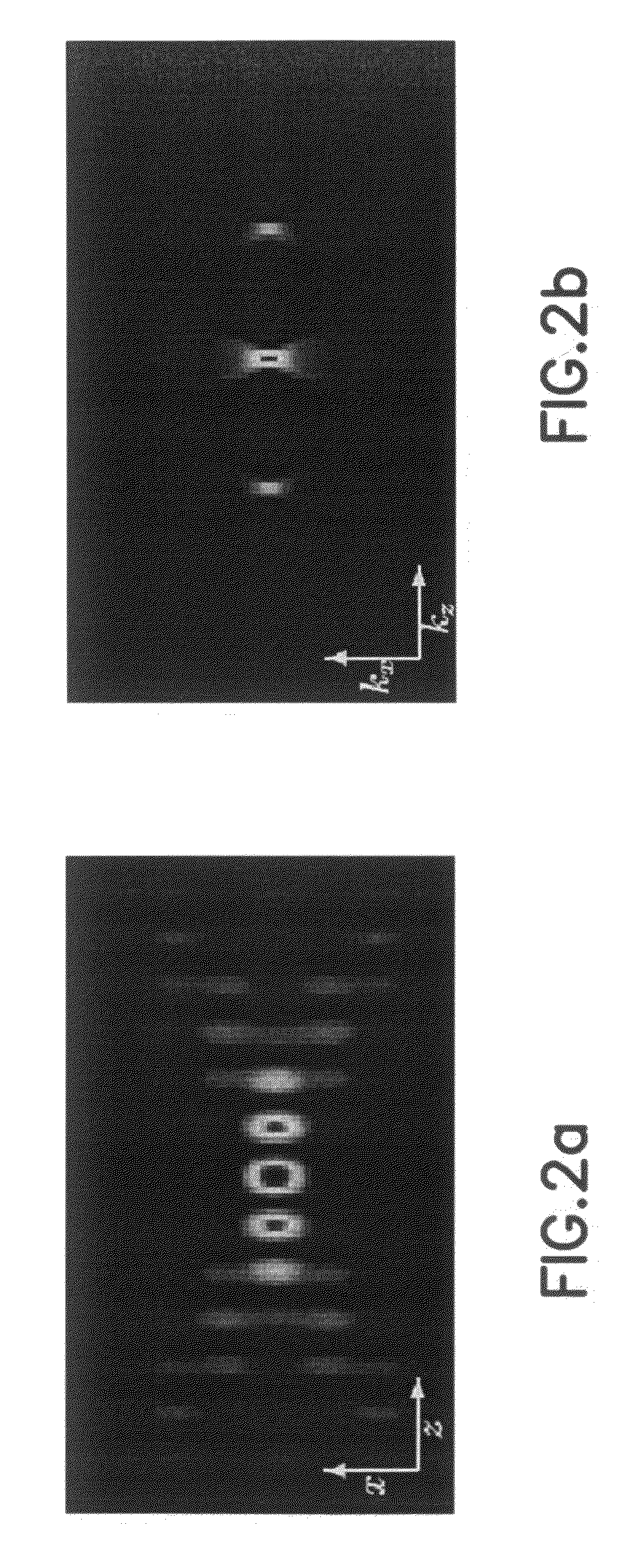
[0083]FIG. 1 shows schematically the optical layout of a SMI microscope (as an example of a microscopical system) with a horizontal arrangement according to one example of the invention.

[0084]SMI microscope uses structured widefield illumination by means of an interference pattern, in combination with a widefield detection.

[0085]The illumination system comprises two or optionally three lasers for 488 nm (laser source 10), 568 nm (laser source 12) and optionally 647 nm (laser source 14) excitation as light sources. For example Lexel 95-4, Lexel 95L-K and Lexel 95-K from Lexel Laser, USA may be used as laser sources. In one embodiment the laser sources may be additionally independently switched with shutters before being combined with the respective dichroic mirrors 16 and 18 (for example from AHF Analysentechnik AG, Tübingen, Germany). The dichroic mirror 20 works as a cleanup filter and reflects the three laser lines into the collimator, consisting of two achromats 22 and 24 (for ex...


PUM


Abstract

The invention relates to a computer implemented method for processing of microscopic images to detect objects of interest. The method includes subjecting the microscopic image to a bandpass filtering to obtain a filtered image, wherein the bandpass filtering is such as to suppress the noise and any objects which are larger than a predetermined size; and processing the filtered image at a plurality of progressively decreasing threshold levels. The processing at each threshold level includes detecting the objects of interest using an object labelling algorithm and removing the detected objects detected at a given threshold level from the working image before proceeding to the next threshold level.

Description

BACKGROUND OF THE INVENTION[0001]Spatially Modulated Illumination (SMI) microscopy is a method of wide field fluorescence microscopy featuring structured, respectively interferometric illumination, which delivers structural information about nanoscale architecture in fluorescently labelled cells. The structural information may be for example information about sizes of and distances between fluorescently marked target regions. To generate the illumination pattern two counter propagating laser beams are brought to interference, establishing a standing wave field. For the analysis of three-dimensional (3D) nanostructures SMI microscopy applies methods of point spread function (PSF) engineering, enabling the quantitative characterisation of the sizes of fluorescent objects in a range of about 30-200 nm in axial direction. Using structured wide field illumination in combination with wide field detection, this technique provides additional information about the analysed objects than conve...

Claims


Application Information

Patent Timeline
IPC IPC(8): G06K9/00G06K9/40
CPCB82Y20/00B82Y35/00G06K9/00127G06T2207/10056G06T5/20G06T7/0002G06T5/002G06V20/69
Inventor CREMER, CHRISTOPHBADDELEY, DAVIDREYMANN, JURGENBIRK, UDO
Owner UNIVERSITY OF HEIDELBERG
Why Eureka
Try Eureka