Regenerating and enhancing development of muscle tissue
a technology of muscle tissue and regenerative mechanism, which is applied in the direction of biocide, drug composition, peptide/protein ingredients, etc., can solve the problems of increasing the impaired muscle regenerative mechanism, but without completely salvaging the altered phenotype, etc., and achieves the effects of regenerating muscle tissue, and enhancing muscle tissue developmen
- Summary
- Abstract
- Description
- Claims
- Application Information
AI Technical Summary
Benefits of technology
Problems solved by technology
Method used
Image
Examples
example 1
Cell Culture
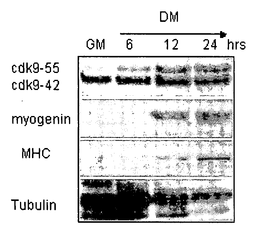
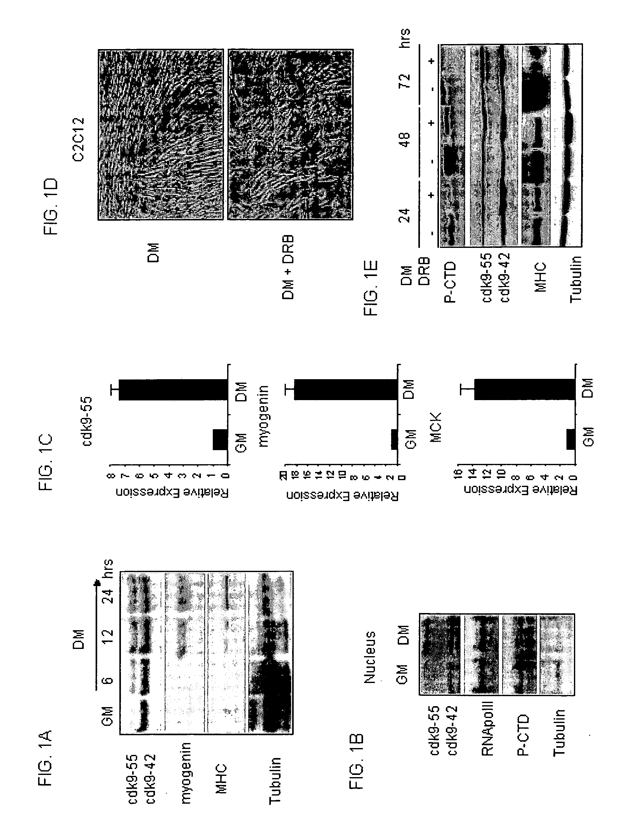
[0067]C2C12 cells were grown in DMEM supplemented with 20% FBS (GM) or with 2% HS (DM).
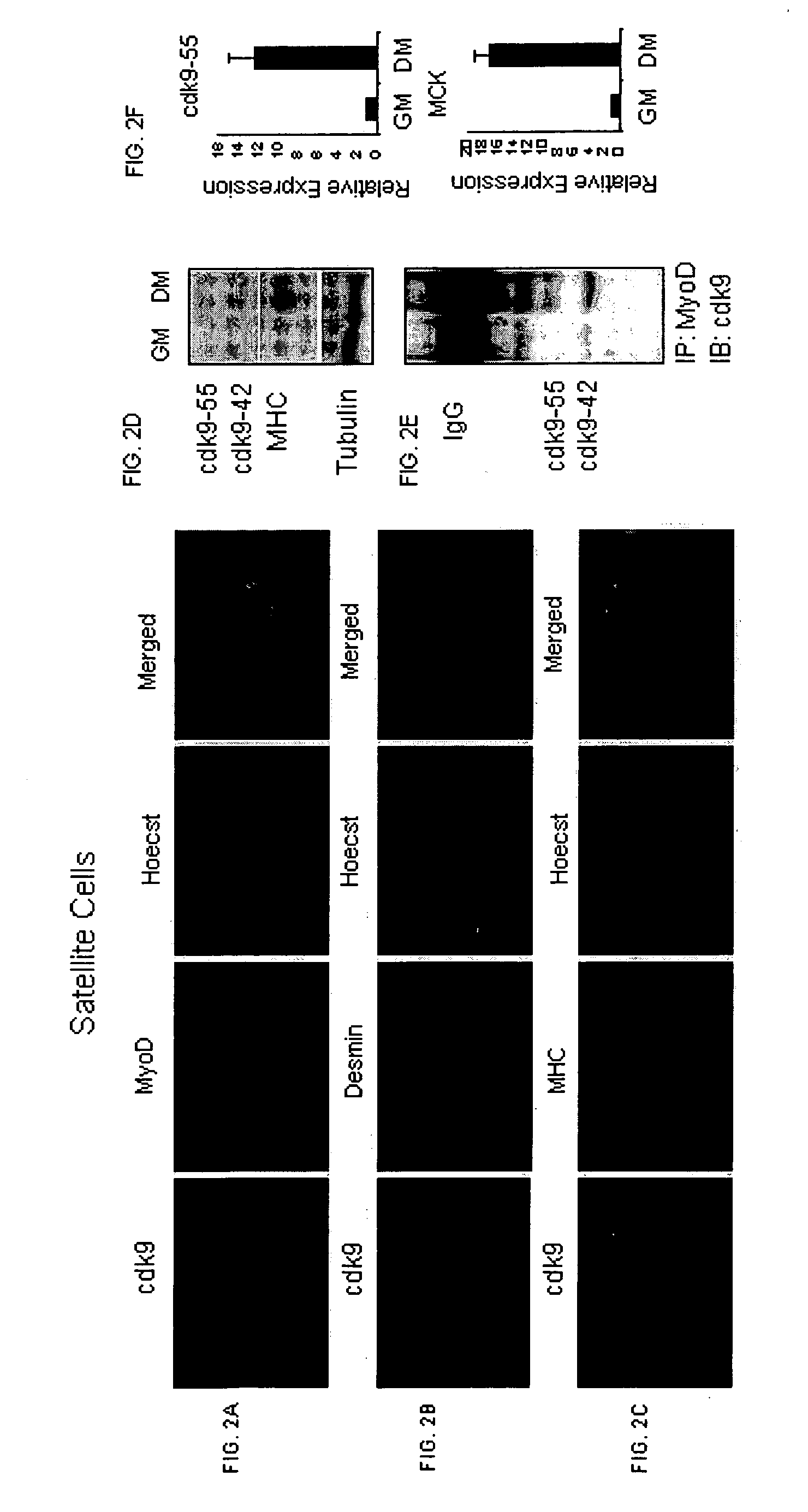
[0068]Single muscle fibers with associated satellite cells were isolated as described in Rosenblatt et al., In Vitro Cell. Dev. Biol. Anim. 31, 773-779, 1995. Briefly, the hind limb muscles were digested for 60 minutes at 37° C. in 0.2% collagenase I (Sigma-Aldrich) and triturated with a wide-bore pipette. Single myofibers were then cultured in 6-well plates (10 fibers / well) pre-coated with ECM gel (Sigma-Aldrich). Fiber cultures were grown in DMEM supplemented with 10% HS and 0.5% chick embryo extract (MP Biomedicals, Solon, Ohio). Three days later, the fibers were removed, and proliferation of detached cells was induced by DMEM supplemented with 20% FBS, 10% HS, and 1% chick embryo extract. After 4-5 days, the cells were allowed to differentiate by DMEM with 2% HS and 0.5% chick embryo extract.
example 2
Muscle Regeneration and Immufluorescence Studies
[0069]Quadriceps and tibialis muscles from C57BL6J mice and 22-month Desmin / nls-LacZ mice were injected with 30 μl of 10 μM cardiotoxin. Mice were anesthetized before cervical dislocation and muscle tissue was separated from bone and most connective tissue and immediately frozen in liquid nitrogen. Frozen sections were fixed in 4% paraformaldehyde 10 min on ice, and then stained with X-gal or pre-incubated in PBS containing 1% BSA, 1:100 goat serum for 1 h at RT, and processed as described in Musarò et al., Nature Genetics 27, 195-200, 2001. Antibodies specific for MyoD, cdk9, Desmin and MHC were used. Nuclei were visualized using Hoechst staining.
example 3
Plasmids and In Vivo Muscle Transfection by Electric Field
[0070]The construct pcDNA3-cdk9DN-HATag expressing dominant negative cdk9 has been described (Simone et al., Oncogene 21, 4137-4148, 2002). In vivo experiments were carried out as described in Dona et al., Biochemical and Biophysical Research Communications 312, 1132-1138, 2003. Briefly, injured quadriceps and tibialis muscles from C57BL6J mice and 22-month Desmin / nls-LacZ mice were exposed by a short incision and DNA (0.06-25 mg) in 50 ml of 0.9% NaCl was injected with a Hamilton syringe in a proximal to distal direction. Then, a pair of spatula-like electrodes (0.5 cm wide, 2 cm long) were placed at each side of the muscle and electric pulses were delivered. Five electric pulses with a fixed pulse duration of 20 ms and an interval of 200 ms were delivered using an electric pulse generator (Electro Square porator ECM 830, BTX, San Diego, Calif.). The ratio of applied voltage to electrode distance was 50 V / cm.
PUM
| Property | Measurement | Unit |
|---|---|---|
| pH | aaaaa | aaaaa |
| elution volume | aaaaa | aaaaa |
| pH | aaaaa | aaaaa |
Abstract
Description
Claims
Application Information
 - R&D
- Intellectual Property
- Life Sciences
- Materials
- Tech Scout
- Unparalleled Data Quality
- Higher Quality Content
- 60% Fewer Hallucinations
Browse by: Latest US Patents, China's latest patents, Technical Efficacy Thesaurus, Application Domain, Technology Topic, Popular Technical Reports.
© 2025 PatSnap. All rights reserved.Legal|Privacy policy|Modern Slavery Act Transparency Statement|Sitemap|About US| Contact US: help@patsnap.com


