Methods for the diagnosis of lung cancer

a lung cancer and method technology, applied in the field of lung cancer diagnosis, can solve the problems of high mortality worldwide, serological types of diagnostic methods, low sensitivity (especially in the early cancer stage), and high cost, and achieve the effect of improving the early detection of lung cancer

Inactive Publication Date: 2009-12-03
PANACEA PHARMA
View PDF7 Cites 16 Cited by
  • Summary
  • Abstract
  • Description
  • Claims
  • Application Information

AI Technical Summary

Benefits of technology

[0009]A potential key to improving the early detection of lung cancer is the identification of an inexpensive, minimally invasive test that could further identify those individuals who might benefit from further diagnostic follow-up. Such a test might be useful either as a guide to the interpretation of CT scan results or as an initial screen suggesting further work-up which might include CT scanning. Application of such a test to the specific case of lung cancer recurrence adds further benefits in that it may identify metastatic disease that has recurred outside of the lung and thus may be missed by chest CT. While a number of tumor markers have been explored, including cytokeratin fragments (CYFRA 21-1), neuron specific enolase (NSE), prog

Problems solved by technology

It is projected that more than 170,000 cases of lung cancer will be diagnosed in the United States in 2007, and such detection will occur by means such as X-ray and CT scanning methods, which have inherently low sensitivity (for early cancer stages especially) and high cost, as compared to serological types of diagnostic methods, generally.
The current detection methods, however, enable such detection in only about 16% of cases overall.
Lung cancer is a leading cause of cancer, and associated with high mortality worldwide.
To date, there are no known blood/serum biomarkers useful for the diagnosis of lung cancer, or at least that are currently approved by regulatory authorities for the detection of such cancer.
Current detection methods are, comparatively, inadequate to address the public health issues concerning lung cancer.
Despite advances made in the diagnosis and treatment of a number of common cancers, lung cancer remains a leading cause of cancer death worldwide.
A primary factor contributing to the high mortality rates associated with lung cancer is the lack of early diagnosis, prior to significant advancement and associated symptoms of the disease.
Thus, the majority of lung cancer patients are not identified until they have developed stage III or IV tumors, which are largely not well treated by surgical resection or by radiation, chemo

Method used


Image

  • Methods for the diagnosis of lung cancer
  • Methods for the diagnosis of lung cancer
  • Methods for the diagnosis of lung cancer

Examples


example i

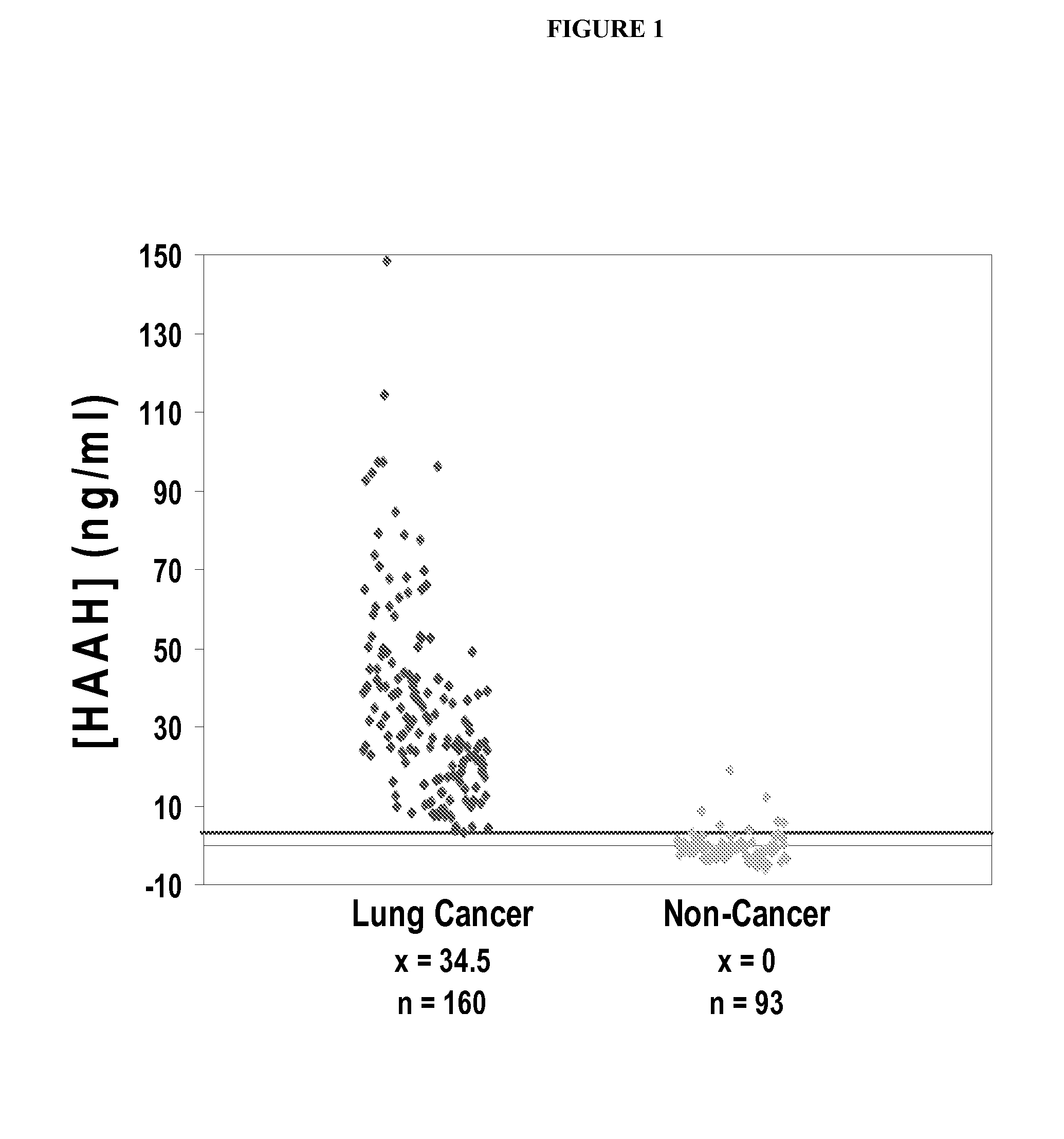
[0056]A large study was conducted using a double monoclonal (FB50 anti-HAAH antibodies) sandwich-type ELISA format with anti-AAH antibodies, providing detection and comparative quantification of HAAH in serum samples obtained from lung cancer patients, and control samples obtained from a pool of non-cancerous subjects (which included samples of cigarette smokers as well—“high risk controls”). The high risk controls were relevant to this study, because 87% of lung cancers are attributable to cigarette smoking, and associative parallels with recent reductions in rates of smoking have been reported in the literature.

Results

[0057]Increased levels of serum HAAH were found in 99% of patients with lung cancer (n=160). Quite strikingly, serum HAAH levels were found to be undetectable in individuals not known to have cancer (normal controls) (n=93, specificity=91%). See FIG. 1.

[0058]In the control subpopulation of 50 smokers not known to have cancer, the mean serum HAAH level was 0 ng / ml, wi...

example 2

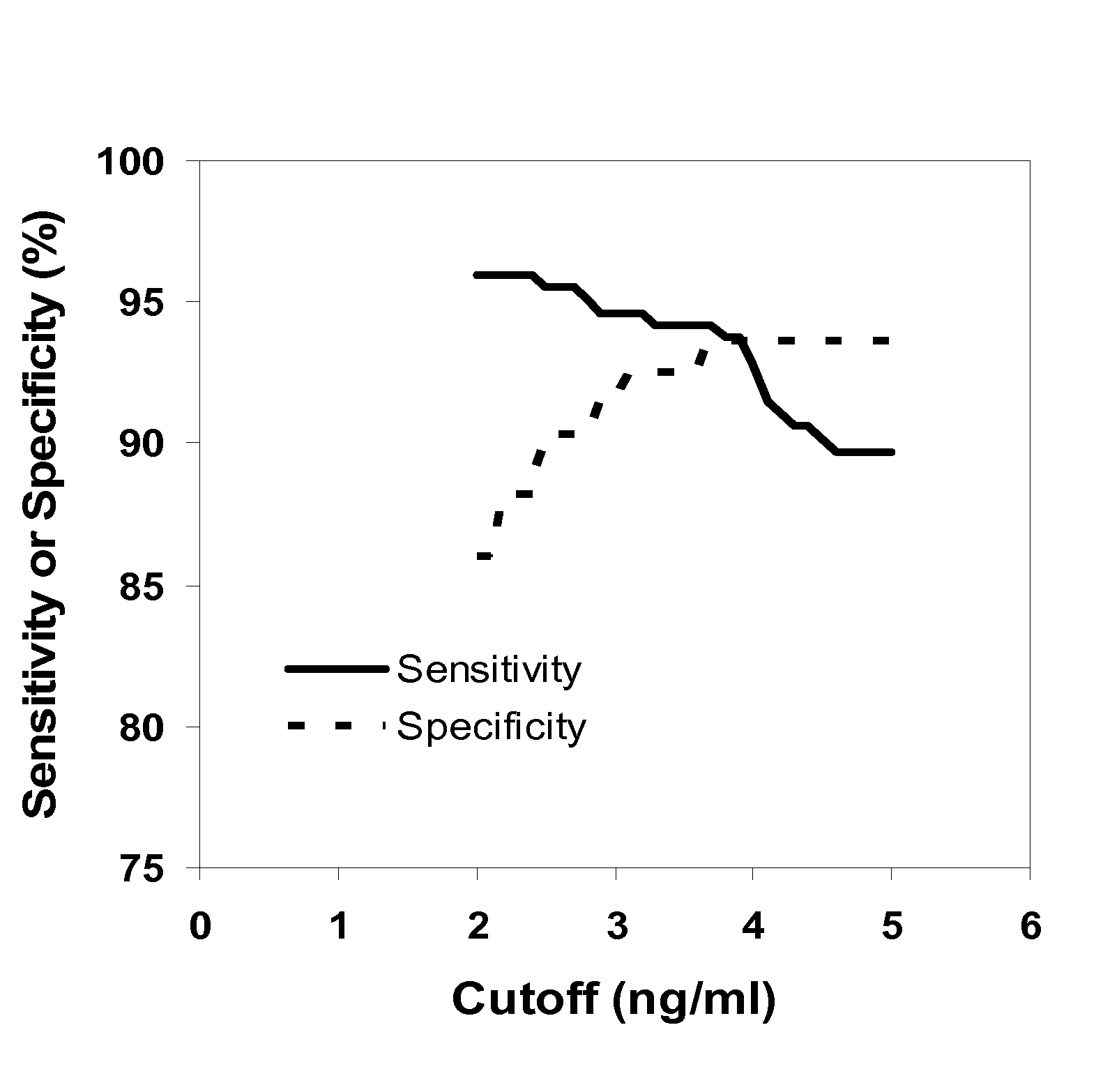
[0060]The goal of this study was to provide further evidence of the clinical sensitivity and specificity of the diagnostic test of the present invention, as well as its unique utility as a screen for NSCLC (non-small cell lung cancer) in patients at increased risk of this disease, due to its ability to detect lung cancer as early as stage I of the disease.

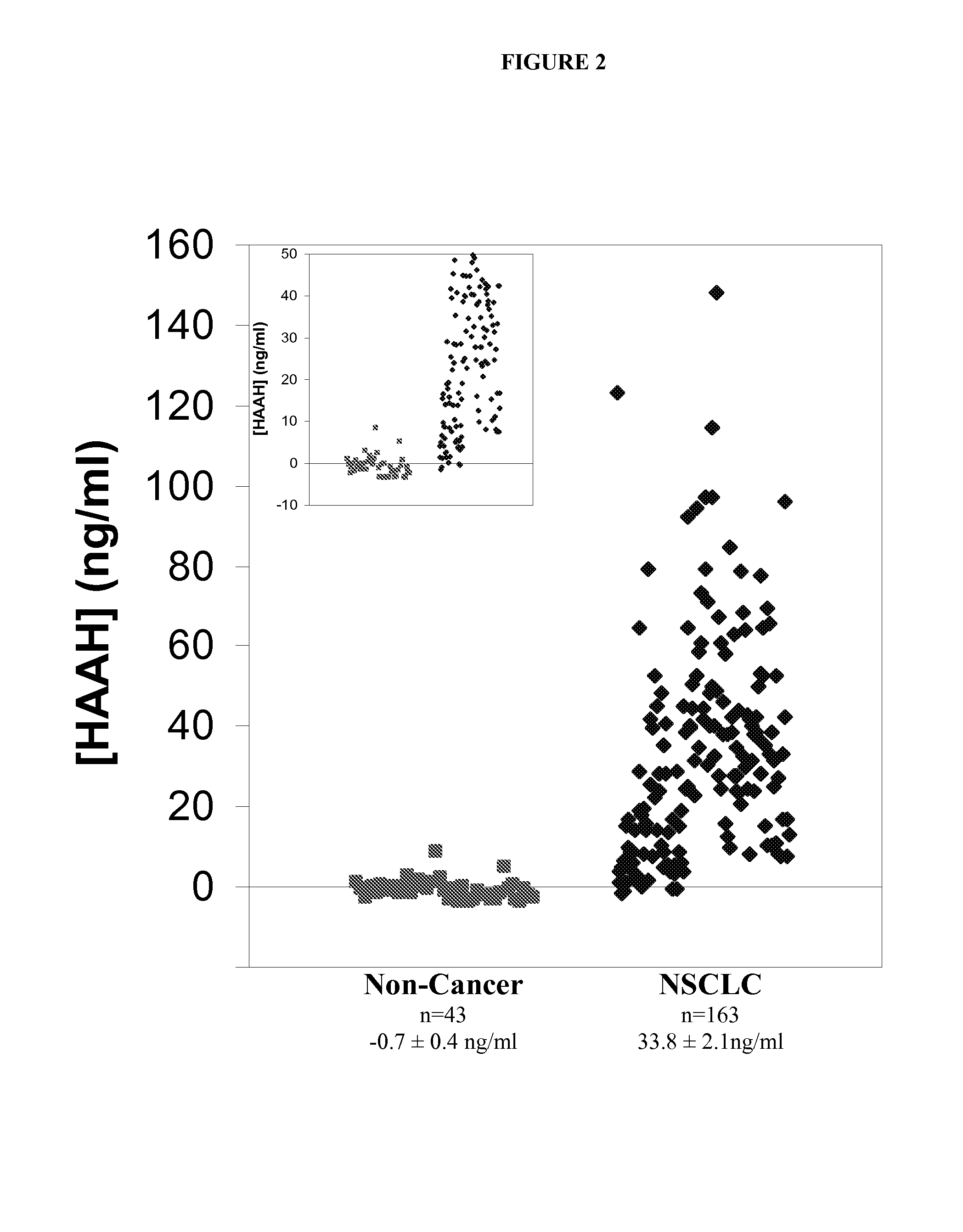
[0061]Briefly, sera obtained from patients with a confirmed diagnosis of NSCLC (n=163) and individuals with no known history of cancer (n=43) were analyzed in a homologous antibody immunoassay for the detection of HAAH and used to determine a threshold value for the test that could serve to discriminate between subjects with and without NSCLC. A second set of patients, including subjects with stages I-IV NSCLC (n=60) and a control group of subjects with a history of moderate to heavy smoking (n=50), was used to further establish this cutoff value.

[0062]Briefly, the resulting data showed that sera from individuals with NSCLC had ele...

example 3

[0076]In this study, we first compared HAAH serum levels in NSCLC patients versus healthy control individuals. Subsequently, a follow-on study of this biomarker was made in order to determine whether levels of the HAAH protein in serum can be correlated to clinical response to treatment.

[0077]Using the double monoclonal antibody sandwich ELISA described above, HAAH levels were measured in the sera of 46 patients with non-small cell lung cancer (NSCLC) and 15 healthy controls. A subsequent study, which measured serum HAAH levels both before and after treatment in 22 of the patients was conducted.

Results

[0078]Serum HAAH was detectable and variable in 92% of NSCLC patients (n=43). Serum HAAH was found to be virtually undetectable in healthy donors. When patients were compared to healthy controls, the pretreatment median level of serum HAAH was significantly higher in NSCLC patients (P=0.00059). It was also noted that HAAH levels did not correlate with gender or age.

[0079]At the end of ...


PUM


Abstract

The present invention is directed to new ways to diagnosis lung cancer, especially at an early clinical stage. In addition, prognosis and the monitoring of therapeutic agents or other treatments, for lung cancer patients, can be accomplished with the disclosed methods. The methods also find use in allowing the assessment by pre-clinical animal efficacy studies to screen for the useful of therapeutic agents for treating lung cancer.

Description

[0001]This application claims priority from U.S. provisional application No. 60 / 959, 869, filed Jul. 17, 2007.FIELD OF THE INVENTION[0002]The present invention is related to lung cancer diagnosis, as well as the collateral aspects of the prognosis and the monitoring of therapeutic agents or other treatments, for lung cancer patients. The methods are also useful in pre-clinical animal efficacy studies to screen for the useful of therapeutic agents for treating lung cancer.BACKGROUND OF THE INVENTION[0003]It is projected that more than 170,000 cases of lung cancer will be diagnosed in the United States in 2007, and such detection will occur by means such as X-ray and CT scanning methods, which have inherently low sensitivity (for early cancer stages especially) and high cost, as compared to serological types of diagnostic methods, generally.[0004]While the five year survival for lung cancer is generally estimated at 15%, a survival rate of about 50% can be achieved when detection is m...

Claims


Application Information

Patent Timeline
IPC IPC(8): G01N33/574
CPCG01N33/57423G01N2800/52G01N2333/90245
Inventor HARRIS, PAMELA J.LEBOWITZ, MICHAEL S.GHANBARI, HOSSEIN A.
Owner PANACEA PHARMA
Why Eureka
Try Eureka