Cumulative time-resolved emission two-dimensional gel electrophoresis

a two-dimensional gel and emission technology, applied in the field of cumulative time-resolved emission two-dimensional gel electrophoresis, can solve the problem of compromising the sensitivity of detection in the cutedgeTM application, and achieve the effect of optimal flexibility

Active Publication Date: 2009-12-24
HIKARI BIO AB
View PDF1 Cites 6 Cited by
  • Summary
  • Abstract
  • Description
  • Claims
  • Application Information

AI Technical Summary

Benefits of technology

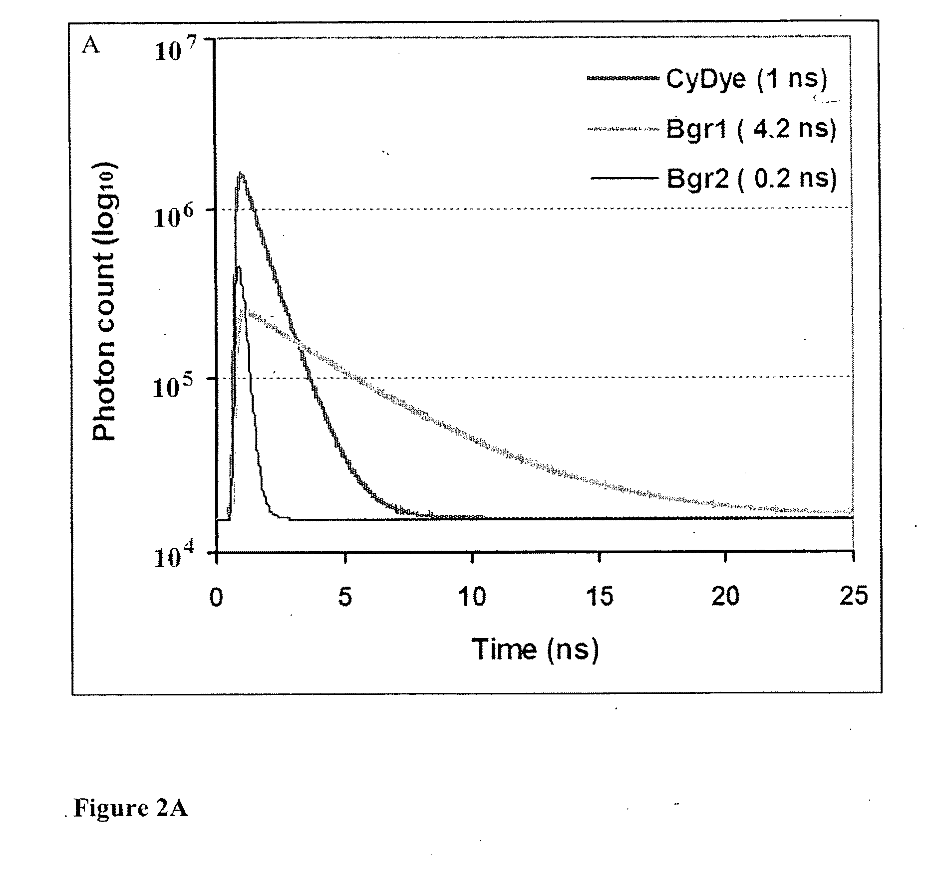
[0016]An additional embodiment of the invention includes the utilization of multi-exponential fitting of the fluorescence decay curve to separate fluorescence originating from the fluorochrome-labeled protein species from fluorescence originating from other sources such as the gel matrix itself, solutions or particles presents in the gel, or scattering effects. The separation of the fluorescent decay curves provides background subtraction on the photon level as illustrated in FIGS. 2A-2C. In addition, this embodiment of the CUTEDGE™ technology reduces the need for software-assisted background subtraction algorithms in the subsequent image analysis, and the increased experimental variance known to be introduced by these algorithms. The distribution of the fluorescent lifetimes in a 2-DGE image is exemplified by FIGS. 4A-4B, which illustrates a pseudocolored lifetime image of a Cy2.
[0017]Yet another embodiment of the invention herein involves sequential use of multiple laser wavelengths for excitation to facilitate a combination of TRF with spectral separation of fluorochromes. Spectral separation is used in current 2-DGE protocols to facilitate correction of inter-gel variations in separation patterns through multiplexing, such as the DIGE (9) and ALIS (10) approaches. Multiplexing protocols for incorporation of an internal standard in 2-DGE are vital for correction of gel-to-gel variations inherent in the 2-DGE method, and are well established in the field of gel-based proteomics. As such, inclusion of this embodiment makes the CUTEDGE™ instrument easy to implement in existing proteomics work flows using current protocols for internal standard in 2-DGE. The use of multiplexing through spectral separation rather than through TRF also decreases the complexity of the life time decay spectra, thus improving the background subtraction capability of the CUTEDGE™ method. However, dual FLIM measurements may also be included in cases where the excitation wavelength and fluorescent lifetimes of the incorporated fluorochromes are compatible, as is the case for the Cy2 / Cy3 dye pair (Example 4).
[0020]Automation and user-friendliness are a core component of the CUTEDGE™ instrument, particularly in terms of graphical and quantitative calculations of acquired 2-DGE image. Automated protocols for FLIM analysis, subtraction of the background components, and export of intensity images for the fluorochrome component(s) are included for the standard fluorochromes used in 2-DGE. In order to facilitate the use of existing 2-DGE image analysis software, the AUC is thus represented as a one-dimensional (Z) intensity in the resulting exported image. The use of a 32 bit Tagged Image File Format (TIFF) or equivalent in replacement of the currently used 16 bit format provides a digital dynamic range of close to 10 orders of magnitude, thus facilitating the full dynamic range of pixel intensities achieved through the CUTEDGE™ technology.
[0021]To maximize automation, the invention also facilitates programming of sequential scanning of multiple different protocols as defined by the user, for example sequential scanning of multiple fluorochromes used for multiplexing.

Problems solved by technology

However, the limited quantum efficiencies (QE) of PMT detectors may compromise the sensitivity of detection in the CUTEDGE™ application.

Method used


Image

  • Cumulative time-resolved emission two-dimensional gel electrophoresis
  • Cumulative time-resolved emission two-dimensional gel electrophoresis
  • Cumulative time-resolved emission two-dimensional gel electrophoresis

Examples


example 1

[0057]In this example, proteins are first isolated from rat airway through lysis-lavage (14), an isolation method that instantly solubilizes the airway epithelial proteome in a 2-DGE-compatible urea-based lysis buffer (7M urea, 2M thiourea, 4% w / v CHAPS, 0.5% Triton-X 100, 2% v / v protease inhibitor cocktail). The protein concentration of the samples is determined using the method of Bradford, and separated using 2-DGE. An aliquot of 400 μg protein / sample is diluted to 350 μl with lysis buffer, and IPG buffer for the appropriate pH range is added to a final concentration of 1% v / v. The protein samples are loaded onto the IPG strip (GE Healthcare) through rehydration at room temperature overnight, and isoelectric focusing (IEF) is performed using a Multiphor II Electrophoresis unit and an EPS 3501 XL power supply at 20° C. with the following gradient protocol: 0-50V, 1 min.; 50V, 1 hr; 50-1000V, 3 hr; 1000-3500V, 3 hr; 3500V, 19 hr (total 74.9 kVh). Following IEF, the strips are incub...

example 2

[0062]Macrophages isolated by bronchoalveolar lavage from smoking and never-smoking human subjects are solubilised using 2-DGE lysis buffer (see Example 1). An internal standard is created through pooling of equal amounts of protein from all the subjects included in the study, and labelled with NHS ester-conjugated Cy2 (minimal DIGE reagent) according to the manufacturer's recommendations (GE Healthcare, Uppsala, Sweden). The protein samples from smoker and never-smoker subjects are randomized into two groups, which are labeled with Cy3 and Cy5 minimal DIGE reagents respectively. The Cy2 labeled internal standard is co-separated with one Cy3 and one Cy5 labeled sample on a 2-DGE gel according to the protocol described in Example 1. The three different protein labels are visualized in an automated sequence of three different scanning protocols using a combination of spectral and time-resolved fluorescence as follows:

[0063]A) The Cy2 fluorochrome (internal standard) is excited through...

example 3

[0067]As an additional step to the protocol described in Example 2, SYPRO™ Ruby staining is performed to facilitate quantification of the total protein content in the DIGE gel. In existing technology, it is not possible to utilize the DIGE and SYPRO™ Ruby fluorochromes in the same 2-DGE gels due to spectral overlap, particularly in terms of the emission curves of SYPRO™ Ruby, Cy3 and Cy5. The embodiments of this invention utilize the temporal dimension to facilitate merging of these to standard protocols (as exemplified in FIG. 3). Following completion of the procedures outlined in Example 2, post-staining with SYPRO™ Ruby staining to quantify total protein content is performed according to steps B and C outlined in Example 1.


PUM


Abstract

A new instrumental design is provided for in-gel detection and quantification of proteins. This new platform, called Cumulative Time-resolved Emission 2-Dimensional Gel Electrophoresis, utilizes differences in fluorescent lifetime imaging to differentiate between fluorescence from specific protein labels and non-specific background fluorescence, resulting in a drastic improvement in both sensitivity and dynamic range compared to existing technology. The platform is primarily for image acquisition of two-dimensional gel electrophoresis, but is also applicable to protein detection in one-dimensional gel systems as well as proteins electroblotted to e.g. PVDF membranes. Given the increase in sensitivity of detection and dynamic range of up to 5-6 orders of magnitude compared to existing approaches, the described invention represents a technological leap in the detection of low abundance cellular proteins, which is desperately needed in the field of biomarker discovery.

Description

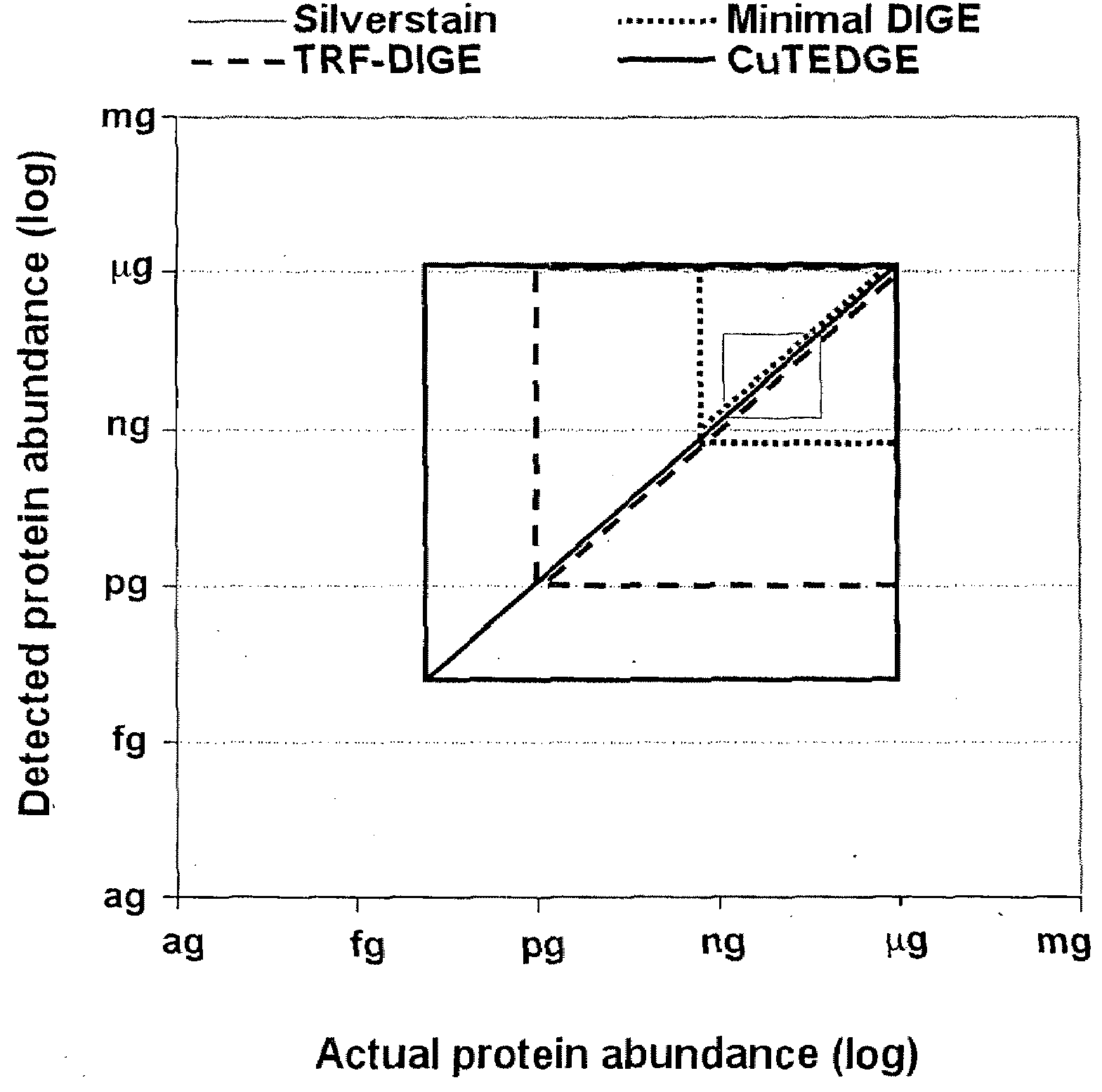
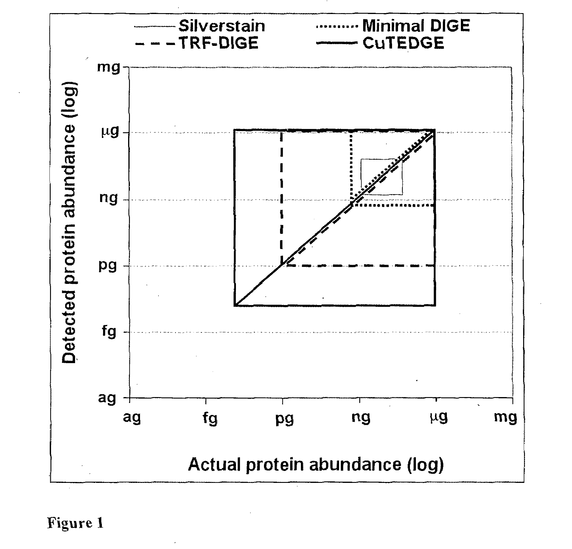
CROSS-REFERENCE TO RELATED APPLICATIONS[0001]This application claims priority from U.S. provisional application Ser. No. 61 / 054,300 filed May 19, 2008.BACKGROUND OF THE INVENTION[0002]1. Field of the Invention[0003]The present invention relates to a method and new image acquisition technology for in-gel detection and quantification of proteins. This new platform, called Cumulative Time-resolved Emission 2-Dimensional Gel Electrophoresis (CUTEDGE™), utilizes differences in fluorescent lifetimes to differentiate between fluorescence from specific protein labels and non-specific background fluorescence, resulting in a drastic improvement in both sensitivity and dynamic range compared to existing technology.[0004]2. Background of the Invention[0005]“Proteomics” refers to the study of the protein complement of the genome (proteome), a term coined by Marc Wilkins in 1994. Over the past decade, many methodologies for simultaneous quantification of thousands of proteins in a cell or tissue ...

Claims


Application Information

Patent Timeline
Patent Type & Authority Applications(United States)
IPC IPC(8): G06K9/46
CPCG01N27/44726G01N27/44778G01N2021/6439G01N21/6428G01N21/6408
Inventor WHEELOCK, ASA
Owner HIKARI BIO AB
Why Eureka
Try Eureka