Neural Stem Cells
Inactive Publication Date: 2009-12-31
STIFTUNG CAESAR
0 Cites 14 Cited by
AI-Extracted Technical Summary
Problems solved by technology
Neurodegenerative diseases are characterized by the loss of specific subsets of neurons, and whilst drug therapies exist for some of these disorders, none of them are curative.
Neural tissue has a limited capacity for repair after injury, and adult neurogenesis is limited to selected regions of the brain (Gage, 2000; ...
Benefits of technology
[0008]The problem underlying the invention is thus to provide an alternative method for obtaining neural stem cells. The stem cells should be easily avail...
Abstract
Subject of the invention is a method for generating neural stem cells in vitro, wherein dental progenitor cells are isolated from soft tissue of tooth or wisdom tooth and cultivated until they form primary spheres which are then dissociated into single cells. These single cells are cultivated until they form spheroids and the spheroid-forming cells are separated to obtain neural stem cells.
Application Domain
BiocideNervous disorder +6
Technology Topic
Neural stem cellProgenitor +4
Image
Examples
- Experimental program(1)
Example
[0044]Cells were enzymatically isolated from dissected soft tissue of wisdom teeth (dental follicle or apical soft tissue) by collagenase/Dispase treatment.
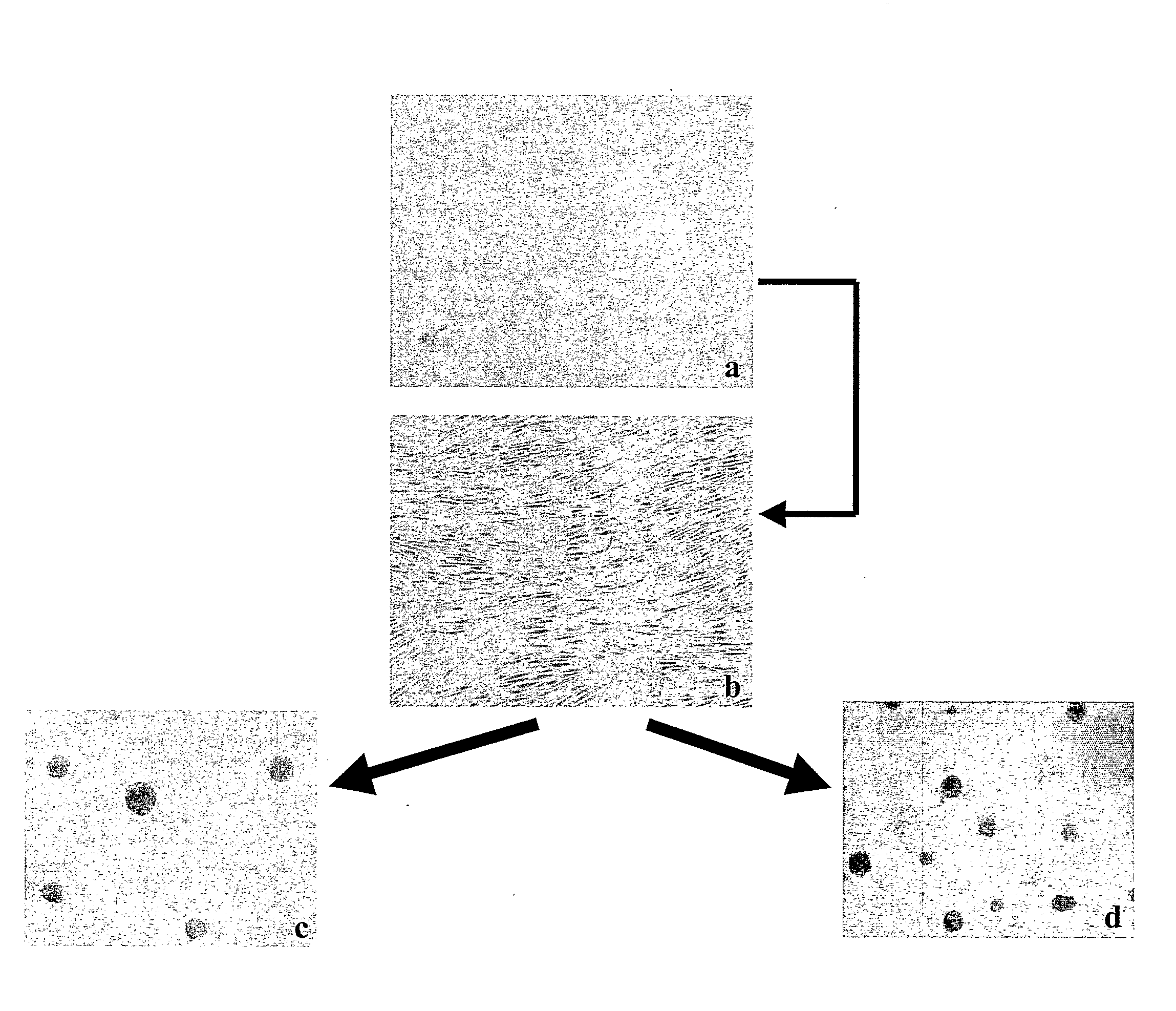
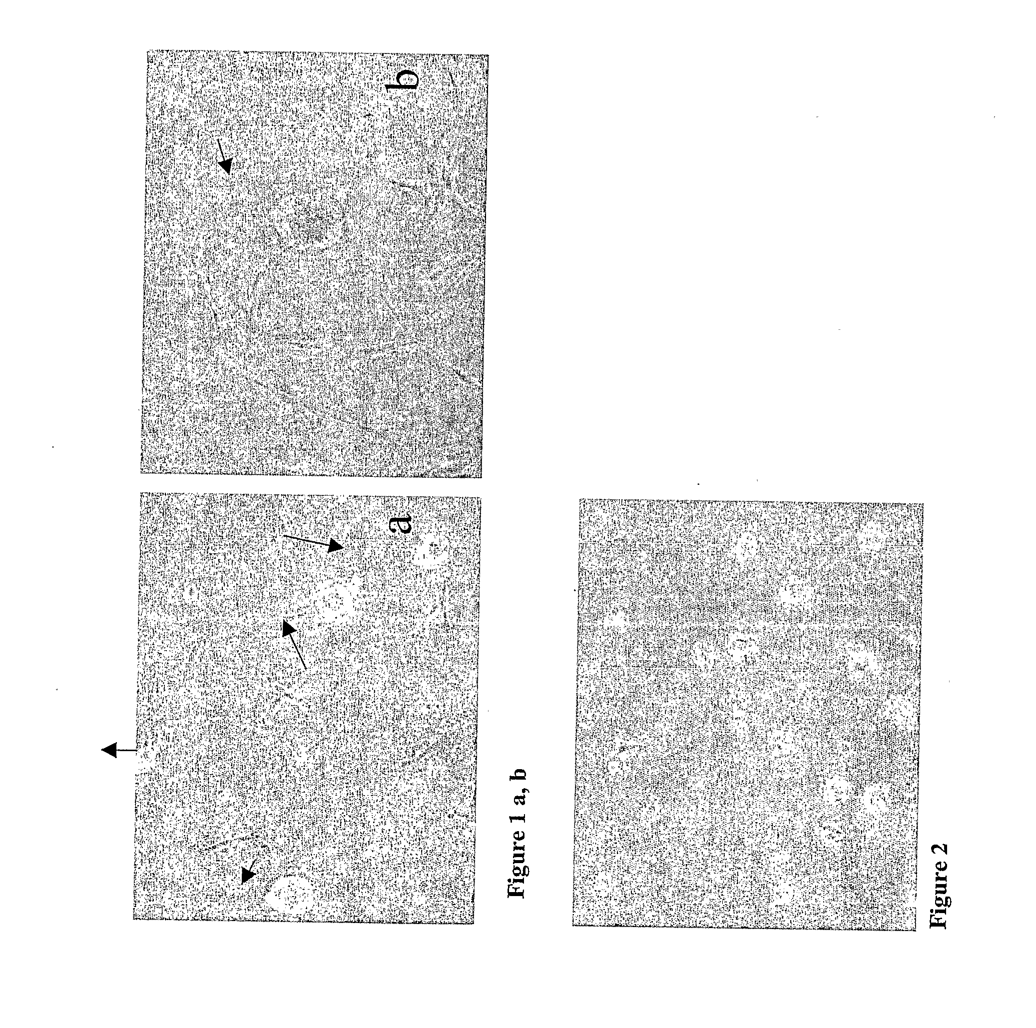
[0045]The ectomesenchymal cells were cultivated in FCS containing medium for 8-12 days. Some of the cells adhered to the plastic culture flask while some died in suspension. However, some of the cells formed spheres (FIG. 1).
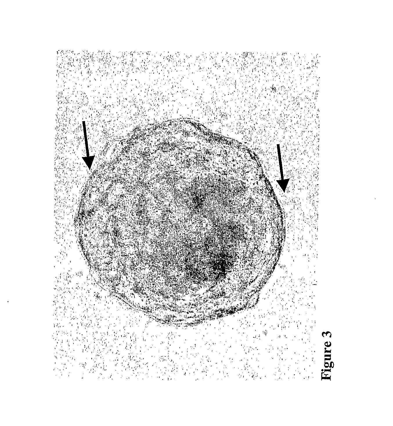
[0046]The floating spheres were transferred to new culture flasks after initial culturing in bFGF (40 ng/ml) and EGF (20 ng/ml), B27 (1:50) and neurobasal medium (Invitrogen) containing medium or bFGF (50 ng/ml), EGF (25 ng/ml), ITS+Premix (1:50) and DMEM High glucose containing medium. Cells in spheres proliferated thereby forming large spheres which were successfully passaged and expanded (FIG. 2). The spheres seemed bright when viewed under a phase contrast microscope and showed cytoplasmic protrusions (cilia) at their surface (FIG. 3).
[0047]The primary spheres were mechanically dissociated into single cell suspensions and cultured again under sphere-forming conditions (ITS+Premix, bFGF, EGF, DMEM HG or bFGF, EGF, B27 and neurobasal medium; FIGS. 4a, b).
[0048]After each passage with both media the number of cells adhering to the plastic flask decreased while the number of cells forming spheroids increased (FIG. 5a). When the spheres grew in DMEM basic medium supplemented with serum, undifferentiated stem cells retained a flat polygonal fibroblast-like morphology (FIG. 5b), in serum-free culture conditions cells formed again spheres (FIGS. 5c, d). Immunocytochemistry with undifferentiated NSCs spheroids revealed that cells were stained for p75, a marker for neural growth factor receptor (FIG. 6).
[0049]The potency of ex vivo expanded wisdom teeth-derived neural stem cells to generate neural cells was analyzed. The stem cells were seeded on fibronectin-coated glass cover slips. Cells were incubated in differentiation-medium containing DMEM, 15% heat inactivated FCS, penicillin/streptomycin/glutamine, 50 ng/ml β-NGF, 20 ng/ml FGF-b, 1 mM dibutyryl cAMP, 0.5 mM 3-isobutyl-1-methylxanthine, 10 μM all trans-retinoic acid. Differentiation-medium was changed every second day. After each day, cells showed neuronal morphology (FIG. 7).
[0050]After one and two weeks, respectively, of differentiation-medium incubation, a subpopulation of neural stem cells was stained for neurofilament, a marker of postmitotic neurons (FIG. 8). Neural stem cells can be differentiated after one and two weeks into the glial lineage, expressing GFAP, a protein of the astrocytic cytoskeleton (FIG. 9). A low percentage of NSCs showed staining for the inhibitory neurotransmitter GABA in basic growth medium after seven days of culture, indicating spontaneous differentiation (FIGS. 10b, c). At day 0, the cells were negative for GABA (FIG. 10a). The staining for the inhibitory neurotransmitter GABA significantly increased by incubation in differentiation-medium (FIGS. 10d, e, f, g). After seven days of stimulation of NSCs with differentiation-medium, immunoreactivity for choline acetyltransferase (CAT) was found indicating the development of a cholinergic neuronal subtype (FIG. 11).
[0051]In order to further analyse the fundamental molecular processes involved in neurogenic differentiation of teeth-derived neural stem cells, a microarray analysis of the critical stages during in vitro stimulation of cells was accomplished. Cells from two volunteers (patient 1: age: 14, gender: w; patient 2: age: 12, gender: w) were isolated from apical, ectomesenchymal soft tissue (apical pad) of tooth and then cultured as described above. Primary spheres were dissociated into single cell suspensions and cultivated in poly-D-lysine and laminin coated plastic flasks for 14 days with neurogenic growth medium (DMEM, 15% heat inactivated FCS, penicilin/streptomycin/glutamin, 50 ng/ml β-NGF, 20 ng/ml FGF-b, 1 mM dibutyryl cAMP, 0.5 mM 3-isobutyl-1-methylxanthine, 10 μM all trans retinoic acid) and with medium omitting growth factors, respectively. RNA isolation was performed on each sample using the RNeasy Mini kit (Qiagen). The mRNA of each sample was isolated and amplified according to the manufacturer's protocol (Qiagen). Each sample was then submitted to an Agilent core facility (caesar), where hybridization of the RNA to the chip probes and fluidics were completed using the standard Agilent gene chip analysis protocol. For analysis a 39.000 oligonucleotide platform was used (Agilent). Raw data were compiled with Agilent software and analysis was achieved using Rosetta software (Agilent).
[0052]A readout of gene chips from patient 1 (with stimulation) shows 6.093 genes upregulated, 6.134 genes downregulated and 28.759 genes unchanged. Patient 2 (with stimulation) shows 4.740 genes upregulated, 5.631 downregulated while 30.671 genes remained unchanged. The matched control (without stimulation) shows 3.837 genes upregulated, 4177 downregulated while 32.944 genes were unchanged. With regard to patient 2, 2.357 genes were regulated in teeth-derived neural stem cells when cultured in neurogenic growth medium for 14 days. See graphic analysis of expression changes in FIG. 12 (patient 1) and FIGS. 13 and 14 (patient 2). Only genes with p values less than 0.05 were used for individual gene analysis to ensure quality of data.
[0053]Next, specifically regulated genes increasing from 1.4-fold to 100-fold (patient 2) and from 3-fold to 100-fold (patient 1) were listed. In particular, transcripts that showed threefold or higher changes in expression included neuronal or proneuronal markers (neurotrophic tyrosine kinase-receptor, neurokinin-1, latexin, neuromedin-U receptor-1, tubulin, beta polypeptide paralog, neurofilament 3, myelin expression factor 2, leukemia inhibitory factor (cholinergic differentiation factor (LIF)) and mRNAs encoding WNT proteins (WNT2, WIF1, WNT5A), which are key regulators of neural stem cell behaviour in embryonic development and with other genes, such as promotor nerve precursor differentiation (Spondin-1), Amphiregulin, a mitogen for adult neural stem cells, other genes such as those involved with the development and differentiation of mature neural cells (IGF1, BMP2, HES1, retinoic acid receptors, TGFβs), also showing upregulation of fivefold or greater. Large numbers of in central nerve system (CNS) expressed transcripts were also upregulated at high levels (i.e., 3- to 36.0-fold change), notably those encoding the proteins brain expressed X-linked proteins (BEX1, BEX2), GABA(A) receptor-associated protein like, GABA-A receptor-associated protein, Selenoprotein P, neurite growth-promoting factor 1, gamma-aminobutyric acid (GABA) A receptor, epsilon (GABRE), solute carrier family 1 (glial high affinity glutamate transporter), member 3 (SLC1A3), serotonin, Dopamine receptor D4, brain-derived neurotrophic factor (BDNF), cerebellar degeneration-related protein 1 (see Table 1 where up-regulations of genes, patient 1+2, are matched against unstimulated cells, patient 2). Only stimulated cells show increase in expression of neural/glial markers while unstimulated cells did not.
[0054]These results demonstrate that the teeth-derived neural stem cells according to the invention may differentiate to cells with characteristics not only of neuronal and glial cells but also of CNS cells such as dopaminergic, serotonergic, and GABA-ergic neurons. Thus, teeth-derived neural stem cells might be an excellent source of cells for treatment of neurodegenerative disorders.
TABLE 1 Comparison of absolute gene expression levels in neurogenic stimulated neural stem cells from tooth (Patient 1 and 2) and 14 days cultivated and untreated neural stem cells from tooth (Patient 2) Fold Fold Fold Change Change Change Patient-1 Patient-2 Patient-2 Accession (neural (neural control Gene Description Number stimulated) stimulated) (unstimulated) amphiregulin (schwannoma-derived NM_001657 100 100 2.9 growth factor) (AREG) spondin 1, extracellular matrix NM_006108 100 92 1.2 protein (SPON1) latexin (LXN) NM_020169 100 100 1.2 retinoic acid receptor responder NM_002888 47 17 2.9 (tazarotene induced) 1 (RARRES1) insulin-like growth factor binding NM_000599 43 28 1 protein 5 (IGFBP5) brain expressed X-linked 2 (BEX2) NM_032621 36 27 2.1 brain expressed, X-linked 1 (BEX1) NM_018476 35 33 1.2 wingless-type MMTV integration NM_003391 33 25 −1.2 site family member 2 (WNT2), selenoprotein P, plasma, 1 NM_005410 32 15 1.005 (SEPP1) WNT inhibitory factor 1 (WIF1) NM_007191 29 19 1.7 chemokine orphan receptor 1 NM_020311 28 92 6.2 (CMKOR1) retinoic acid receptor, beta (RARB) NM_000965 27 19 1.8 chemokine (C-X-C motif) ligand 1 NM_001511 26 14 1.3 (melanoma growth stimulating activity, alpha) (CXCL1), retinol dehydrogenase 10 (all-trans) NM_172037 25 14 −1.06 (RDH10) solute carrier family 1 (glial high NM_004172 21 15 −1.8 affinity glutamate transporter), member 3 (SLC1A3) GABA(A) receptor-associated NM_031412 18 12 1.6 protein like 1 (GABARAPL1) synuclein, alpha interacting protein NM_005460 16 9 1.4 (synphilin) (SNCAIP), Tissue factor pathway inhibitor 2 AK129833 16 9 1.06 precursor (TFPI-2) (Placental protein 5) (PP5) integrin beta 3 S70348 15 14 2.9 wingless-type MMTV integration NM_003392 14 10 1.6 site family, member 5A (WNT5A) Angiopoietin-like 4 (ANGPTL4), NM_139314 12 12 4.1 spermidine/spermine N1- NM_002970 12 9.6 3.6 acetyltransferase (SAT) hairy and enhancer of split 1, NM_005524 12 7.5 25 (Drosophila) (HES1) neurotrophic tyrosine kinase, NM_002529 12 12.5 1.4 receptor, type 1 (NTRK1) interleukin 8 (IL8) NM_000584 11 3.5 1.1 tachykinin, precursor 1 (substance NM_013996 11 12 −1.3 K, substance P, neurokinin 1, neurokinin 2, neuromedin L, neurokinin alpha, neuropeptide K, neuropeptide gamma) (TAC1) pleiotrophin (heparin binding growth NM_002825 10 14 2.2 factor 8, neurite growth-promoting factor 1)(PTN), Voltage-gated calcium channel THC2054079 9 6.5 −1.6 alpha(2)delta-3 subunit angiopoietin-like 1 (ANGPTL1) NM_004673 9 9.2 5.3 GABA-A receptor-associated AF180519 8 6.7 0 protein bone morphogenetic protein 2 NM_001200 7 10 2.4 (BMP2) SRY (sex determining region Y)- NM_003107 7 5 2.1 box 4 (SOX4) transforming growth factor, beta 3 NM_003239 7 2.5 3 (TGFB3) melanoma associated antigen NM_152423 6 17 0 (mutated) 1-like 1 (MUM1L1) neuroblastoma, suppression of NM_182744 6 9 1 tumorigenicity 1 (NBL1) 3′,5′-cyclic AMP phosphodiesterase L12686 6 6 1 cerebellar degeneration-related NM_004065 6 5.6 4.3 protein 1, 34 kDa (CDR1) insulin-like growth factor 1 NM_000618 6 28 −1.09 (somatomedin C) (IGF1) matrix metalloproteinase 10 NM_002425 6 11 4.3 (stromelysin2) (MMP10) transforming growth factor, beta 2 NM_003238 6 5 −1.2 (TGFB2) myeloid leukemia factor 1 (MLF1) NM_022443 6 7 −1.7 gamma-aminobutyric acid (GABA) NM_021990 6 5 −1.2 A receptor, epsilon (GABRE) matrix metalloproteinase 3 NM_002422 5 1.4 −16.8 (stromelysin 1, progelatinase) (MMP3) neuromedin U receptor 1 (NMUR1) NM_006056 5 5 1.3 platelet derived growth factor D NM_025208 5 3 3.4 (PDGFD) platelet-derived growth factor NM_006207 5 3 2.7 receptor-like (PDGFRL) interleukin 11 (IL11) NM_000641 5 11 −1.8 meteorin, glial cell differentiation NM_001004431 5 4.4 −1.22 regulator-like (METRNL) neuropilin 2 (NRP2) NM_201266 5 4 −1.14 Dopamine receptor D4 (Fragment) THC2173240 5 2.8 1.1 leukemia inhibitory factor NM_002309 3.7 3.8 2.4 (cholinergic differentiation factor) (LIF) tubulin, beta polypeptide paralog NM_178012 3.5 2.7 −1.6 (MGC8685) brain-derived neurotrophic factor NM_170735 3 3 −1.5 (BDNF) 5-hydroxytryptamine (serotonin) NM_019859 3.2 2.3 −1.3 receptor 7 SRY (sex determining region Y)- NM_007084 3 1.8 1.4 box 21 (SOX21) snail homolog 1 (Drosophila) NM_005985 3 3.3 2.6 (SNAI1) neurofilament 3 (150 kDa medium) NM_005382 3 8 −1.3 (NEF3) fibroblast growth factor 7 NM_002009 3 1.9 1.4 (keratinocyte growth factor) (FGF7) myelin expression factor 2 (MYEF2) NM_016132 3 1.7 −1.3 Purkinje cell protein 4 (PCP4) NM_006198 1.4 2.8 1.1 nerve growth factor receptor NM_014380 2 1.5 −1.6 (TNFRSF16) associated protein 1 (NGFRAP1) nestin (NES) NM_006617 −4.2 −4.1 −1.4 fibroblast growth factor 5 (FGF5) NM_004464 −33 −10 −2
REFERENCES
[0055]Gage F H: Mammalian neural stem cells, Science Feb. 25, 2000, 287(5457):1433-8
[0056]Magavi S S, Leavitt B R, Macklis J D: Induction of neurogenesis in the neocortex of adult mice, Nature. Jun. 22, 2000 405(6789):951-5
[0057]Rakic S, Zecevic N: Programmed cell death in the developing human telencephalon, Eur J Neurosci August 2000, 12(8):2721-34
[0058]Temple S, Alvarez-Buylla A: Stem cells in the adult mammalian central nervous system, Curr Opin Neurobiol February 1999, 9(1):135-41
[0059]Bain G, Kitchens D, Yao M, Huettner J E, Gottlieb D I: Embryonic stem cells express neuronal properties in vitro, Dev Biol. April 1995, 168(2):342-57
[0060]Brustle 0, Jones K N, Learish R D, Karram K, Choudhary K, Wiestler O D, Duncan I D, McKay R D: Embryonic stem cell-derived glial precursors: a source of myelinating transplants, Science Jul. 30, 1999, 285(5428):754- 6
[0061]Lindvall O, Brundin P, Widner H, Rehncrona S, Gustavii B, Frackowiak R, Leenders K L, Sawle G, Rothwell J C, Marsden C D, et al.: Grafts of fetal dopamine neurons survive and improve motor function in Parkinson's disease, Science Feb. 2, 1990, 247(4942):574-7
[0062]McKay R: Stem cells in the central nervous system, Science Apr. 4, 1997, 276(5309):66-71
[0063]Freed C R, Greene P E, Breeze R E, Tsai W Y, DuMouchel W, Kao R, Dillon S, Winfield H, Culver S, Trojanowski J Q, Eidelberg D, Fahn S: Transplantation of embryonic dopamine neurons for severe Parkinson's disease, N Engl J Med Mar, 8, 2001, 344(10):710-9
[0064]Brazelton T R, Rossi F M, Keshet G I, Blau H M: From marrow to brain: expression of neuronal phenotypes in adult mice, Science Dec. 1, 2000, 290(5497):1775-9
[0065]Mezey E, Chandross K J, Harta G, Maki R A, McKercher S R: Turning blood into brain: cells bearing neuronal antigens generated in vivo from bone marrow, Science Dec. 1, 2000, 290(5497):1779-82
[0066]Sanchez-Ramos J R: Neural cells derived from adult bone marrow and umbilical cord blood, J Neurosci Res Sep. 15, 2002, 69(6):880-93
[0067]Jiang Y, Jahagirdar B N, Reinhardt R L, Schwartz R E, Keene C D, Ortiz-Gonzalez X R, Reyes M, Lenvik T, Lund T, Blackstad M, Du J, Aldrich S, Lisberg A, Low W C, Largaespada D A, Verfaillie C M: Pluripotency of mesenchymal stem cells derived from adult marrow, Nature Jul. 4, 2002, 418(6893):41-9; Epub Jun. 20, 2002
[0068]Svendsen C N, Caldwell M A, Shen J, ter Borg M G, Rosser A E, Tyers P, Karmiol S, Dunnett S B: Long-term survival of human central nervous system progenitor cells transplanted into a rat model of Parkinson's disease, Exp Neurol November 1997, 148(1):135-46
[0069]Vescovi A L, Parati E A, Gritti A, Poulin P, Ferrario M, Wanke E, Frolichsthal-Schoeller P, Cova L, Arcellana-Panlilio M, Colombo A, Galli R: Isolation and cloning of multipotential stem cells from the embryonic human CNS and establishment of transplantable human neural stem cell lines by epigenetic stimulation, Exp Neurol March 1999, 156(1):71-83
[0070]Studer L, Tabar V, McKay R D: Transplantation of expanded mesencephalic precursors leads to recovery in parkinsonian rats, Nat Neurosci August 1998, 1 (4):290-5
[0071]Wu P, Tarasenko Y I, Gu Y, Huang L Y, Coggeshall R E, Yu Y: Region-specific generation of cholinergic neurons from fetal human neural stem cells grafted in adult rat, Nat Neurosci December 2002, 5(12):1271-8
[0072]Daadi M M, Weiss S: Generation of tyrosine hydroxylase-producing neurons from precursors of the embryonic and adult forebrain, J Neurosci Jun, 1, 1999, 19(11):4484-97
[0073]Vicario-Abejon C, Collin C, Tsoulfas P, McKay R D: Hippocampal stem cells differentiate into excitatory and inhibitory neurons, Eur J Neurosci February 2000, 12(2):677-88
[0074]Hofstetter C P, Schwarz E J, Hess D, Widenfalk J, El Manira A, Prockop D J, Olson L: Marrow stromal cells form guiding strands in the injured spinal cord and promote recovery, Proc Natl Acad Sci USA Feb. 19, 2002, 99(4):2199-204
[0075]Prockop D J, Azizi S A, Colter D, Digirolamo C, Kopen G, Phinney D G: Potential use of stem cells from bone marrow to repair the extracellular matrix and the central nervous system, Biochem Soc Trans 2000, 28(4):341-5
[0076]Toma J G, Akhavan M, Fernandes K J, Barnabe-Heider F, Sadikot A, Kaplan D R, Miller FD: Isolation of multipotent adult stem cells from the dermis of mammalian skin, Nat Cell Biol September 2001, 3(9):778-84
[0077]Kruger G M, Mosher J T, Bixby S, Joseph N, Iwashita T, Morrison S J: Neural crest stem cells persist in the adult gut but undergo changes in self-renewal, neuronal subtype potential, and factor responsiveness, Neuron Aug. 15, 2002, 35(4):657-69
[0078]Bixby S, Kruger G M, Mosher J T, Joseph N M, Morrison S J: Cell-intrinsic differences between stem cells from different regions of the peripheral nervous system regulate the generation of neural diversity, Neuron Aug, 15, 2002, 35(4):643-56
[0079]Miura M, Gronthos S, Zhao M, Lu B, Fisher L W, Robey P G, Shi S. SHED: Stem cells from human exfoliated deciduous teeth, Proc Natl Acad Sci USA May 12, 2003, 100(10):5807-12; Epub Apr. 25, 2003
[0080]Gronthos S, Brahim J, Li W, Fisher L W, Cherma n N, Boyde A, DenBesten P, Robey P G, Shi S: Stem cell properties of human dental pulp stem cells, J Dent Res August 2002, 81(8):531-5
[0081]Schierholz J, Brenner N, Zeilhofer F, Hoffmann K. H, Morsczeck C: Pluripotent embryonic-like stem cells derived from teeth and uses thereof, 2003 PCT: WO 02/066840
PUM


Description & Claims & Application Information
We can also present the details of the Description, Claims and Application information to help users get a comprehensive understanding of the technical details of the patent, such as background art, summary of invention, brief description of drawings, description of embodiments, and other original content. On the other hand, users can also determine the specific scope of protection of the technology through the list of claims; as well as understand the changes in the life cycle of the technology with the presentation of the patent timeline. Login to view more.
Similar technology patents
Agent for Alleviating Vascular Failure
Owner:FUJI CHEM IND CO LTD +1
Reinforcement tape and an assembly of the reinforcement tape and a substrate
Owner:JOE GREEN PTE LTD
Information processing apparatus and print control apparatus, data processing method, storage medium and program
InactiveUS7283266B2easily available
Owner:CANON KK
Classification and recommendation of technical efficacy words
- high yield
- easily available
Nitride semiconductor device
Owner:KK TOSHIBA
Polymerizable adamantane derivatives and process for producing the same
Owner:DAICEL CHEM IND LTD
Lower alkane oxidative dehydrogenation catalysts and a process for producing olefins
Owner:NIPPON SHOKUBAI CO LTD
Information processing apparatus and print control apparatus, data processing method, storage medium and program
InactiveUS7283266B2easily available
Owner:CANON KK
Reinforcement tape and an assembly of the reinforcement tape and a substrate
Owner:JOE GREEN PTE LTD
Agent for Alleviating Vascular Failure
Owner:FUJI CHEM IND CO LTD +1