Device and method for mitral valve repair

Inactive Publication Date: 2010-02-04
EDWARDS LIFESCIENCES CORP
View PDF6 Cites 107 Cited by
  • Summary
  • Abstract
  • Description
  • Claims
  • Application Information

AI Technical Summary

Benefits of technology

[0021]Locating the proximal anchor outside of the coronary sinus can offer various advantages: The P3 commissure can be completely surrounded by the cinching mechanism, thereby improving the opportunities for reduction and/or elimination of any regurgitant orifice adjacent the P3 scallop; The securing ability of the anchors can be enhanced because the bridging element can be significantly longer and the bridges can be secured to areas having improved “holding” abilities; A one-size-fits-all device is possible because the right atrium, inferior vena cava, and superior vena cava exist entirely outside of the target area for cinching. Accurate placement of the proximal anchor is thus both easier to achieve and less

Problems solved by technology

These geometric alterations result in incomplete leaflet coaptation during ventricular systole, thereby producing regurgitation.
Because the cinching action typically occurs distally of the proximal anchor, an implant thus deployed may have limited ability to reduce regurgitant a

Method used


Image

  • Device and method for mitral valve repair
  • Device and method for mitral valve repair
  • Device and method for mitral valve repair

Examples


Embodiment Construction

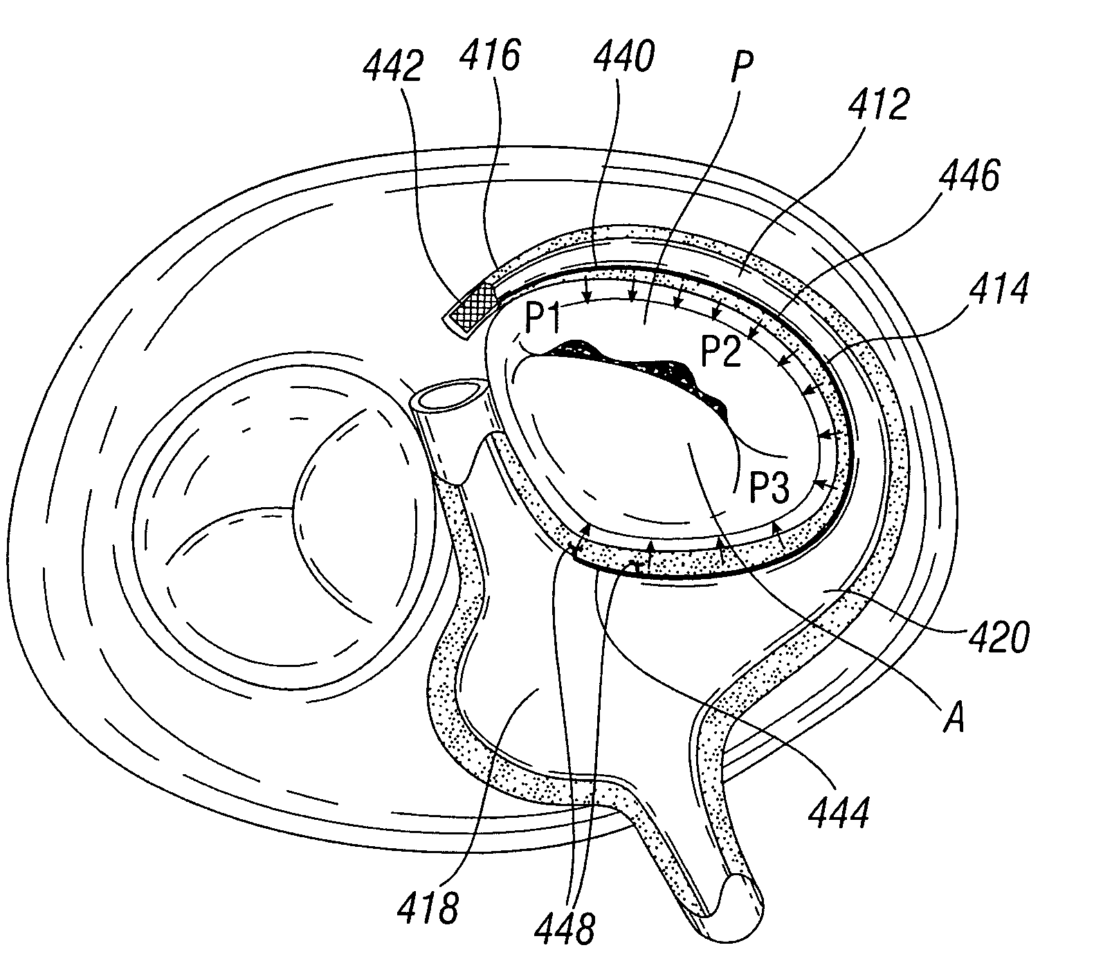
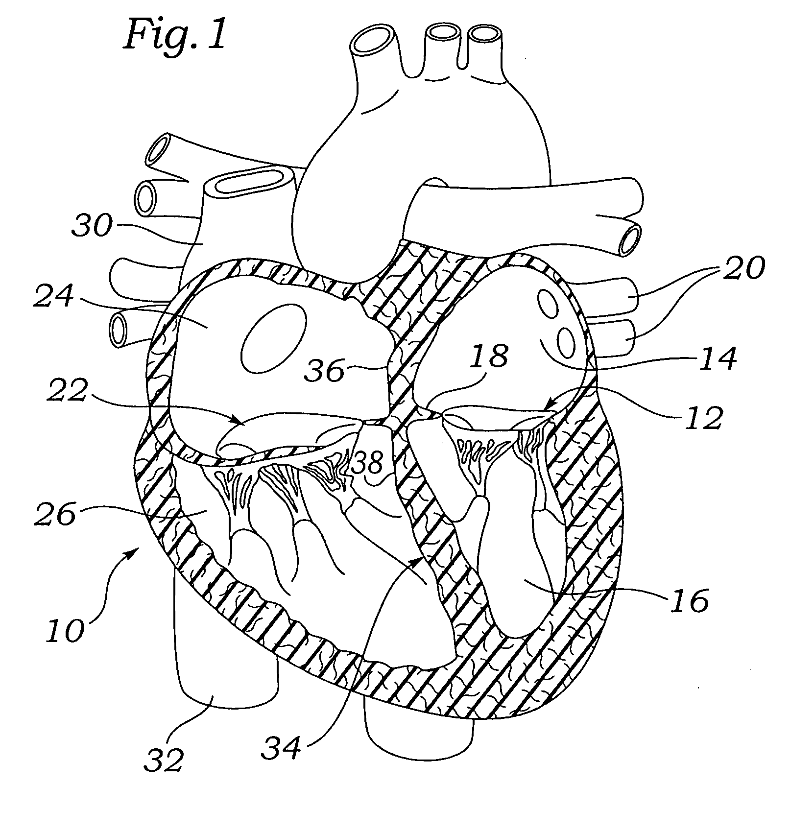
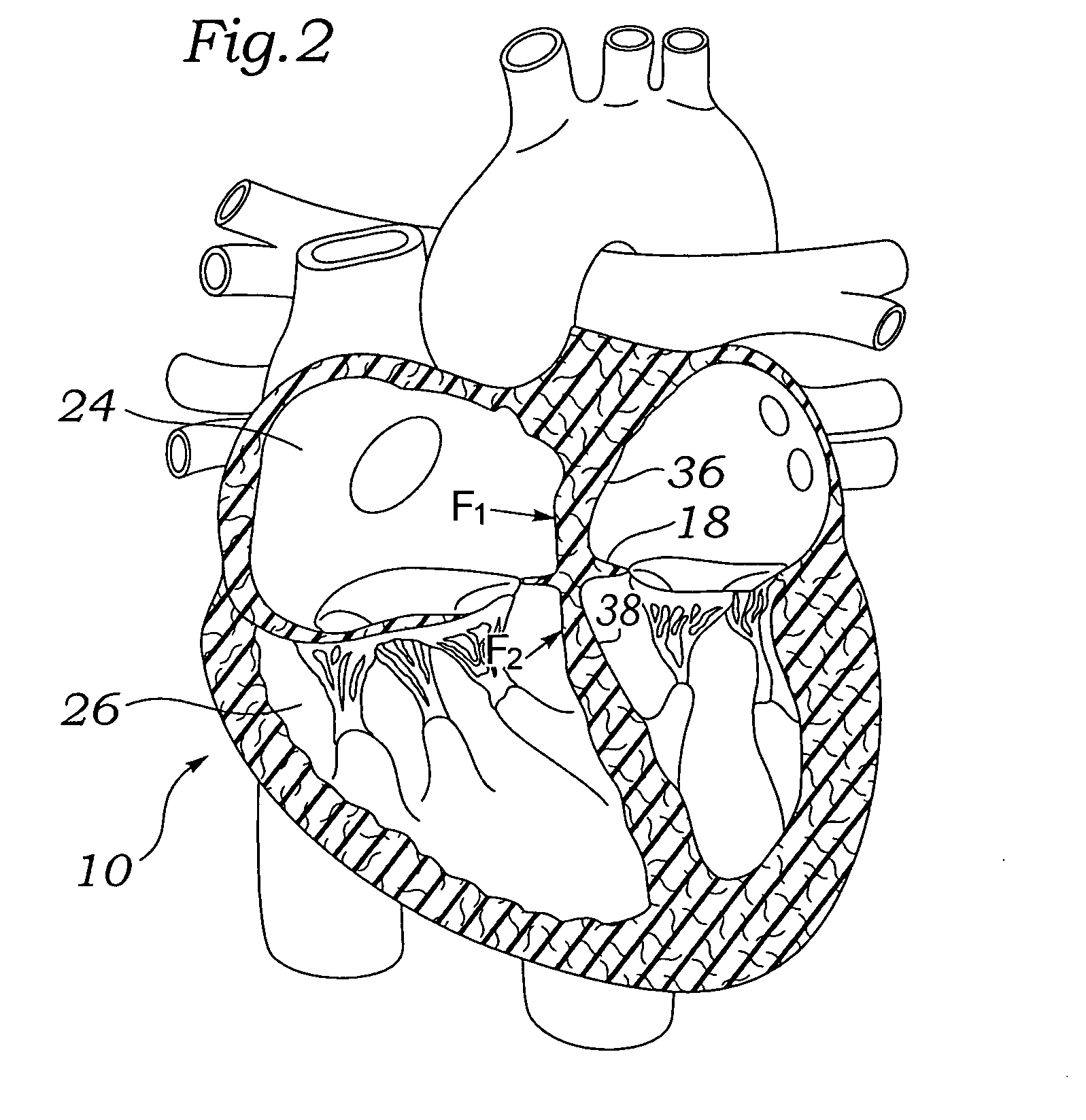
[0060]Various embodiments of the present invention depict medical implants and methods of use that are well-suited for treating mitral valve regurgitation. It should be appreciated that the principles and aspects of the embodiments disclosed and discussed herein are also applicable to other devices having different structures and functionalities. For example, certain structures and methods disclosed herein may also be applicable to the treatment of other heart valves or other body organs. Furthermore, certain embodiments may also be used in conjunction with other medical devices or other procedures not explicitly disclosed. However, the manner of adapting the embodiments described herein to various other devices and functionalities will become apparent to those of skill in the art in view of the description that follows.

[0061]As used herein, “distal” means the direction of a device as it is being inserted into a patient's body or a point of reference closer to the leading end of the...


PUM


Abstract

Devices and methods for reshaping a mitral valve annulus are provided. One device according to the invention is configured for deployment in the right atrium and is shaped to apply a force along the atrial septum. The device causes the atrial septum to deform and push the anterior leaflet of the mitral valve in a posterior direction for reducing mitral valve regurgitation. Another embodiment of a device is deployed in the left ventricular outflow tract at a location adjacent the aortic valve. The device may be expandable for urging the anterior leaflet toward the posterior leaflet. Another embodiment of the device includes a first anchor, a second anchor, and a bridge, with the bridge having sufficient length to reach from the coronary sinus to the right atrium and/or superior or inferior vena cava. In a further embodiment a device includes a middle anchor positioned on the bridge between the distal and proximal anchors.

Description

FIELD OF THE INVENTION[0001]The present invention relates to medical devices and methods and, more particularly, to medical devices and methods for repairing a defective mitral valve in a human heart.BACKGROUND[0002]Heart valve regurgitation, or leakage from the outflow to the inflow side of a heart valve, occurs when a heart valve fails to close properly. Regurgitation often occurs in the mitral valve, located between the left atrium and left ventricle, or in the tricuspid valve, located between the right atrium and right ventricle. Regurgitation through the mitral valve is often caused by changes in the geometric configurations of the left ventricle, papillary muscles, and / or mitral valve annulus. Similarly, regurgitation through the tricuspid valve is often caused by changes in the geometric configurations of the right ventricle, papillary muscles, and / or tricuspid valve annulus. These geometric alterations result in incomplete leaflet coaptation during ventricular systole, there...

Claims


Application Information

Patent Timeline
IPC IPC(8): A61F2/24A61B19/00
CPCA61B19/54A61F2/2412A61F2/2454A61F2/2442A61F2/2451A61F2/2418A61B90/39A61F2/2487
Inventor BOBO, DONALD E.ROWE, STANTONPOPP, MICHAELYI, SEUNG
Owner EDWARDS LIFESCIENCES CORP
Why Eureka
Try Eureka