

Detection of Specific Binding Reactions Using Magnetic Labels
a specific binding and magnetic label technology, applied in the field of specific binding reactions, can solve the problems of affecting the specificity of the fret pair of these new fluorescent proteins, altering or losing biologically relevant information,
- Summary
- Abstract
- Description
- Claims
- Application Information
AI Technical Summary
Problems solved by technology
Method used
Image

Examples
example 1
mOrange2 / mCherry Protease Biosensor in Vitro
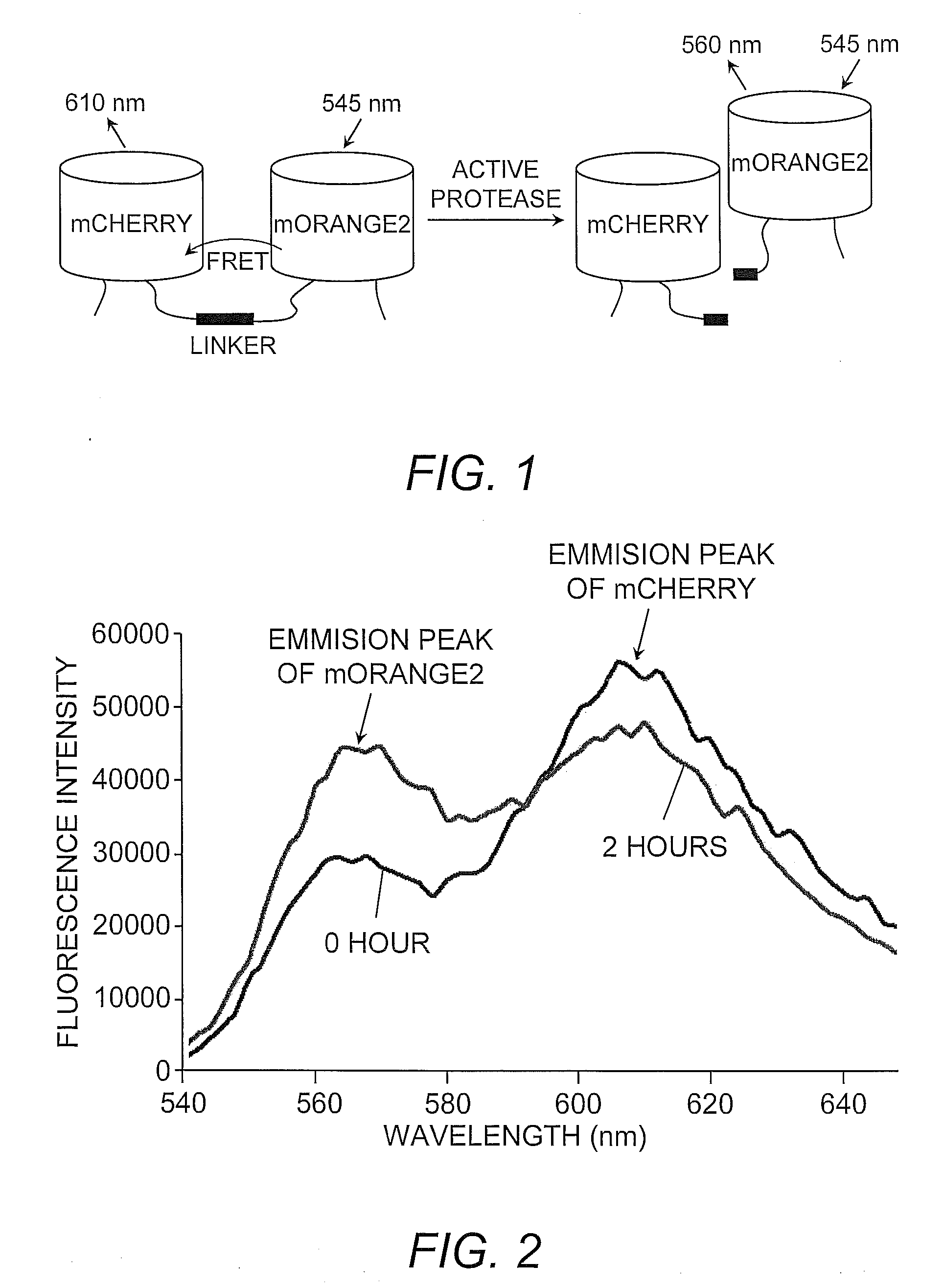
[0025]By way of illustration, membrane-type 1-metalloproteinase (MT1-MMP) activity was monitored using the mOrange2 and mCherry FRET pair. MT1-MMP is a membrane-anchored enzyme belonging to matrix metalloproteinase (MMP) family. MT1-MMP is known to digest MMP-2, another member of MMP family, as well as extracellular matrix protein collagen. A specific and sensitive MT1-MMP biosensor based on mOrange2 and mCherry was generated utilizing mCherry at the N-terminus and mOrange2 at the C-terminus with a substrate sequence derived from MMP-2 (Cys-Pro-Lys-Glu-Ser-Cys-Asn-Leu-Phe-Val-Leu-Lys-Asp, SEQ ID NO:3, wherein the underlined amino acids Asn-Leu form the cutting site recognizable by MT1-MMP) located between mCherry and mOrange2.
[0026]The basis of this assay is that when MT1-MMP is inactive, the mCherry and mOrange2 are positioned in proximity and favor a strong FRET between the two moieties (FIG. 1). Therefore, the excitation of mOrange2 at ...
example 2
mOrange2 / mCherry Protease Biosensor in Vivo
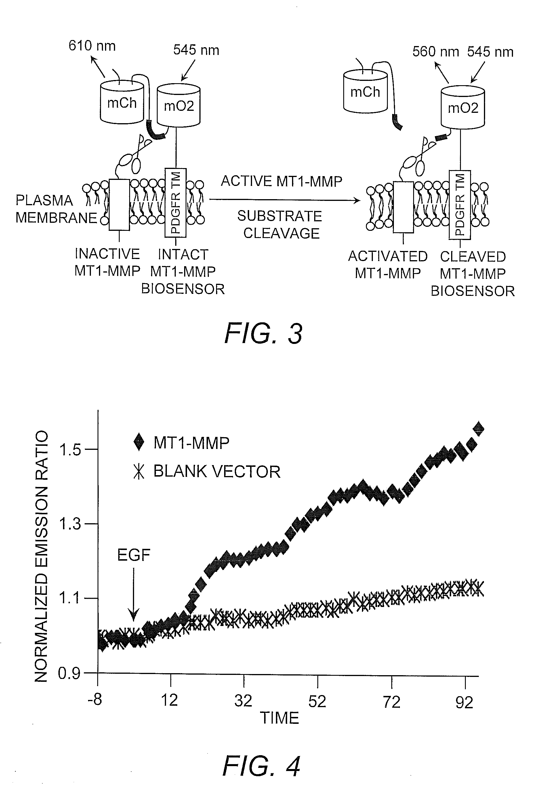
[0030]The catalytic domain of MT1-MMP and the transmembrane domain of the platelet-derived growth factor receptor beta (i.e., PDGFRβ) are known to co-localize at the cell surface. Accordingly, a cDNA encoding the MT1-MMP biosensor described herein (see Example 1) was fused with a nucleic acid molecule encoding the transmembrane domain of PDGFRβ, resulting in the fusion construct designated “PDGFR™”. This construct was designed so that the linker of the biosensor was located in the extracellular space, but in close proximity to the MT1-MMP catalytic domain (FIG. 3).
[0031]Nucleic acid molecules encoding the mOrange2 / mCherry MT1-MMP biosensor construct and MT1-MMP were introduced into mammalian HeLa cells. Epidermal growth factor (EGF) is known in the art to induce MT1-MMP, therefore EGF was added to the recombinant HeLa cells. The results indicate that EGF induced a significant FRET change of the MT1-MMP biosensor in HeLa cells co-transfected...
example 3
mOrange2 / mCherry and CFP / YFP-Based Biosensors
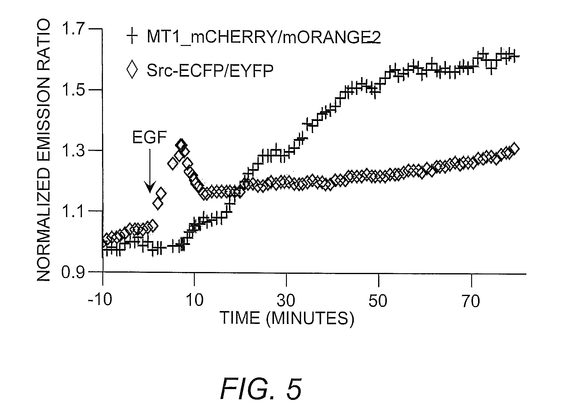
[0032]To demonstrate the utility of the instant biosensor in monitoring multiple molecule events in vivo, the mOrange2 / mCherry-based MT1-MMP biosensor was combined with a CFP / YFP-based Src biosensor to simultaneously visualize MT1-MMP and Src activity in the same live mammalian cells. The results indicated that EGF induced a fast and global activation of Src, while the MT1-MMP activation upon EGF stimulation was relatively slow and concentrated at the cell periphery. Normalized time courses of averaged FRET ratios also indicated that the activation of Src was fast with a transient peak at around 5 minutes of EGF stimulation, while the MT1-MMP activation was delayed with sustained increase (FIG. 5). These results indicated that the mOrange2 / mCherry biosensor could be combined with CFP / YFP-based biosensor to visualize two active signaling events simultaneously in the same live cells.
PUM
| Property | Measurement | Unit |
|---|---|---|
| Color | aaaaa | aaaaa |
| Resonance energy | aaaaa | aaaaa |
| Fluorescence | aaaaa | aaaaa |
Abstract
Description
Claims
Application Information