
Needle Guides for Catheter Delivery

a catheter and needle guide technology, applied in the field of ultrasound imaging systems and methods, can solve the problems of small error margin, severe damage or death of nerves, and patient death due to cardiac or respiratory collapse, and achieve the effect of easy visualization

Inactive Publication Date: 2010-02-18
ANES VENTURES
View PDF14 Cites 70 Cited by
  • Summary
  • Abstract
  • Description
  • Claims
  • Application Information

AI Technical Summary

Benefits of technology

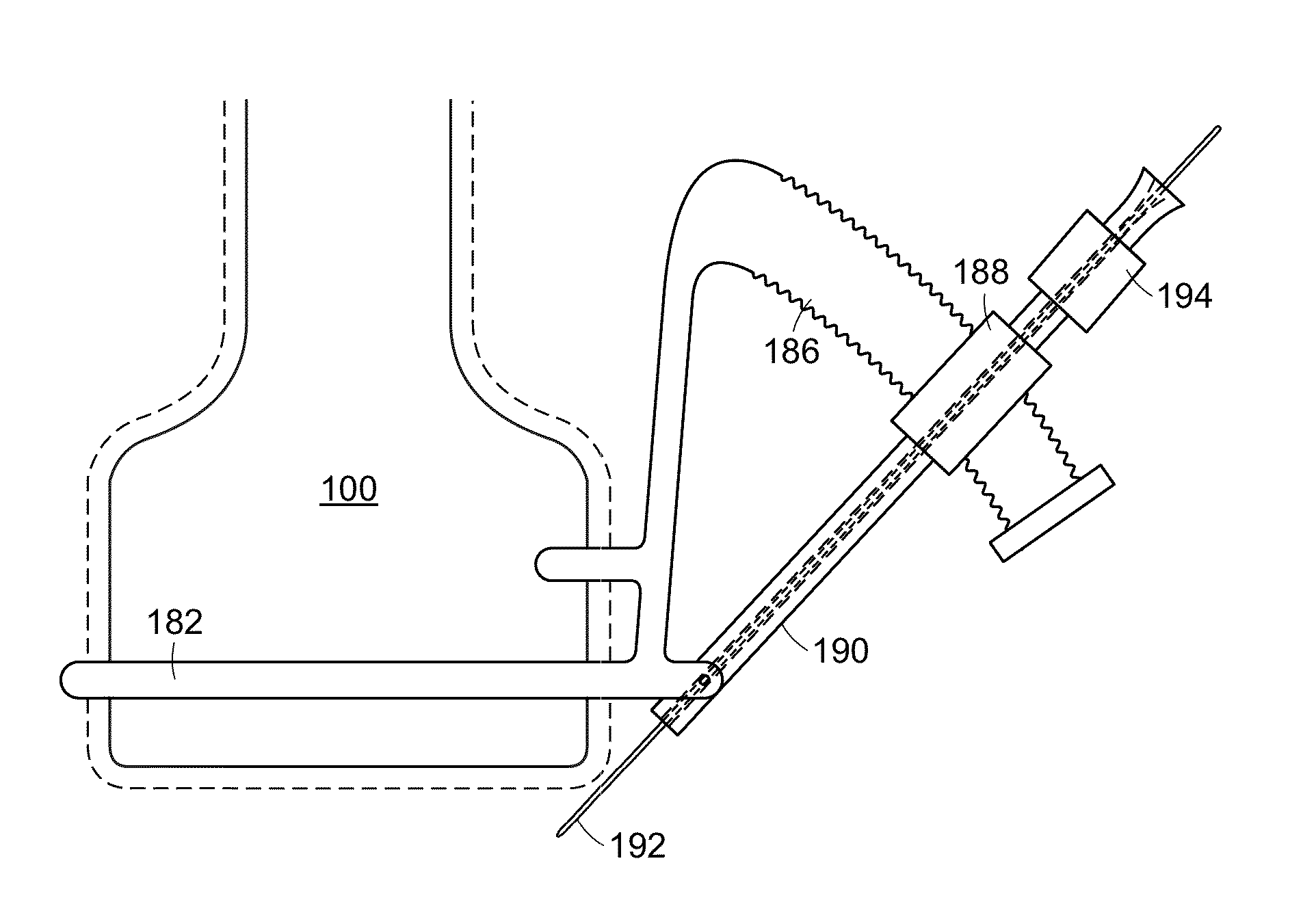
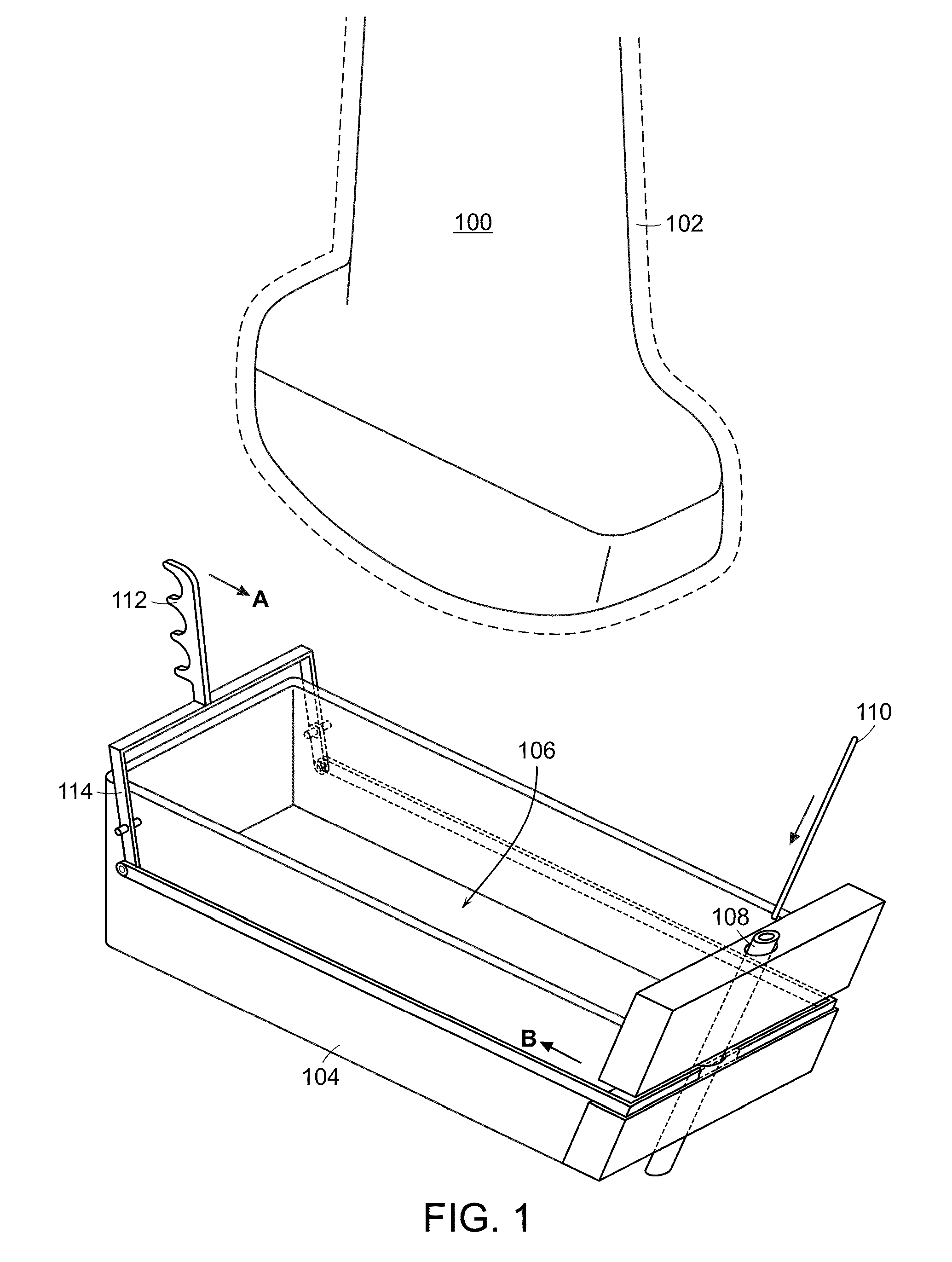
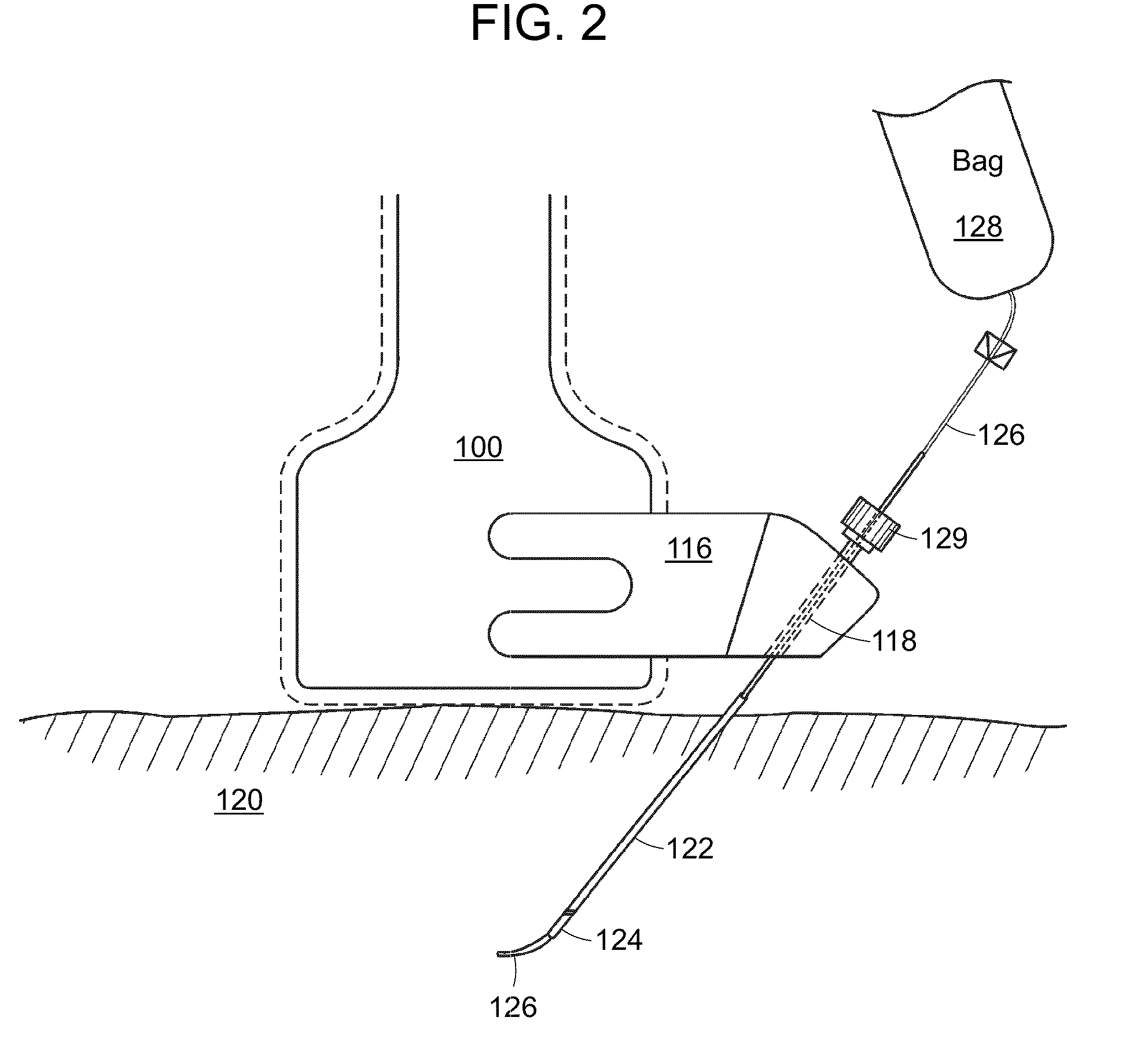
[0017]The present invention overcomes limitations in the prior art by providing improved methods and apparatuses for the placement of needles, catheters or other indwelling medical devices using ultrasound guidance. In certain aspects, the present invention provides a needle-locking device that may be attached or coupled to an ultrasound probe. The needle-locking device can allow for the substantial immobilization of a needle during an ultrasound guided needle insertion; this immobilization can allow a practitioner to insert a catheter through the needle while maintaining ultrasound visualization of the needle and catheter. In various embodiments, the tip of the catheter may be coated with a metal or other substance that allows for easy visualization of the catheter tip during catheter placement. In certain embodiments, the needle-locking device may be locked with the same hand that is used by the practitioner to hold the ultrasound probe.
[0022]In yet further aspects, the present invention provides kits comprising a needle lock of the present invention in a container means. The kit may further comprise a catheter. The catheter may be coated with a substance which may be easily visualized via ultrasound. The substance may be a metal or a polymer. In various embodiments, the needle lock may incorporate, attach to, or be integral with a needle guide. The kit may further comprise a needle. The needle may be coated with a substance which may be easily visualized via ultrasound.
[0024]Catheters which are contemplated by the present invention include catheters wherein a portion of one end of the catheter is coated with a ultrasound visualization agent. Said coating may be on the internal and / or external portion of the catheter. The catheter may be made via a variety of methods (e.g., dip-coating or spray coating) which would be appreciated by one of skill. In various embodiments, the catheter tip may contain a coil or piece of metal which would be easily visualized with the ultrasound machine.

Problems solved by technology

Damage to a nerve or injection of an anesthetic solution into a nerve during a nerve block procedure can severely damage or kill the nerve.
Delivery of a local anesthetic into an artery can result in patient death due to cardiac or respiratory collapse.
This allows only a very small margin for error on the part of the anesthesiologist, and obviously to those skilled in the art, any movement of the needle could reduce the effect of the regional anesthetic, damage the nerve, or both.
Several serious limitations exist regarding ultrasound visualization techniques for guided needle or catheter placement.
During this portion of time it is not possible to know with any certainty where the tip of the catheter is relative to the tip of the needle.
During the threading of a catheter it is typically not possible to visualize the catheter as it exits the needle as the ultrasound probe has been set down.
Since the catheter is threaded into a sterile field, this is often difficult to achieve and an assistant may not be available.
The inability of one to visualize the catheter tip during catheter placement presents a significant risk.
For example, during a nerve block procedure, placement of a catheter too far through a needle could insult, damage, or kill a nerve or puncture a lung rather than remain at a location appropriate for bathing the nerve in local anesthetic.
Placement of the catheter not far enough into a patient could result in an incomplete nerve block and / or fail to establish an acceptable level of anesthesia.
Accidental puncturing of a blood vessel could result in central neural toxicity and / or cardiac toxicity due to, e.g., transport of an anesthetic via an artery.
Failure to appropriately place a catheter accounts for a significant portion of morbidity and mortality.
This often proves quite difficult, as the“beam” or “field of view” emitted by the probe is only a few millimeters wide.
The current limitation on ultrasound needle guides is their inability to hold a needle still while pressure greater than gravity is applied.
However, when solution is injected through a needle, or when a medical device is passed into a patient through a needle, a great deal of force can be generated thereby placing the needle at risk for unwanted movement.
There is currently no needle guide on the market that takes this need into account.
Another limitation of current needle guides is their inability to accommodate a variety of needle insertion sites.
This is often not the optimal needle insertion site to thread a foreign body into a patient.
A final limitation of current needle guides is the inability of the user to “freehand” when necessary.
Current guides do not usually allow for this type of free movement of a needle.

Method used


Image

  • Needle Guides for Catheter Delivery
  • Needle Guides for Catheter Delivery
  • Needle Guides for Catheter Delivery

Examples


Embodiment Construction

[0040]The present invention overcomes limitations in the prior art by providing improved methods and apparatus for the placement of catheters using ultrasound guidance. In certain aspects, the present invention provides a needle locking device which may be attached or coupled to an ultrasound probe. The needle locking device can allow for the immobilization of a needle during an ultrasound guided needle insertion; this immobilization can allow a practitioner to insert a catheter or other medical device through the needle while maintaining ultrasound visualization of the needle. In various embodiments, an area near the tip of the catheter may be coated, e.g., dip coated or spray coated, with a metal or other substance, which allows for easy ultrasound visualization of the catheter tip during catheter placement. In some embodiments, a region just prior to the tip of the catheter may be coated with an ultrasound visualizing substance. Alternately, a catheter with a metal coil inserted ...


PUM


Abstract

Provided are needle locks and methods of using the same for ultrasound guided catheter insertion. The needle locks hereof substantially immobilizes a needle relative to an ultrasound probe to permit manipulation of a catheter or other device through the needle. The needle locks of the present invention may be used in nerve block, venous catheter insertion procedures or other invasive procedures.

Description

PRIORITY CLAIM[0001]The present application claims priority to U.S. Provisional Application No. 61 / 188,833 filed Aug. 13, 2008 and is incorporated by reference herein.BACKGROUND OF THE INVENTION[0002]1. Field of the Invention[0003]The present invention relates generally to systems and methods for ultrasound imaging, and in particular, to systems and methods for correct placement of needles, catheters, and other medical devices using an ultrasound probe.[0004]2. Description of Related Art[0005]Ultrasound imaging is a medically important technique that may be used for a variety of applications including regional nerve block, line placement, tumor imaging, epidural placement, emergency room diagnosis, and catheter delivery to name a few. Present methods for catheter delivery using ultrasound guidance normally require the practitioner to hold an ultrasound probe with one hand and insert a needle with the other hand. The ultrasound probe allows the practitioner to visualize the internal ...

Claims


Application Information

Patent Timeline
IPC IPC(8): A61B8/00A61M19/00
CPCA61B17/3403A61B2017/3405A61M25/0662A61B2017/347A61M5/427A61B2017/3413A61B8/4209
Inventor SCHLITT, JOHNFINDLAY, GEOFFREY S.
Owner ANES VENTURES
Learn More