Image generation based on limited data set

a technology of limited data set and image generation, applied in image analysis, image enhancement, instruments, etc., can solve the problems of insufficient use and prolonged acquisition time, and achieve the effect of increasing patient comfor

Inactive Publication Date: 2010-03-04
KONINKLIJKE PHILIPS ELECTRONICS NV
View PDF3 Cites 12 Cited by
  • Summary
  • Abstract
  • Description
  • Claims
  • Application Information

AI Technical Summary

Benefits of technology

[0011]Based on an input sequence of only a limited amount of biological process images, the method is capable of providing an image, such as a parametric, functional or molecular image, that maps the underlying biological process. Thus, for example, given only a few late-time tracer uptake images from a scanner, the method can be used to estimate reliably a parametric image which is close to the corresponding parametric image that would have been derived had the entire time sequence of images been available. The method thus renders it possible to increase patient comfort since the patient only needs to be scanned for a limited period of time in a dynamic scanning sequence after injection of a radio tracer or contrast agent, rather than having to spend a long time in the scanner in order to record a complete sequence of images covering a long period of time.

Problems solved by technology

One issue with acquiring four dimensional (4D) dynamic scans is the prolonged acquisition times, which may amount to 3 hours in some cases (e.g. 18F-FMISO for PET hypoxia imaging).
These late-time images, which represent the tracer distribution at a particular point in time, while useful may not convey the entire picture.
However, many of the tracers employed in PET and SPECT to probe biological processes display complex tracer kinetics, necessitating the need for pharmacokinetic modeling and analysis.

Method used


Image

  • Image generation based on limited data set
  • Image generation based on limited data set
  • Image generation based on limited data set

Examples


Embodiment Construction

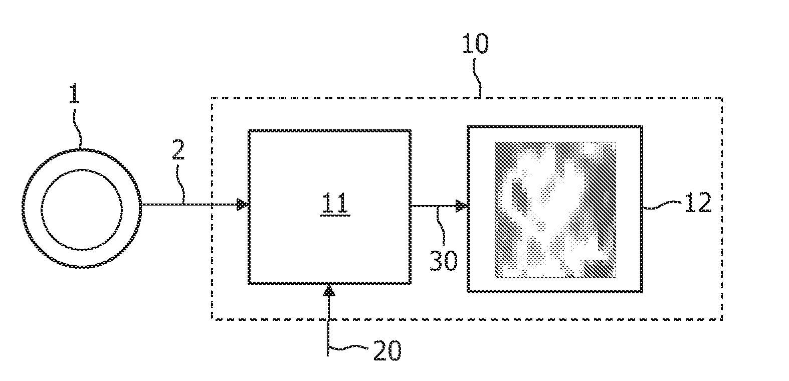
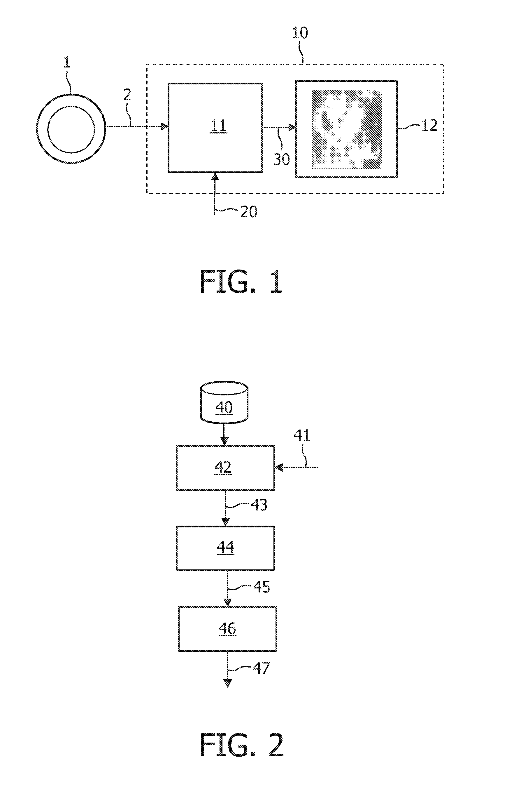
[0042]FIG. 1 illustrates a device 10 arranged for operation in connection with a scanner 1, e.g. a PET scanner, which can record a sequence of images 2 as a function of time, or data representing such images 2. The sequence of images 2 represents a scanning of a regional part of a human body after injection of a radio tracer or contrast agent. The sequence of images 2 may represent, for example, FMISO data with missing time points. It may be that images in a particular time range (e.g. 0-90 minutes) after injection are missing. The incomplete sequence of images 2 is then processed by a signal processor 11, either directly from the scanner 1 or after being stored. The signal processor 11 also receives additional data 20, such as literature-based data regarding a kinetic parameter range, and optionally an input function, such as blood clearance functional data. The signal processor 11 then performs an iterative algorithm on the data 2, 20 comprising the application of a pharmacokineti...


PUM


Abstract

A method, signal processor, device, and system for estimating a parametric or functional image 47 mapping a biological process on the basis of a limited or incomplete sequence of biological process images 40 recorded as a function of time, e.g. by a PET or SPECT scanner after injection of a radio tracer. One or more kinetic parameters 43 are first extracted by applying a pharmacokinetic model 42 (compartmental model of the underlying tracer kinetics) to the sequence of biological process images 40. Additional data 41 are used in the model, comprising at least a predetermined kinetic parameter range (e.g. from the literature), and optionally an input function or a blood clearance function. Next, an iterative algorithm 44 is applied to arrive at a modified sequence of images 45, e.g. by inserting an estimated image into the incomplete sequence of images, utilizing the one or more kinetic parameters 43. After a stop criterion has been fulfilled, the resulting image 47 is finally estimated 46 from the modified sequence of images 45. The method can be used e.g. to estimate a hypoxia parameter k3 image in the case of a FMISO data set where only late-time images are available. The method may be implemented as part of existing PET, SPECT, CT, MR or Ultrasound scanner software, and since only a limited amount of late time post injection images are necessary to provide a reliable result, the method helps to increase patient comfort and clinical throughput.

Description

FIELD OF THE INVENTION[0001]The present invention relates to the field of generating images mapping a biological process, such as that used within clinical medical applications to assist therapy and diagnosis. More specifically, the invention provides a method, a signal processor, a device, and a system for generating an image that maps a biological process and that is to be used in combination with an output unit of a scanner for mapping tracer kinetics. Especially, the invention is capable of providing an image based on a limited or incomplete set of data, such as an incomplete time sequence of images of a biological process.BACKGROUND OF THE INVENTION[0002]Molecular imaging modalities such as positron-emission tomography (PET) and single-photon emission tomography (SPECT) are unique in employing radioactively labeled biological molecules as tracers for studying and visualizing pathophysiological mechanisms in vivo. In principle, functional imaging modalities would allow early vis...

Claims


Application Information

Patent Timeline
Patent Type & Authority Applications(United States)
IPC IPC(8): G06K9/00
CPCG06T7/2033G06T2207/10072A61B6/037A61B6/507G06T2207/30004G06T7/0016G06T7/246
Inventor NARAYANAN, MANOJ
Owner KONINKLIJKE PHILIPS ELECTRONICS NV
Why Eureka
Try Eureka