Non-invasive monitoring cancer using integrated microfluidic profiling of circulating microvesicles

a technology of microfluidic profiling and cancer, applied in the field of non-invasive monitoring of cancer using integrated microfluidic profiling of circulating microvesicles, can solve the problems of pressure-caused damage of vesicles, inconvenient use, and severe constraints on exosome analysis, and achieve the effect of improving sensitivity and specificity

Active Publication Date: 2019-07-16
UNIVERSITY OF KANSAS +1
View PDF6 Cites 1 Cited by
  • Summary
  • Abstract
  • Description
  • Claims
  • Application Information

AI Technical Summary

Benefits of technology

[0005]This disclosure, which is more fully described under the Detailed Description section below, provides in certain embodiments a “sample-in-answer-out” microfluidic exosome profiling platform which integrates exosome isolation and targeted proteomic analysis in one rapid assay. We demonstrate utility of two microfluidic devices and methods of using them for quantitative profiling of surface exosomal biomarker and intravesicle biomarkers individually, directly from 30 μL plasma samples within ˜100 min, with markedly enhanced sensitivity and specificity compared to previously available approaches.
[0007]Microfluidic chips provide multiple advantages over conventional testing. The presently provided microfluidic chips use smaller volumes of fluids, provide faster results, are more compact and require less manual operation and lab space, increase testing throughput, reduce costs, reduce technician exposure to fluids due to the smaller amounts of fluids that are required, and can increase patient comfort due to the smaller amounts of body fluids that are required for testing. The design of these microfluidic chips can vary by applications, and each variation can use a form of microfluidics to enable fluid flow to various regions in the chip.

Problems solved by technology

Despite the potential for cancer research, exosome analyses have been severely constrained by the daunting technical difficulties in isolation and molecular analysis of such nano-scale and molecularly diverse vesicles.
Current isolation protocols largely rely on multiple ultracentrifugation steps, which are tedious, time-consuming (>10 h) and inefficient.
Size exclusion methods normally do not concentrate exosomes and are prone to pressure-caused damage of vesicles and contaminations.
Molecular analysis of isolated microvesicles is primarily performed using Western blot, ELISA and mass spectrometry, which require lengthy processes and large sample sizes (purified from more than 3 mL plasma or 300 mL cell culture media), limiting the progress in clinical investigation and utilities of exosomes.
To date, no well-defined protocols and markers for quantitative evaluation of extracellular vesicle proteins exist.

Method used


Image

  • Non-invasive monitoring cancer using integrated microfluidic profiling of circulating microvesicles
  • Non-invasive monitoring cancer using integrated microfluidic profiling of circulating microvesicles
  • Non-invasive monitoring cancer using integrated microfluidic profiling of circulating microvesicles

Examples


example 1

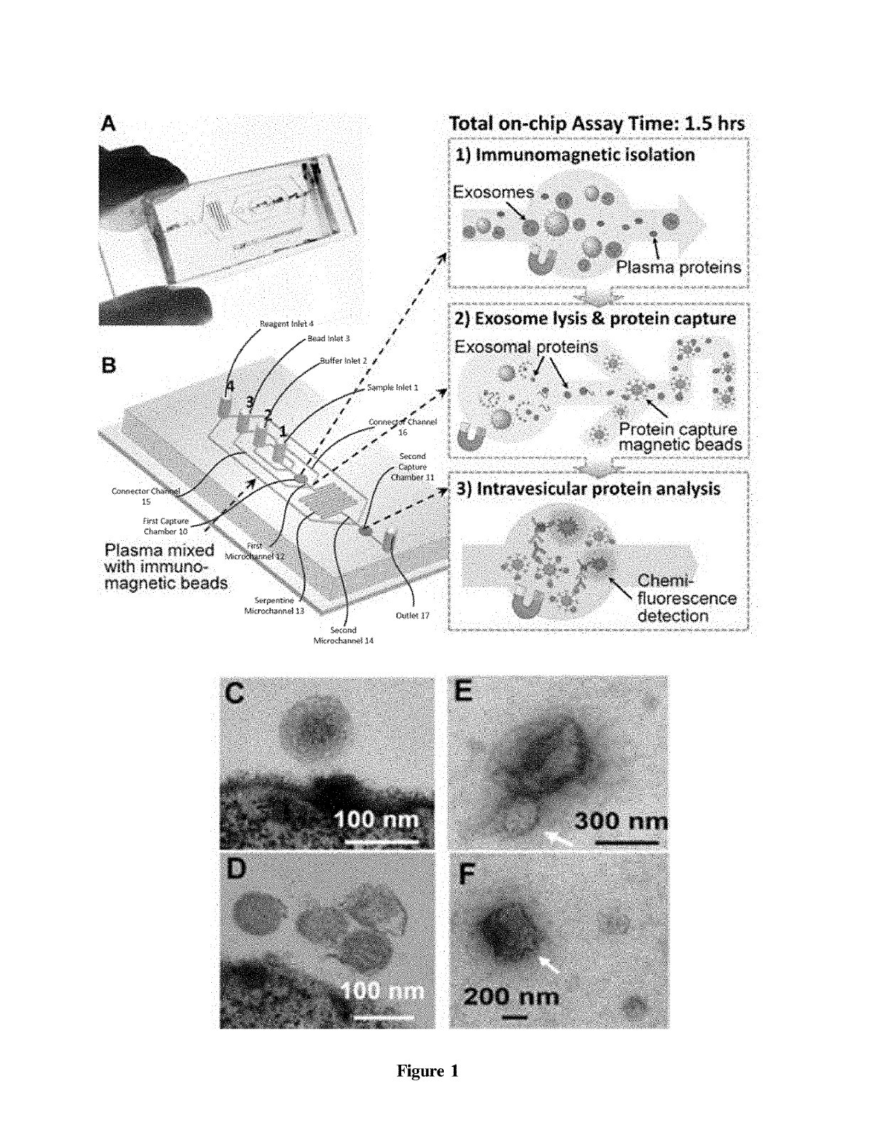
[0062]This and the following Examples relate to integrated microfluidic microvesicle profiling using an illustrative device which contains a cascading microchannel circuit to integrate and streamline the multi-step profiling of exosomes directly from human plasma, including exosome isolation and enrichment (1st stage capture), exosome lysis, fluidic mixing and immunomagnetic capture of intravesicular targets (2nd stage capture), and protein assays (FIG. 1). A representative device architecture is detailed in FIG. 1A. The two cascading magnetic-bead capture chambers (first capture chamber 10 and second capture chamber 11) are of 4-mm diameter and are capable of capturing up to 109 2.8 μm microbeads each. A plasma sample pre-mixed with antibody labeled magnetic beads is introduced through sample inlet 1 into the first capture chamber 10 where the magnetic beads with bound exosomes are isolated from the sample and washed by PBS washing buffer (FIG. 1B). The lysis buffer is introduced t...

example 2

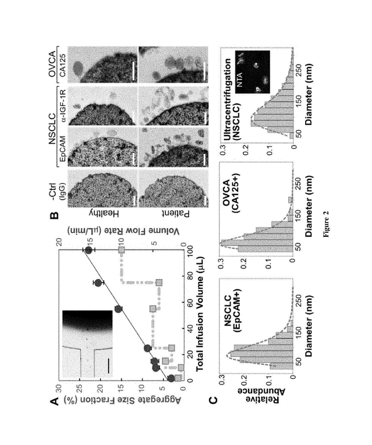
[0065]This Example provides a description of on-chip immunomagnetic isolation of circulating exosomes. We first investigated magnetic capture of beads as it dictates the overall performance of exosome isolation and analysis. The beads suspended in a buffer solution were retained by a magnet placed underneath the capture chamber (FIG. 2A, inset), forming an aggregate (FIG. 8) induced by the dipolar interactions between the beads. It was reported that the amount of magnetically captured beads in a microchannel can be represented by the size of the bead aggregate, which increases linearly with time at a constant flow rate. We adopted this approach to conveniently assess the bead capture a function of flow conditions (FIG. 2A). It was found that the aggregate size was linearly dependent on the total sample infusion volume regardless of the flow rates applied to reach certain infusion volumes (1-10 μL / min, FIG. 2A). The independence on flow rate indicates a high bead capture efficiency a...

example 3

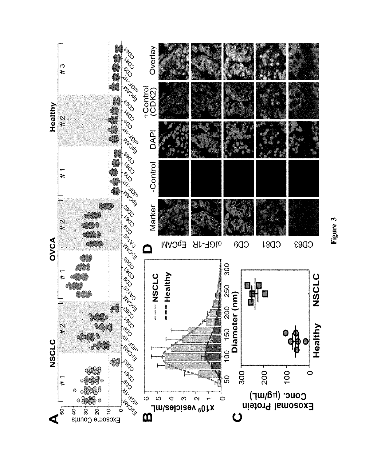
[0068]This Example provides a description of profiling of exosome subpopulations by protein phenotype. Surface protein composition of exosomes plays an important role in exosome-mediated effects and may provide tumor fingerprints. To demonstrate the ability to detect exosomal expression patterns associated with cancer, we conducted relative quantification of five exosome subpopulations defined by individual surface markers using TEM. FIG. 3A exemplifies the results for two of the NSCLC samples that we have tested. Distinct subpopulation landscapes were observed, as compared to the healthy controls with a 3- to 5-fold increase in abundance for the surface markers except CD63. We further demonstrated the adaptability of our method for other cancers by testing on OVCA with the tumor markers (EpCAM and CA125) and exosomal markers (CD9, CD81, and CD63). The OVCA samples also provide a positive control for the NSCLC studies, as CD63 was found to be highly expressed in OVCA exosomes. High ...


PUM

PropertyMeasurementUnit
diameteraaaaaaaaaa
volumesaaaaaaaaaa
flow rateaaaaaaaaaa

Abstract

A microfluidic exosome profiling platform integrating exosome isolation and targeted proteomic analysis is disclosed. This platform is capable of quantitative exosomal biomarker profiling directly from plasma samples with markedly enhanced sensitivity and specificity. Identification of distinct subpopulation of patient-derived exosomes is demonstrated by probing surface proteins and multiparameter analyzes of intravesicular biomarkers in the selected subpopulation. The expression of IGF-1R and its phosphorylation level in non-small cell lung cancer (NSCLC) patient plasma is assessed as a non-invasive alternative to the conventional biopsy and immunohistochemistry. Detection of ovarian cancer also is assessed. The microfluidic chip, which may be fabricated of a glass substrate and a layer of poly(dimethylsiloxane), includes a serpentine microchannel to mix a fluid and a microchamber for the collection and detection of exosomes.

Description

CROSS-REFERENCE TO RELATED APPLICATIONS[0001]This application is a continuation-in-part of U.S. application Ser. No. 15 / 125,822, filed Sep. 13, 2016, which is a national phase application of PCT / US15 / 20683 filed Mar. 16, 2015, and which claims priority to the provisional patent application filed Mar. 14, 2014 and assigned U.S. App. No. 61 / 953,109 and to the provisional patent application filed Jul. 16, 2014 and assigned U.S. App. No. 62 / 025,151. This application also claims priority to the provisional patent application filed Nov. 20, 2015 and assigned U.S. App. No. 62 / 258,258. The disclosures of each of these applications is hereby incorporated by reference.STATEMENT REGARDING FEDERALLY-SPONSORED RESEARCH OR DEVELOPMENT[0002]This invention was made with government support under grant numbers CA186846 and GM103638 awarded by the National Institutes of Health. The government has certain rights in the invention.BACKGROUND OF THE DISCLOSURE[0003]Developing non-invasive blood-based test...

Claims


Application Information

Patent Timeline
Patent Type & Authority Patents(United States)
IPC IPC(8): B01F13/00B01L3/00G01N33/574B01F5/06
CPCB01L3/502761B01F5/0647B01F13/0059G01N33/574B01L2400/0487B01L2300/0816B01L2300/0864B01L2300/0883B01L2400/043B01L2200/0668B01F25/4331B01F33/30
Inventor ZHAO, ZHENGHE, MEIZENG, YONG
Owner UNIVERSITY OF KANSAS
Why Eureka
Try Eureka