
Therapeutic composite for cartilage disorder using extracellular matrix (ECM) scaffold

a technology of extracellular matrix and composite, which is applied in the direction of skeletal/connective tissue cells, prosthesis, peptide/protein ingredients, etc., can solve the problems of fibrocartilage which lacks the biomechanical characteristics of normal articular cartilage, cannot produce high-quality tissue engineered cartilage to be used in clinical practice, and oa. large problem

Inactive Publication Date: 2010-05-13
MIN BYOUNG HYUN
View PDF6 Cites 33 Cited by
  • Summary
  • Abstract
  • Description
  • Claims
  • Application Information

AI Technical Summary

Benefits of technology

[0011]Another object of the present invention is to provide a therapeutic composition for treating cartilage disorder, which contains the ECM scaffold having chondrocytes or stem cells attached thereto as an effective component.

Problems solved by technology

Although some successful applications in vivo and in vitro were reported with some scaffolds, there was still a problem that they could not produce high-quality tissue engineered cartilage enough to be used in clinical practices.
Articular cartilage damage is difficult to treat because it has limited intrinsic healing ability, and this easily causes degeneration of surrounding cartilage, thus resulting in extensive osteoarthritis (OA).
However, the tissue regenerated using these methods generally has characteristics of fibrocartilage which lacks biomechanical characteristics of normal articular cartilage.
Due to incongruence of interface between transplant and host cartilage and restrictions in the donor source, there has been a limitation in applying the osteochondral transplantation techniques.
However, there are still some remaining problems with the method, which include loss of chondrocytes function during in vitro culture, the possibility of transplanted chondrocytes leaking from the transplant site, heterogeneous distribution of chondrocytes, and periosteal hypertrophy.
Regrettably, however, no biocompatible substance has been reported that showed satisfying results in the production of mature cartilage in vivo.

Method used


Image

  • Therapeutic composite for cartilage disorder using extracellular matrix (ECM) scaffold
  • Therapeutic composite for cartilage disorder using extracellular matrix (ECM) scaffold
  • Therapeutic composite for cartilage disorder using extracellular matrix (ECM) scaffold

Examples


example 1

Construction of Cell-Derived ECM Scaffold

[0046]A cell-derived ECM scaffold was constructed using chondrocytes isolated from knee joints of 2 to 4-week-old pigs.

[0047]After culturing pig chondrocytes for 3˜4 days, cell layers having ECM components, were carefully separated, and transformed into a pellet-type construct through centrifugation. The pellet-type structure was adjusted for 3 weeks for the growth of new cartilage tissue, after repetition of freeze-thawing 3 times every 12 hours, ECM was obtained by freeze-drying at −56° C. for 48 hours at a pressure of 5mTorr.

[0048]The constructed ECM was decellularized through a step of removing nuclear and cytoplasmic components by treating it with a proteolytic enzyme, detergent or ultrasound. 0.05% trypsin was used as a proteolytic enzyme and ionic and non-ionic detergents such as SDS, triton X, deoxycholate were used as a detergent. Nuclear components, such as DNA, etc. were removed by treating it with DNase.

[0049]The final decellulari...

example 2

Construction of Tissue-Engineered Cartilage Using Rabbit Chondrocytes and ECM Scaffold

[0050]The cell-derived ECM scaffold constructed in Example 1 was socked in 70% ethanol for 1 hour to wash with PBS several times, and left to stand overnight in DMEM medium without serum before inoculating cells. Chondrocytes were isolated from a 2 week-old New Zealand white rabbit and cells at passage 1 were inoculated into the ECM scaffold at a concentration of 3×10⁶ cells / ml for 1.5 hours. The ECM scaffold inoculated with chondrocytes was cultured in a 6-well plate for 2 days, 2 weeks and 4 weeks, before transplantation.

example 3

Maturity Measurement of Transplant In Vitro

3-1: Histological and Immunohistochemical Analysis

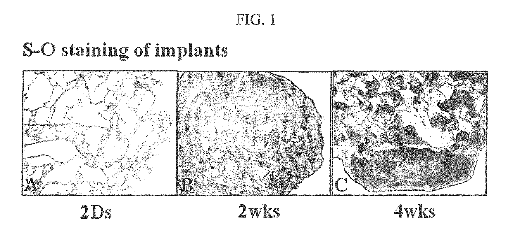
[0051]The cultured ECM scaffold containing chondrocytes, constructed in Example 2, was fixed in 4% formalin fixative solution for 24 hours. Then, the sample was embedded in paraffin, sectioned into a thickness of 4 μm, and stained with safranin-O. As a result, it was observed that accumulation of sulfated proteoglycan was increased gradually with time, which filled the pores of the scaffold (FIG. 1).

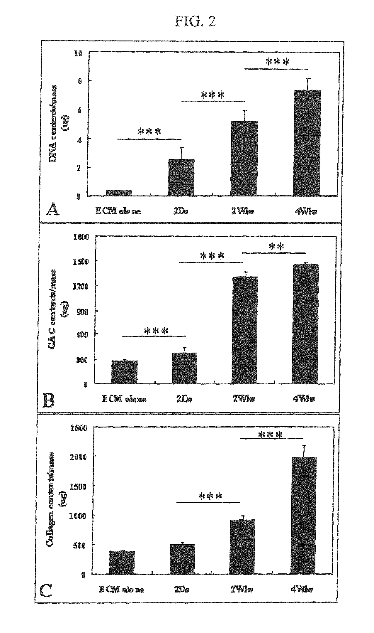
[0052]For chemical analysis, the transplant (the cultured ECM scaffold containing chondrocytes) was decomposed with a papain solution (125 μg / ml of papain, 5 mM of L-cystein, 100 mM of Na2HPO4, 5 mM of EDTA, pH 6.4) at 60° C. for 24 hours and centrifuged at 12,000 g for 10 minutes. For the measurement of total GAG (glycosaminoglycan) content, culture supernatant was analyzed using DMB assay (1,9-dimethylmethylene blue). Each sample was mixed with DMB solution to measure the absorbance at 225 nm. ...


PUM

PropertyMeasurementUnit
Temperatureaaaaaaaaaa
Timeaaaaaaaaaa
Timeaaaaaaaaaa

Abstract

The present invention relates to a method for preparing a cell-derived ECM scaffold to which chondrocytes or stem cells are attached, a method for cartilage regeneration by tissue engineering, which comprises using the cell-derived ECM scaffold, and a therapeutic composition for treating cartilage disorder, which contains the ECM scaffold as an effective component. More specifically, the present invention relates to a method for cartilage regeneration by tissue engineering, which comprises transplanting ECM scaffold, having chondrocytes or stem cells attached thereto, into cartilage defects, and a therapeutic composition for treating cartilage disorder, which contains the ECM scaffold, having chondrocytes or stem cells attached thereto, as an effective component. According to the present invention, when the inventive ECM scaffold having chondrocytes or stem cells attached thereto is transplanted into a cartilage defect, mature articular cartilage having the same appearance and characteristics as those of natural cartilage tissue, can be regenerated without side effects such as inflammatory responses.

Description

TECHNICAL FIELD[0001]The present invention relates to a method for preparing a cell-derived ECM scaffold to which chondrocytes or stem cells are attached, a method for cartilage regeneration by tissue engineering, which comprises the cell-derived ECM scaffold to which chondrocytes or stem cells are attached, and a therapeutic composition for treating cartilage disorder, which contains the ECM scaffold to which chondrocytes or stem cells are attached as an effective component.BACKGROUND ART[0002]The method of autologous chondrocyte implantation (ACI) used for treating damaged cartilage is a clinically approved therapeutic method that can regenerate hyaline cartilage tissues in the defect (Brittberg, M. et al., New Eng. J. Med., 331:889, 1994). However, along with the development of studies on chondrocytes or mesenchymal stem cells (MSCs), more advanced methods are currently being developed including cell transplantation using various scaffolds and production of artificial cartilage t...

Claims


Application Information

Patent Timeline
IPC IPC(8): A61F2/00C12N11/02C12N13/00A61K35/12
CPCA61L27/3633A61L27/3817A61L2430/06A61L27/3852A61L27/56A61L27/3834A61F2/30A61K35/28A61K35/32A61K38/30C12N5/0655
Inventor MIN, BYOUNG-HYUNPARK, SO RAJIN, CHENG ZHECHOI, BYUNG HYUNE
Owner MIN BYOUNG HYUN
Learn More