
Fluorescent neutralization and adherence inhibition assays

a technology of fluorescence neutralization and inhibition assays, applied in biochemistry apparatus and processes, instruments, peptide sources, etc., can solve the problem of increasing the efficiency of quenching surface-bound fluorescence, and achieve the effect of inexpensive assays

Active Publication Date: 2010-05-13
SANOFI PASTEUR VAX DESIGN
View PDF4 Cites 6 Cited by
  • Summary
  • Abstract
  • Description
  • Claims
  • Application Information

AI Technical Summary

Benefits of technology

[0020]The assay is inexpensive, using, for example, the rugged BioPlex bead array platform as a simplified flow cytometer at a cost ˜20% of a regular flow cytometer such as, for example, a BD LSR II (BD Biosciences).
[0022]In another embodiment, for example, a bead array reader, such as a BioPlex, is used as a simplified flow cytometer to detect fluorescence of the labeled virus engulfed by, or attached to, target cells. Using a bead array reader, such as a BioPlex, instead of a flow cytometer can reduce the cost of the assay by ˜5-fold and allows working with lower numbers of target cells in the sample.
[0023]Another embodiment of the present invention involves “addressed” affinity quenching of the phycoerythrin fluorescence using an anti-phycoerythrin antibody coupled with the QSY-9 quenching dye. This method increases the efficiency of quenching surface-bound fluorescence, which is undesirable in fluorescence-based microneutralization (fmNt) experiments.

Problems solved by technology

This method increases the efficiency of quenching surface-bound fluorescence, which is undesirable in fluorescence-based microneutralization (fmNt) experiments.

Method used


Image

  • Fluorescent neutralization and adherence inhibition assays
  • Fluorescent neutralization and adherence inhibition assays
  • Fluorescent neutralization and adherence inhibition assays

Examples


example 1

Influenza Virus as a Model for Developing the Neutralization Assay: Affinity Fluorescent Labeling

[0108]BPL-inactivated influenza virus standards of various strains are readily available, for example, from the US Centers for Disease Control and Prevention (CDC). Solomon Islands H1N1, New Caledonia H1N1, and Wisconsin H3N2 strains containing ˜109 viral particles / mL were used in most of the experiments, as examples.

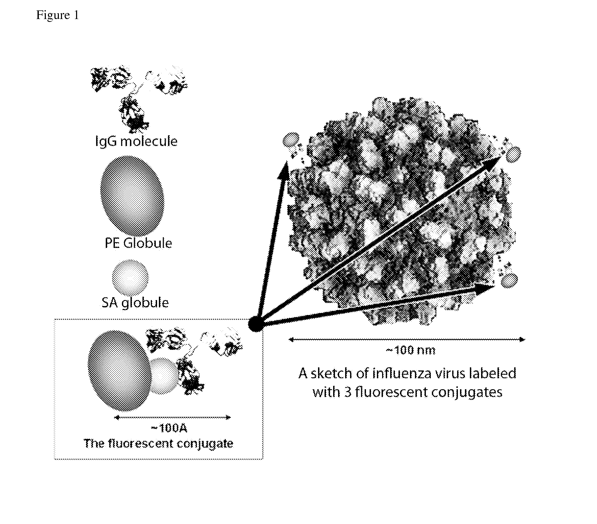
[0109]Experiments were conducted with various labels, including direct biotinylation, quantum dots and fluorescent nanoparticles. Affinity labeling with biotinylated influenza A-specific antibodies with subsequent attachment of the fluorescent streptavidin-phycoerythrin (SA-PE) conjugate provided the brightest labeling of the influenza A virus. Direct biotinylation of the virus also provided an acceptable level of signal.

[0110]It was previously found that the goat polyclonal anti-influenza A H1 specific antibody # 1301 (ViroStat) had a high affinity for H1N1 viruses, but low...

example 2

Assessing Replacement of the Affinity Label by Other Virus-Specific Antibodies

[0115]An important question needed to be addressed before using the described affinity labeling in the neutralization experiments. Testing the neutralizing capacity of a test sera or other fluids requires incubating the labeled virus with a significant excess of other virus-specific antibodies in the tested sample, many of which may be cross-reactive with the viral epitopes recognized by the labeling antibody. It was therefore important to check whether other virus-specific antibodies would displace the labeling antibodies on the virus surface.

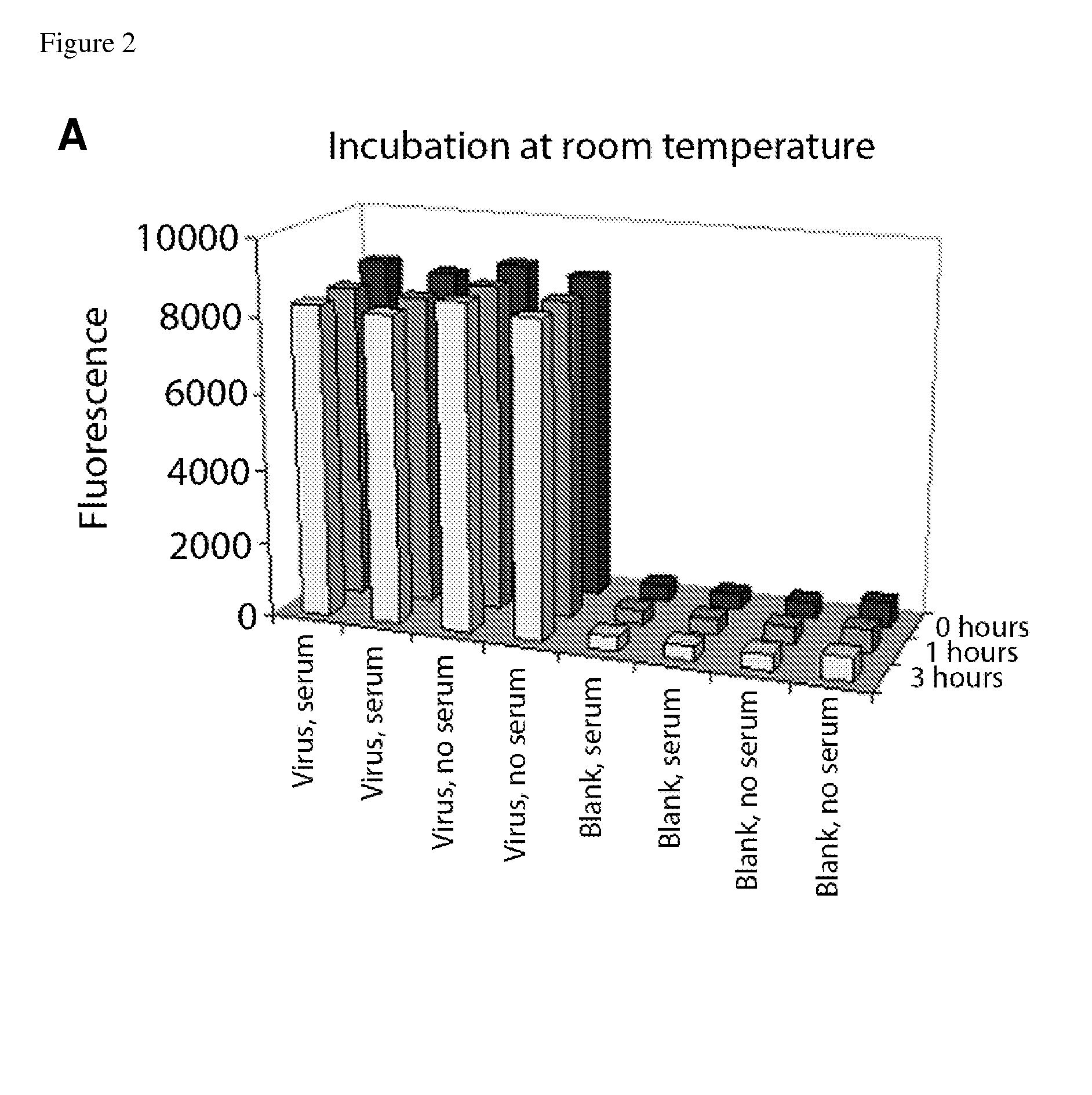
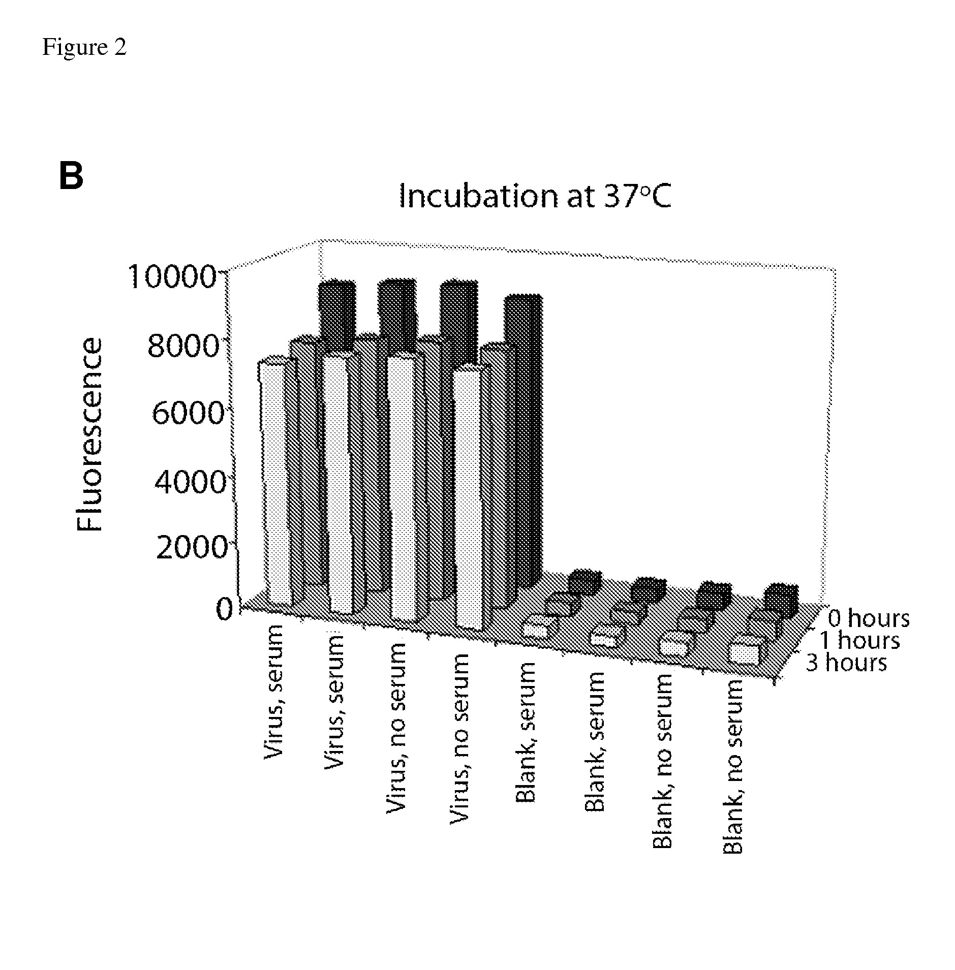
[0116]Label replacement tests were performed at the same concentrations, volumes, temperatures, and times of incubation as the prospective neutralization assays (FIG. 2). In the first test, four wells of the ELISA plates were coated with inactivated Solomon Islands virus (“Virus”), and the other four wells were left blank (“Blank”), as negative controls. After blocki...

example 3

Quenching of Surface-Bound Phycoerythrin Fluorescence

[0120]Monitoring fluorescently labeled virus engulfed by target cells inevitably requires subduing the fluorescence of surface-adherent virus. This can be done via extensive protease treatment of the cell surface, or (preferably) quenching of the surface-bound fluorescence using non- or low-fluorescence quenching agents. Trypan blue (TB) and crystal violet have been used successfully to quenching surface-bound fluorescein (Nichols et al. (1993) Arch. Virol. 130, 441; Collins & Buchholz (2005) J. Virol. Meth. 128, 192-197). It was found, however, that these non-specific quenchers were less effective in quenching the fluorescence of phycoerythrin (PE), likely because some of the phycobilin fluorescent clusters of PE are buried deep in the protein globule and inaccessible to the occasional contacts with quenching molecules.

[0121]To achieve more efficient quenching, an “addressed” affinity quencher was prepared, based on anti-PE antib...


PUM


Abstract

The present invention comprises rugged, inexpensive, reliable, and sensitive laboratory assays of antibody-based viral neutralization activity and antibody-based viral adherence inhibition activity. The assays use inactivated, fluorescently-labeled virus, allowing the tests to be performed without extensive safety precautions. The interaction of the labeled virus with target cells is monitored using flow cytometric methods. A preferred embodiment uses simple and inexpensive flow cytometry methodologies and equipment, such as bead array readers used as simplified flow cytometers. The assays are rapid, taking no longer than a few hours and are readily conducted by a trained technician. The assays are sensitive because they use labeled viruses at low concentrations and determine neutralizing and blocking capacity of sera and antibody at low concentrations. The methods are appropriate for high-throughput screening of large panels of samples.

Description

CROSS-REFERENCE TO RELATED APPLICATIONS[0001]This application claims benefit under 35 U.S.C. §119(e) of U.S. Patent Application No. 61 / 113,263, filed Nov. 11, 2008, which is hereby incorporated herein by reference in its entirety.BACKGROUND OF THE INVENTION[0002]The ability of an anti-viral pathogen vaccine to produce an effective antibody response is typically evaluated in various types of virus neutralization tests or assays. Common features of such tests include monitoring the level of infectivity of the virus (natural or attenuated) in a standardized target cell culture, and evaluating the reduction in infectivity of the virus after incubation with the tested serum / sera or antibody solution(s) of interest.[0003]The dilution of a serum or antibody (Ab) solution that provides 50% or more reduction of infectivity is referred to as the ‘neutralization titer’ (Niedrig et al. (2008) Clin. Vaccine Immunol. 15, 177; Guidelines for Plaque-Reduction Neutralization Testing of Human Antibod...

Claims


Application Information

Patent Timeline
IPC IPC(8): C12Q1/70
CPCC12Q1/18G01N33/6854G01N33/56983A61K49/085C07K14/005G01N33/502G01N2333/11G01N2500/02G01N2500/10
Inventor KACHURIN, ANATOLYKACHURINA, OLGAWITTMAN, VAUGHANTAPIA, TENEKUA
Owner SANOFI PASTEUR VAX DESIGN
Learn More