

Expression of foxp3 by cancer cells
a cancer cell and expression technology, applied in the field of cancer diagnosis and prophylaxis based on the expression of foxp3 in cancer cells, can solve the problems of unclear primary suppressive mechanisms and the negative impact of treg on health, and achieve the effect of treating or preventing ptld
- Summary
- Abstract
- Description
- Claims
- Application Information
AI Technical Summary
Benefits of technology
Problems solved by technology
Method used
Image

Examples
example 1
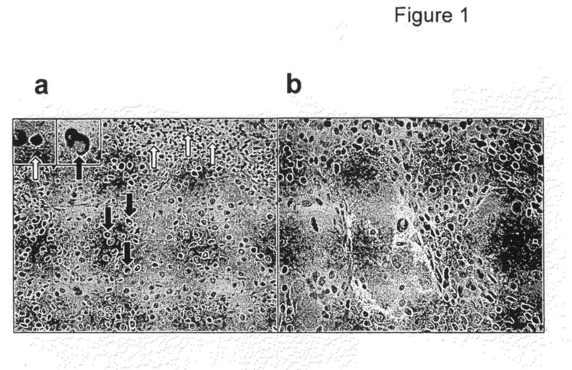
FoxP3 is Expressed by Both Lymphocytes and Tumor Cells in Metastatic Melanoma Tissue
[0233]With the aim of characterizing the frequency and localization of Treg in melanoma, we performed immunohistochemistry (IHC) on metastatic melanoma tissue sections using a polyclonal anti-FoxP3 antibody. Surprisingly, in addition to the expected staining of cells with lymphocyte morphology (small, round, dense nucleus and little cytoplasm), staining was also occasionally apparent in cells which had several morphological features of tumor cells, such as a large, irregular nucleus and abundant cytoplasm (FIG. 1a). Although unexpected, this staining appears to be specific, given the nuclear localization of the signal and the lack of staining with control rabbit IgG (not shown). Furthermore, this is not an isolated phenomenon, as FoxP3+ tumor cells were detectable in four additional biopsy specimens from different patients and various sites of metastasis.
[0234]This preliminary analysis therefore sugg...
example 2
FoxP3 is Widely Expressed by Melanoma Cell Lines
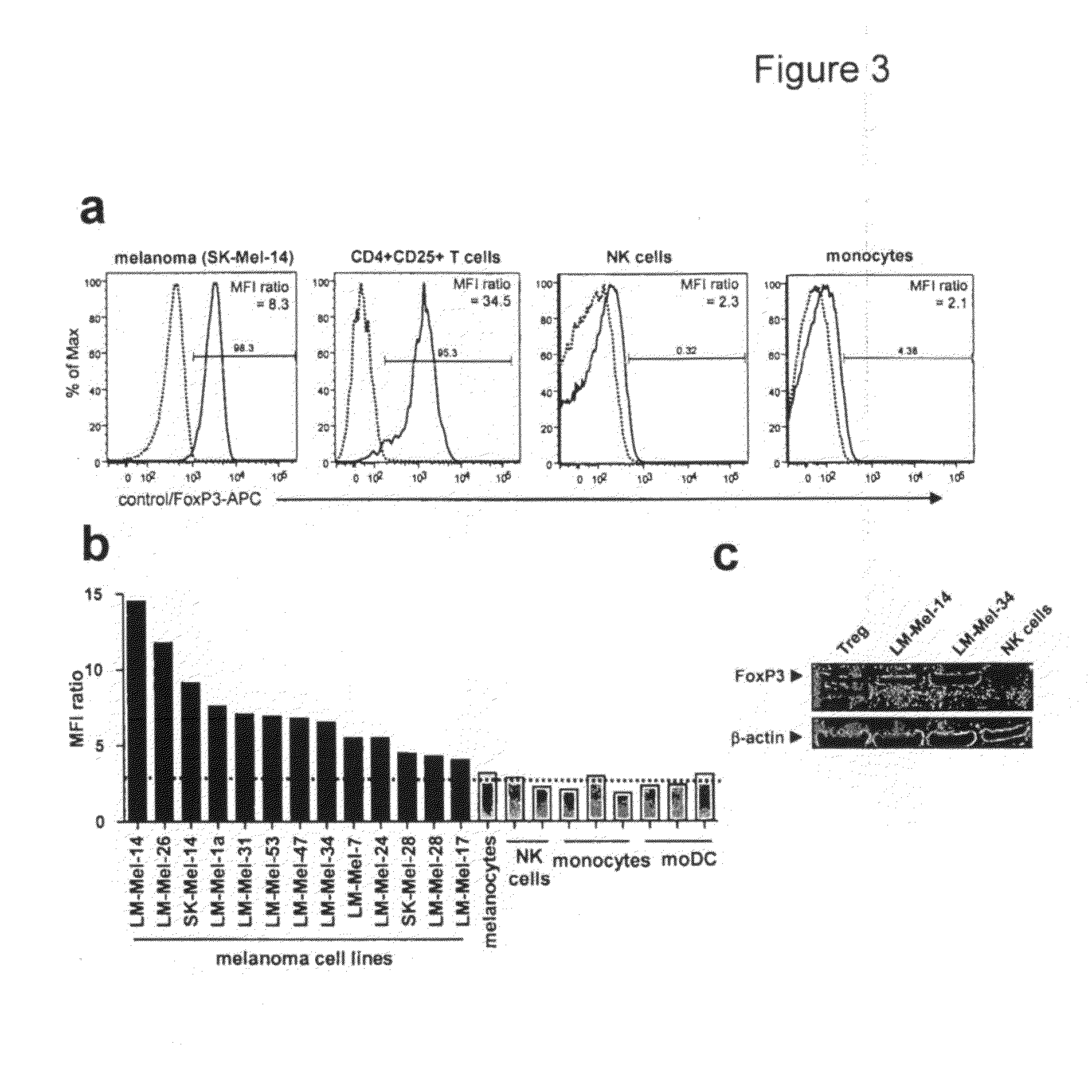
[0239]The results presented thus far demonstrate, using a variety of methods, that cells with the morphology and surface marker phenotype of melanoma can express FoxP3. However, the identification of melanoma cells within a mixed population using these criteria is not unequivocal. Further confirmation of FoxP3 expression in melanoma cells was obtained by analyzing a panel of established melanoma cell lines by flow cytometry (FIG. 3) after staining with anti-FoxP3 mAb. The melanoma cell line shown in FIG. 3a (SK-Mel-14) was uniformly positive for FoxP3, although the intensity of staining was not quite as high as for CD4+ CD25+ T cells. Staining of freshly isolated blood NK cells and monocytes was negative, as expected.
[0240]Staining for FoxP3 on additional melanoma cell lines is summarized in FIG. 3b. For all cell lines analyzed, staining with anti-FoxP3 resulted in a shift in the fluorescence of the entire population compared to the is...
example 3
The FOXP3 Gene is Expressed in Melanoma Cells as Three Distinct mRNA Variants, One of which is Predicted to Encode a Novel Protein
[0241]To confirm FOXP3 gene expression in melanoma cells, RNA was extracted from a panel of melanoma cell lines and used to generate cDNA by reverse transcription, which was then subject to PCR using primers specific for FOXP3 or the housekeeping gene CYP-A (cyclophilin-A). PCR products could be readily detected following amplification using FOXP3-specific primers (35 cycles), confirming that melanoma cell lines express FOXP3 mRNA (FIG. 4a).
[0242]Previous reports have demonstrated that, in Treg, FOXP3 mRNA is expressed as two variants: full-length and an alternatively spliced version lacking exon 2 (Δ2) (11, 15, 23). Using our primers, these two products would be expected to result in band sizes of 608 and 503 bp, respectively, and for CD4+ T cells these two bands were observed as expected (FIG. 4a). For melanoma cells, however, an additional band (˜100 b...
PUM
| Property | Measurement | Unit |
|---|---|---|
| Fraction | aaaaa | aaaaa |
| Composition | aaaaa | aaaaa |
Abstract
Description
Claims
Application Information