
Temporal release of growth factors from 3D micro rod scaffolds for tissue regeneration

a technology of growth factors and scaffolds, applied in the direction of prosthesis, peptide/protein ingredients, drug compositions, etc., can solve the problems of poor force production, failure of random seeding of cardiac cells into matrices or hydrogels, and insufficient function of artificial tissue, so as to promote cell survival and promote cell survival. the effect of differentiation

Inactive Publication Date: 2010-06-24
THE BOARD OF TRUSTEES OF THE UNIV OF ILLINOIS
View PDF20 Cites 7 Cited by
  • Summary
  • Abstract
  • Description
  • Claims
  • Application Information

AI Technical Summary

Benefits of technology

[0024]Methods are also contemplated by the present invention. In one embodiment, a method for repairing damaged tissue comprising the step of administering the microrod scaffold to damaged tissue in an amount and over a time effective to repair damaged tissue is provided.

Problems solved by technology

A major challenge in engineering tissues is mimicking the complex cellular organization and function of the native tissues of the human body.
Without proper cellular organization, an artificial tissue does not function adequately.
Seeding of cardiac cells randomly into matrices or hydrogels fails because the cells are isolated from each other promoting cellular atrophy and apoptosis.
The surviving cells are few in number and not electrically connected resulting in poor force production (Langer and Vacanti, 1993).
Rat neonatal cells from primary culture have been grown on surfaces adhering to peptides stamped in parallel lines but are not all electrically connected laterally (Reinecke, 1999) and have all the disadvantages of two-dimensional culture.
Repeat surgeries at daily intervals would not be acceptable for human application.
However, it is not easy to see how this approach could be scaled up for surgery.
The recent exterior “chain-mail jacket” approach (Zimmerman, 2006) is fraught with practical difficulties for use in human patients.
The cell source also remains an unresolved challenge.
There are inadequate local repair mechanisms within the heart to deal with physical and free radical damage following ischemic injury or mechanical overload.
This problem is exacerbated by a wave of programmed death (apoptosis) of susceptible myocytes resulting in contractile dysfunction (Cheng, 1996; Kajstura, 1996).
However, data from animal studies have been equivocal, and data from human clinical trials have not demonstrated any significant effects of either administration of the mature IGF-1 or growth hormone (Deurr, 1996; Fazio, 1996).
This result may not be too surprising since there is no evidence showing increased secretion of growth hormone or elevation of systemic IGF-I levels following myocardial infarcts.
Furthermore, there are limitations to this approach due to the bioavailability in the heart because IGF-1 binds very rapidly to proteins in the systemic circulation.

Method used


Image

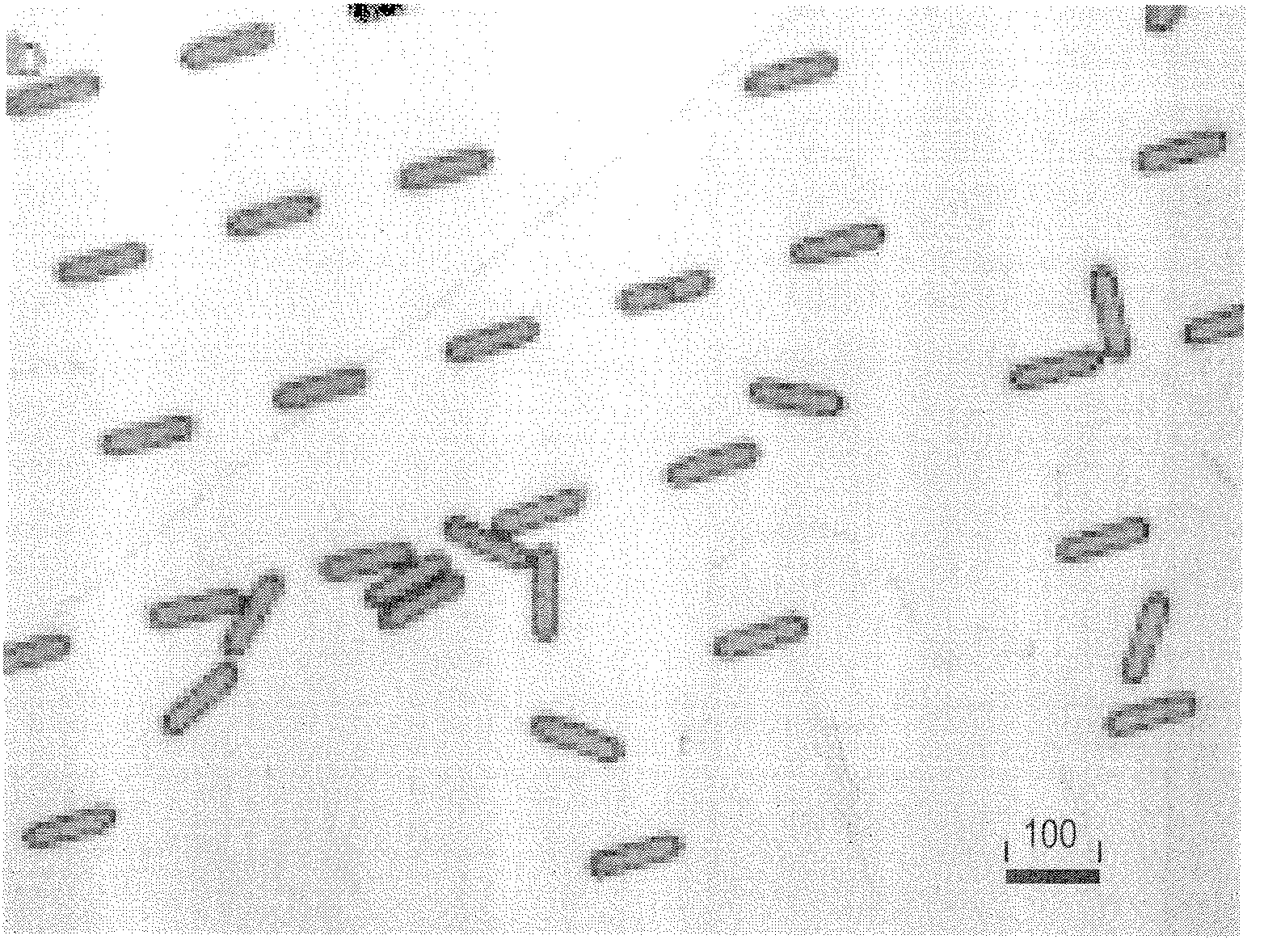
  • Temporal release of growth factors from 3D micro rod scaffolds for tissue regeneration
  • Temporal release of growth factors from 3D micro rod scaffolds for tissue regeneration
  • Temporal release of growth factors from 3D micro rod scaffolds for tissue regeneration

Examples


example 1

Effect of Biodegradable Microrod Scaffolds on Myocyte Organization and Function

[0085]Microstructure Size

[0086]Microstructures with the volume (cross-sectional area 15×15 μm2, length 100 μm) but varying in stiffness (tensile moduli 0.01 kPa to 1 GPa) are used and fabricated as described below. All experiments are repeated with 5 primary cultures for statistical analysis.

[0087]Microstructure Stiffness

[0088]PLGA / PCL blends are initially chosen because this material can easily be microfabricated, is biocompatible, has a tunable modulus, and can degrade and release drug over a suitable time period (−2 weeks). Specifically, the effects of MRS made from polymer blends of varying stiffness are compared. The stiffness of the polymer MRS can be modulated in the proposed range by varying the PLGA co-polymer ratio and incorporation of a less stiff PCL component. This systematic tuning in mechanical properties was recently demonstrated by Sung (2005). The following blends are initially used: 50 / ...

example 2

MRS Stiffness and NRVM Retention of the Organization and Cell Connectivity as the 3D-Architecture Degrades

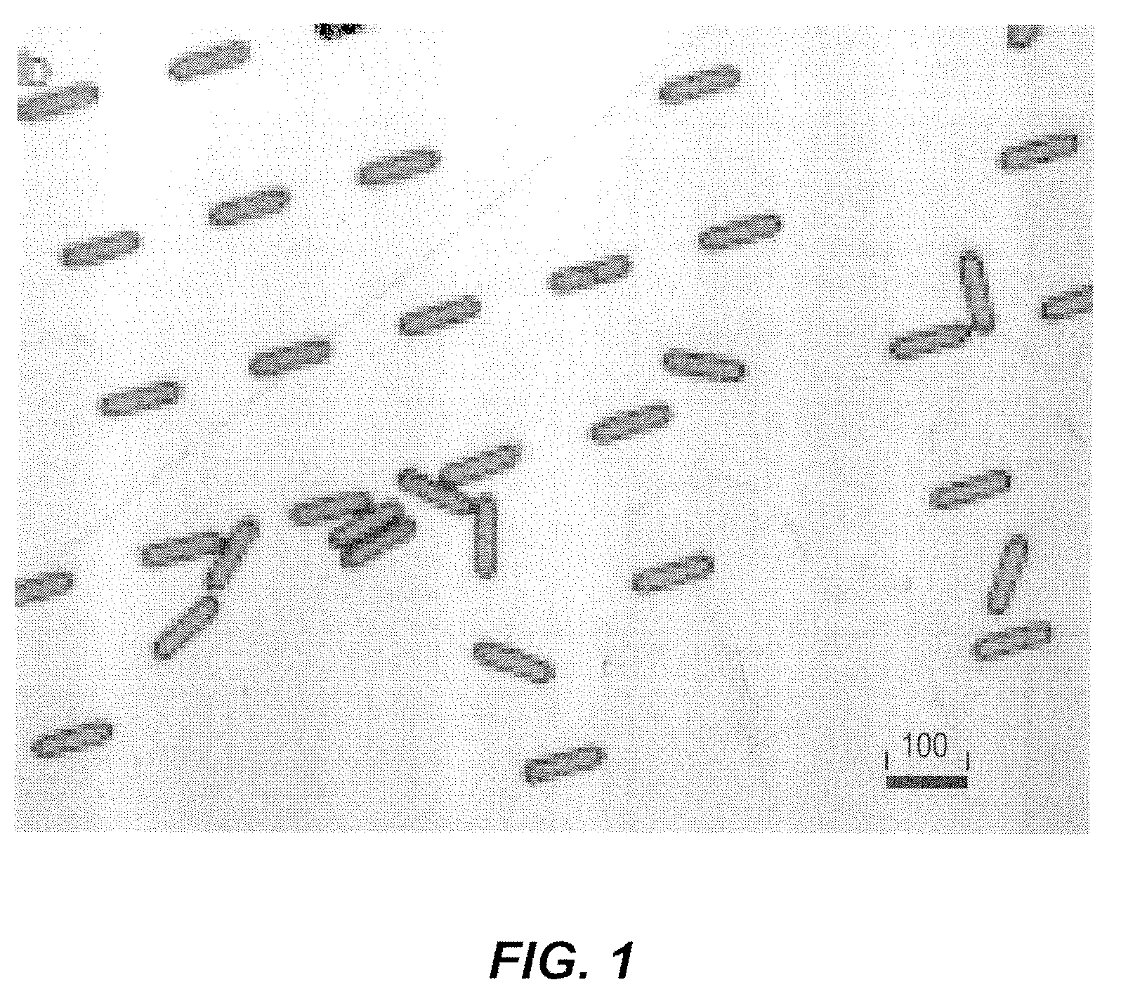
[0111]Microengineered systems can be made to degrade over an intended time period. Given the appropriate biomaterial one can observe long-term maintenance of cellular organization with and without structural cues. The ability to microfabricate structures out of biodegradable materials (a PLGA co-polymer blend) (FIGS. 1 & 2; Snyder and Desai, 2001) and have achieved short-term cell organization has been demonstrated.

[0112]Control of Degradation by Selection Materials

[0113]MRS are manufactured from biodegradable materials instead of traditional microfabrication polymers such as PDMS or SU-8 to assess survival of tissue architecture after the topography disappears. Factors such as molecular weight, exposed surface area and crystallinity affect degradation rates (Lanza, 2000). Also, processing conditions have can alter degradation characteristics of a material. For instance, heat mo...

example 3

Effects of Microscaffolds Contractile Maturation and Proliferation on Mouse Embryonic Stem Cell Derived Cardiomyocytes (ESCM)

[0121]ESCM

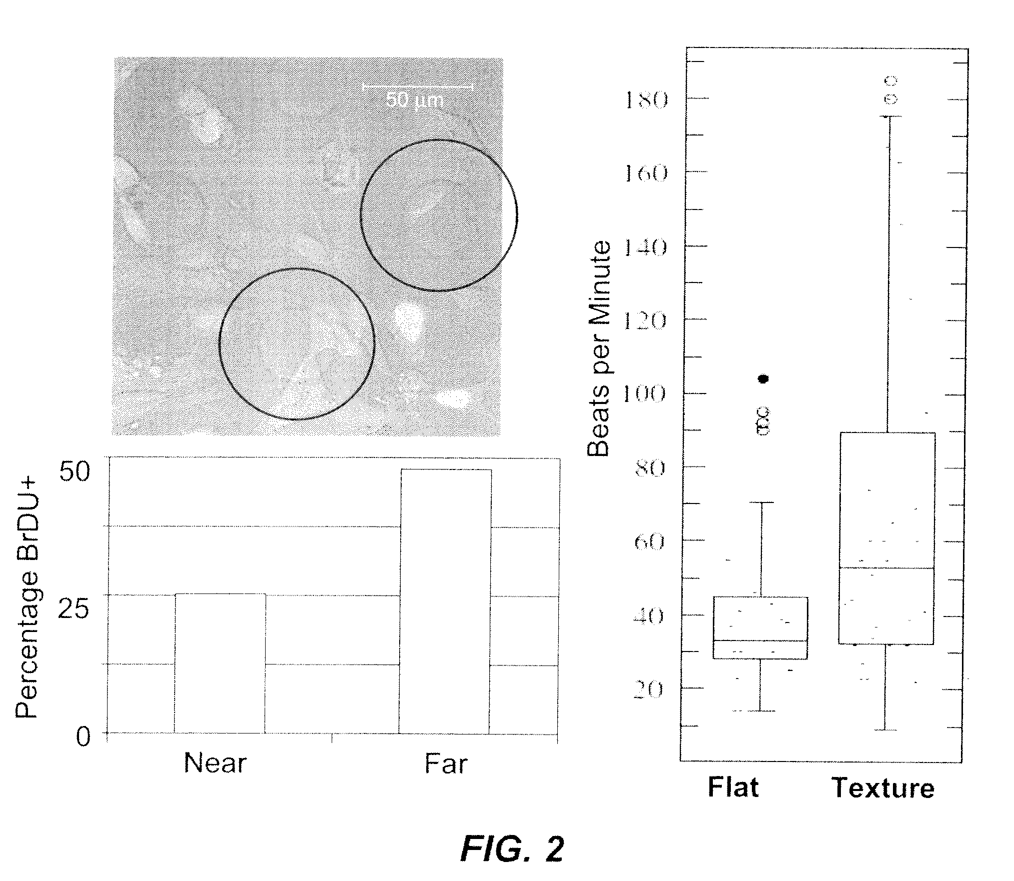
[0122]Although cell-cell or cell-ECM interactions are important, how changes in the physical niches affect the differentiation capacity of these cells and their potential to form viable, robust cardiomyocytes remain unknown. The niche in which stem cells reside and differentiate is a complex physicochemical microenvironment that regulates cell function. The role played by three-dimensional physical contours was studied on cell progeny derived from mouse embryonic stem cells using microtopographies created on PDMS (poly-dimethylsiloxane) membranes. While markers of differentiation were not affected, the proliferation of heterogeneous mouse embryonic stem cell-derived progeny was attenuated by 15 m-, but not 5 m-high microprojections. This reduction was reversed by Rho kinase and myosin light chain kinase inhibition, which diminishes the tension genera...


PUM

PropertyMeasurementUnit
lengthaaaaaaaaaa
diameteraaaaaaaaaa
sizeaaaaaaaaaa

Abstract

The present invention relates to the use of three-dimensional microrod scaffolds for the temporal release of growth factors useful in tissue regeneration, engineering and treatment of disorders.

Description

[0001]This application is a continuation in part application of PCT application number PCT / US2008 / 070471, which was filed Jul. 18, 2008, claiming the benefit of priority of U.S. Provisional Application No. 60 / 950,454, which was filed Jul. 18, 2007. The present application also claims the benefit of priority of U.S. Provisional Application No. 61 / 179,584, which was filed May 19, 2009. The entire text of each of these priority documents is specifically incorporated herein by reference.FIELD OF THE INVENTION[0002]The present invention relates to the use of three-dimensional microrod scaffolds for the temporal release of growth factors useful in tissue regeneration, engineering, and treatment of disorders.BACKGROUND OF THE INVENTION[0003]A major challenge in engineering tissues is mimicking the complex cellular organization and function of the native tissues of the human body. Tissue structure and function are very highly interrelated so that cellular and macromolecular organization of ...

Claims


Application Information

Patent Timeline
Patent Type & Authority Applications(United States)
IPC IPC(8): A61K9/00A61K38/18A61P9/10
CPCA61K9/0019A61P9/10
Inventor RUSSELL, BRENDADESAI, TEJAL A.GOLDSPINK, PAUL
Owner THE BOARD OF TRUSTEES OF THE UNIV OF ILLINOIS
Learn More