
Application of synovium-derived mesenchymal stem cells (MSCS) for cartilage or meniscus regeneration

a technology of mesenchymal stem cells and synovium, which is applied in the direction of skeletal/connective tissue cells, drug compositions, biocide, etc., can solve the problems of unpredictable therapeutic effect damage to healthy cartilage tissue, etc., and achieves greater ex vivo expansion and chondrogenic ability

Inactive Publication Date: 2010-07-15
NAT UNIV CORP TOKYO MEDICAL & DENTAL UNIV
View PDF2 Cites 12 Cited by
  • Summary
  • Abstract
  • Description
  • Claims
  • Application Information

AI Technical Summary

Benefits of technology

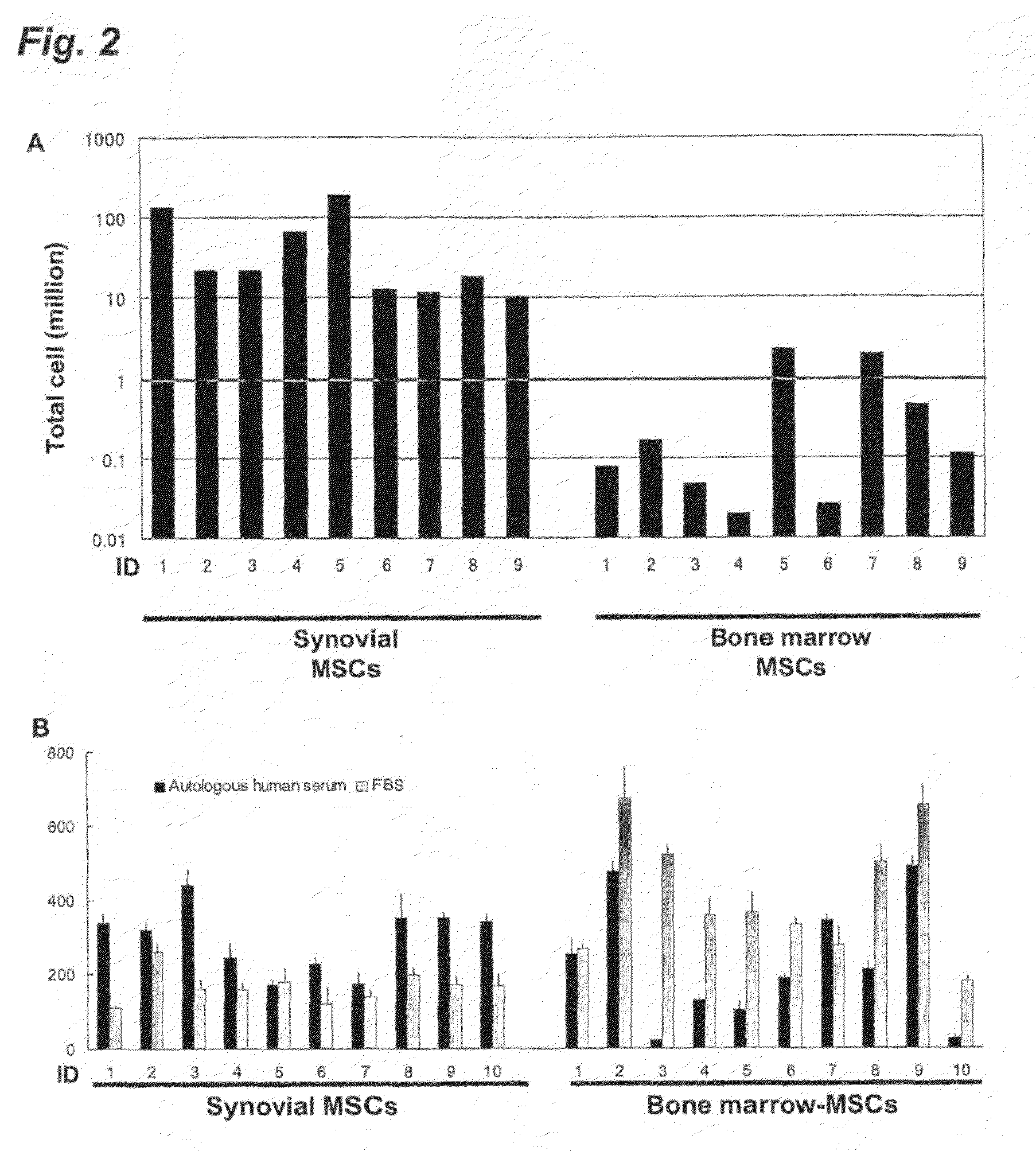
[0014]The inventors previously compared human MSCs derived from a variety of mesenchymal tissues including bone marrow and determined that synovium-derived MSCs had greater ex vivo expansion and chondrogenic ability than MSCs from other tissues (Sakaguchi et al., 2005, Arthritis Rheum. 52:2521-9). This indicates that synovium-derived MSCs may be an optimal cell source for cartilage regeneration.

Problems solved by technology

This procedure is advantageous in that it is simple and can be performed arthroscopically, but is disadvantageous in that defects are repaired by fibrous cartilage rather than hyaline cartilage, which makes a therapeutic effect unpredictable.
However, it is still disadvantageous in that it causes damages to the healthy cartilage tissue.
However, ACI is considered to be disadvantageous in that it causes damage to healthy cartilage tissue.
Furthermore, since removed cartilage cells (chondrocytes) have to be cultured in vitro, and primary chondrocytes do not proliferate well with human serum, only by around 10 fold, it is necessary to use materials derived from an animal other than human (such as fetal bovine serum) or to use artificial materials (such as collagen gel derived from bovine skin); otherwise, it is possible to treat only a relatively small defect, though the surgical procedure is both a little bit invasive and complex.
Despite a diverse and growing body of information regarding MSCs and their use in cell-based strategies, the mechanisms that govern MSC self-renewal and multilineage differentiation are not well understood and remain an active area of investigation.
10:199-206), a number of questions such as whether the donor cells differentiated into chondrocytes or how donor cells contributed to chondrogenesis still exist, limiting clinical applications for cartilage injury.

Method used


Image

  • Application of synovium-derived mesenchymal stem cells (MSCS) for cartilage or meniscus regeneration
  • Application of synovium-derived mesenchymal stem cells (MSCS) for cartilage or meniscus regeneration
  • Application of synovium-derived mesenchymal stem cells (MSCS) for cartilage or meniscus regeneration

Examples


example 1

Isolation of Rabbit Synovium-Derived MSCs

[0068]This example is provided to describe a method for obtaining synovium-derived MSCs from rabbits.

[0069]Skeletally mature Japanese White Rabbits weighing about 3.2 kg (ranging 2.8-3.6 kg) were used in the experiments. Animal care was strictly in accordance with the guidelines of the animal committee of Tokyo Medical and Dental University. Synovium was harvested under anesthesia induced by intramuscular injection of 25 mg / kg of ketamine hydrochloride and intravenous injection of 45 mg / kg of sodium pentobarbital.

[0070]Harvested rabbit synovium was digested in a 3 mg / ml collagenase D solution (Roche Diagnostics, Mannheim, Germany) in αMEM (Invitrogen, Carlsbad, Calif., USA) at 37° C. After 3 hours digestion, digested cells were filtered through a 70-μm nylon filter (Becton Dickinson, Franklin Lakes, N.J., USA) and the remaining tissues were discarded.

[0071]The obtained cells were plated at 5×10⁴ cells / cm2 in 60-cm2 culture dishes (Nalge Nunc ...

example 2

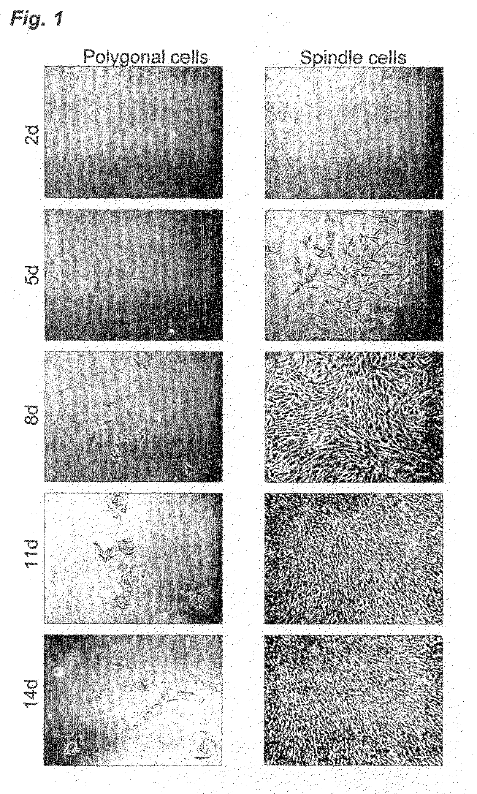
Isolation and Characterization of Human Synovium-Derived MSCs with Autologous Human Serum

[0073]In this example, the inventors isolated human synovium-derived MSCs and bone marrow-derived MSCs and identified the characterization thereof.

(i) Isolation of Human MSCs and Proliferative Effect Thereof

[0074]The study was approved by the local institutional review board, and all human study subjects provided informed consent. Human synovium and bone marrow were harvested during the operation of anterior cruciate ligament (ACL) reconstruction of the knee from 8 donors (27±5 years old).

[0075]Bone marrow from the tibia was obtained with an 18-gauge needle just before drilling for the insertion of reconstructed ligaments. Synovium with subsynovial tissue from the inner side of the medial joint capsule, which overlies the noncartilaginous areas of the medial condyles of the femur, was harvested with a pituitary rongeur, under arthroscopic observation. One day before the operation of ACL reconstr...

example 3

Chondrogenic Ability of Synovium-Derived MSCs

[0088]This example is provided to identify the chondrogenic ability of rabbit synovium-derived MSCs ex vivo.

[0089]For ex vivo chondrogenesis, 250,000 cells were placed in a 15-ml polypropylene tube (Becton Dickinson, Franklin Lakes, N.J., USA) and were centrifuged at 450 g for 10 min. The pellet was cultured in a chondrogenesis medium consisting of high-glucose Dulbecco's modified Eagle's medium (DMEM high glucose; Invitrogen Corp, Carlsbad, Calif., USA) supplemented with 500 ng / ml BMP-2 (bone morphogenetic protein-2; Yamanouchi Pharmaceutical, Tokyo, Japan), 10 ng / ml TGF-β3 (transforming growth factor-β3; R&D Systems. Minneapolis, Minn., USA), 10−7M dexamethasone (Sigma-Aldrich Corp. St. Louis, Mo., USA), 50 μg / ml ascorbate-2-phosphate, 40 μg / ml proline, 100 μg / ml pyruvate, and 1:100 diluted ITS+Premix (BD Biosciences. Bedford, Mass., USA; 6.25 μg / ml insulin, 6.25 μg / ml transferrin, 6.25 ng / ml selenious acid, 1.25 mg / ml BSA, and 5.35 mg / ...


PUM


Abstract

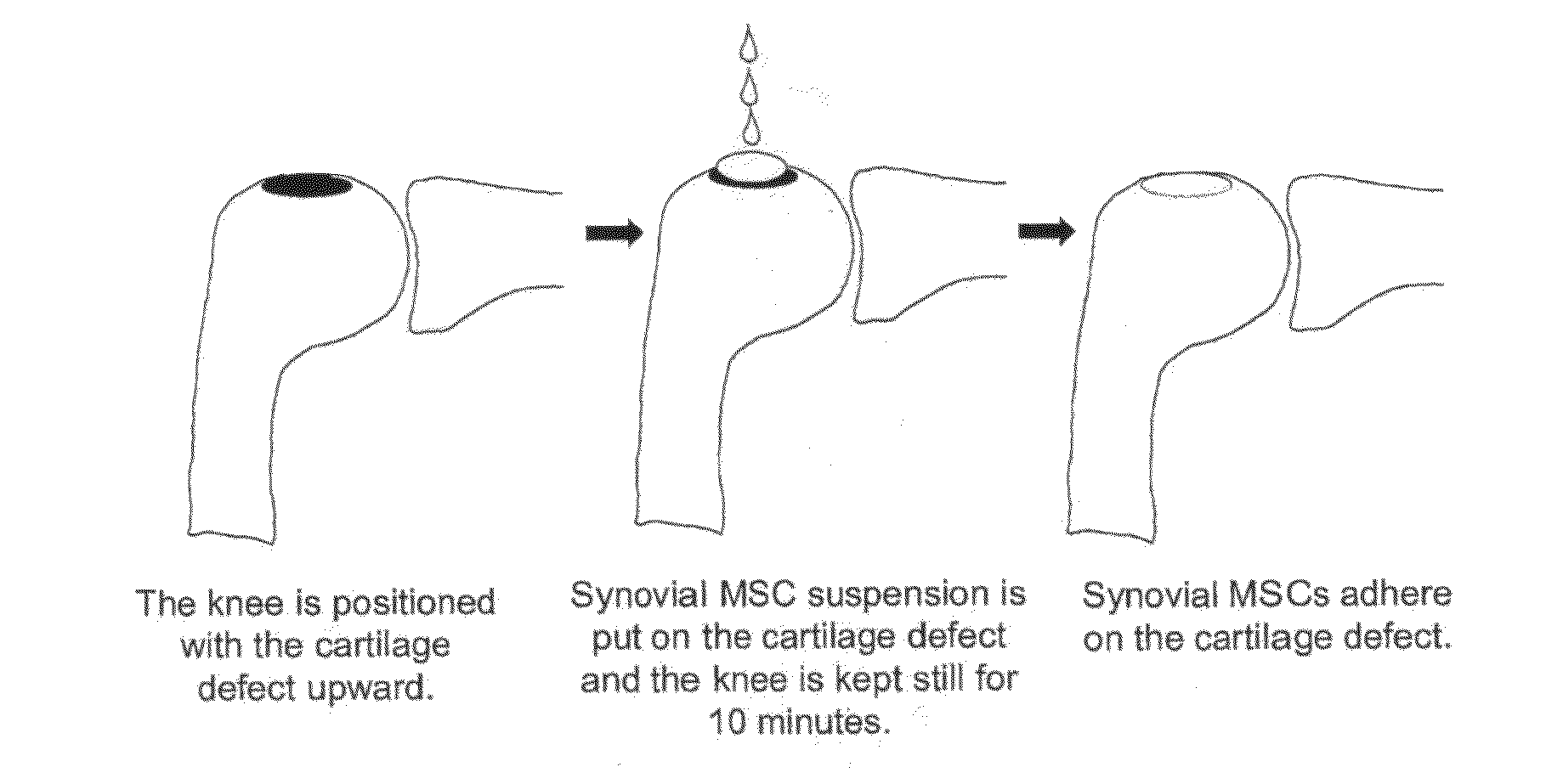
An object of the present invention is to provide a method for treating defects of articular cartilage or meniscus of a patient using in vivo chondrogenesis of synovium-derived MSCs. The present invention provides a method for treating a disease associated with defects of cartilage or meniscus. In the present invention, the method for treating a disease associated with defects of cartilage or meniscus comprises the following steps: culturing ex vivo autologous synovium-derived mesenchymal stem cells (MSCs); implanting the MSCs such that said cartilage defect site or meniscal defect site is covered by the MSCs; and regenerating cartilage tissue at the cartilage defect site or meniscal defect site in situ by differentiating the MSCs into cartilage cells.

Description

BACKGROUND ART[0001]This invention relates to a method for treating defects of the articular cartilage or meniscus of a patient using in vivo chondrogenesis of synovium-derived MSCs.[0002]Articular cartilage defects and meniscal defects are associated with articular pain, decrease in range of motion, hydrarthrosis, mobility impairment and so on. A patient suffering from articular cartilage defects or meniscal defects caused by trauma is usually treated by an orthopaedic surgeon. Surgical repair of cartilage defects or meniscal defects aims to eliminate any loose debris that could cause further damage to the joint, and to restore function to the affected joint.[0003]Some procedures are often recommended by orthopaedic surgeons for articular cartilage injury, depending on the severity of damage. Examples of procedures employed by orthopaedic surgeons include marrow stimulation, mosaicplasty (also referred to as osteochondral autograft, or bone / cartilage plug implantation), autologous ...

Claims


Application Information

Patent Timeline
Patent Type & Authority Applications(United States)
IPC IPC(8): A61K35/12A61P19/00
CPCA61K35/12C12N5/0668C12N2533/54C12N5/0655A61P19/00A61P19/02
Inventor SEKIYA, ICHIROMUNETA, TAKESHIMORIO, TOMOHIROSHIMIZU, NORIOKUROIWA, YASUYUKI
Owner NAT UNIV CORP TOKYO MEDICAL & DENTAL UNIV
Learn More